INTRODUCTION
Gastric cancer is one of the leading causes of death in the world. Metastasis to the regional lymph node is an indicator of tumor progression as well as an important prognostic fac- tor in gastric cancer. Recent evidence suggests that tumor lymphangiogenesis promotes lymphatic metastasis (1-3). How- ever, little is known about the mechanism of lymphangio- genesis in gastric carcinoma.
Vascular endothelial growth factor (VEGF)-C and VEGF- D are the best-characterized lymphangiogenic growth factors.
These growth factors stimulate lymphangiogenesis by acti- vating VEGF receptor (VEGFR)-3, also known as Flt (fms- like tyrosine kinase)-4, a receptor which is expressed in the lymphatic endothelium (4-7). VEGFR-3 was once thought to be a marker of lymphatic endothelial cells because it is mainly expressed in the lymphatic endothelium of adult tis- sue (8); however, VEGFR-3 has also been detected in blood vessels within tumors and wounds that are healing (9, 10).
Several studies have correlated VEGF-D expression with lymph node metastasis in a variety of cancers including col- orectal (11), breast (12), pancreatic (13), ovarian (14), and endometrial (15). Furthermore, a high VEGFR-3-positive
vessel density has been correlated with poor prognosis in breast cancer (16) and non-small cell lung cancer (17).
The role of VEGF-D and VEGFR-3 in gastric carcinoma has not been fully determined. Recently, Jutter et al. report- ed that VEGF-D and VEGFR-3 are novel independent prog- nostic marker molecules for reduced survival after the cura- tive resection of gastric adenocarcinoma (18). The goal of our study was to investigate the clinical value of VEGF-D expres- sion and VEGFR-3-positive vessel density in gastric carcino- ma with regard to lymphangiogenesis.
MATERIALS AND METHODS Study population and tissue samples
This study comprised 104 patients who underwent surgi- cal resection for gastric adenocarcinoma at Hanyang Univer- sity Guri Hospital between April 2000 and November 2003.
Of those, 84 patients had advanced gastric cancers and 20 patients had early gastric cancers. Well-documented clinical data were collected from all patients. Information concerning the date of initial diagnosis, clinical characteristics, relapse,
592
Jung-Hye Choi, Young-Ha Oh*, Yong-Wook Park*, Hong-Kyu Baik�, Young-Yiul Lee, and In-Soon Kim
Departments of Internal Medicine, Pathology*, and Surgery�, College of Medicine, Hanyang University, Seoul, Korea
Address for correspondence Jung-Hye Choi, M.D.
Department of Internal Medicine, Hanyang University Guri Hospital, 249-1 Gyomoon-dong, Guri 471-701, Korea
Tel : +82.31-560-2236, Fax : +82.31-553-7369 E-mail : [email protected]
*This work was supported by the research fund of Hanyang University (HY-2006-C).
DOI: 10.3346/jkms.2008.23.4.592
Correlation of Vascular Endothelial Growth Factor-D Expression and VEGFR-3-Positive Vessel Density with Lymph Node Metastasis in Gastric Carcinoma
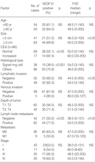
Lymph node metastasis is an important prognostic factor in gastric cancer. Vascu- lar endothelial growth factor-D (VEGF-D) is a lymphangiogenic growth factor that activates VEGF receptor (VEGFR)-3, a receptor expressed in the lymphatic endothe- lium. We investigated the clinical value of VEGF-D expression and VEGFR-3 posi- tive vessel density in gastric carcinoma with regard to lymphangiogenesis. Immuno- histochemical staining was used to determine the expression of VEGF-D and VEGFR- 3 in specimens from 104 cases of resected gastric cancer. VEGF-D expression was observed in 62.5% of the gastric cancers and in 9.6% of the non-neoplastic gastric tissue. The VEGFR-3-positive vessel density was significantly greater in the VEGF- D positive group than the negative group. VEGF-D expression was significantly associated with lymph node metastasis, increased serum CEA levels, and the non- signet ring cell type. The VEGFR-3-positive vessel density was correlated with tumor size, lymphatic invasion, and lymph node metastasis. The VEGF-D expression and high VEGFR-3-positive vessel density were significant poor prognostic factors for relapse-free survival. These results suggest that VEGF-D and VEGFR-3-positive vessel density are potential molecular markers that predict lymphatic involvement in gastric carcinoma.
Key Words : Vascular Endothelial Growth Factor D; Vascular Endothelial Growth Factor Receptor-3; Stom- ach Neoplasms
Received : 24 April 2007 Accepted : 18 December 2007
. .
and death were retrospectively obtained. In addition, adjacent non-neoplastic stomach tissue samples as confirmed by Hema- toxylin and Eosin staining were used as controls. This study was approved by the institutional review board of Hanyang University Guri Hospital.
Immunohistochemistry
The avidin-biotin complex (ABC) method was used for immunostaining. Formalin-fixed, paraffin-embedded tissue blocks were sectioned at a 4- m thickness. The tissue sections were deparaffinized by three, 10-min incubations in xylene and then rehydrated in serial graded alcohol. For antigen re- trieval, the sections were heated in a microwave oven for 10 min in 10 mM/L sodium citrate buffer (pH 6). Endogenous peroxidase activity was eliminated by preincubation in 3%
hydrogen peroxide and 10% methanol for 15 min followed by three washes in phosphate-buffered saline. All slides were pre-incubated at 37℃for 20 min with two drops of normal blocking solution (goat serum, 100 L/slide). The slides were then incubated with either a goat polyclonal anti-VEGF-D antibody (R&D Systems, Minneapolis, MN) at a 1:100 dilu- tion overnight at 4℃or a rabbit polyclonal anti-VEGFR-3 antibody (Zymed Laboratories, San Francisco, CA, U.S.A.) at a 1:200 dilution for 2 hr at room temperature. Biotinylat- ed secondary antibody was added to each slide and incubat- ed for 30 min at 37℃. The slides were then treated with the avidin-biotinylated peroxidase complex (Immunotech, Cedex, France) for an additional 30 min at room temperature. 3, 3’- diaminobenzidine tetrahydrochloride (Immunotech, Cedex, France) was used for color development. Finally, the sections
were counterstained with hematoxylin.
Evaluation of staining
All slides were coded and evaluated by two experienced pathologists without knowledge of patient identity or clini- cal status. Each experiment was performed twice independent- ly. In the discrepant cases, two pathologists reviewed the cases together and reached a consensus. The VEGF-D staining intensity was scored as 0 (negative), 1 (weak), 2 (medium), and 3 (strong). The extent of staining was scored according to the percentage of areas with positive VEGF-D staining as follows: 0 (0%), 1 (1 to 25%), 2 (26 to 50%), 3 (51 to 75%), and 4 (76 to 100%). The final staining score was derived from the sum of the intensity score and the extent score. Tumors with a final staining score of ≥5 were considered as positive for VEGF-D expression.
The VEGFR-3-positive vessel density was assessed accord- ing to the method described by Weidner et al. (19). Microves- sel counting was performed twice. Each slide was first scanned at 100× magnification to determine three “hot spots” defined as areas with the maximum number of VEGFR-3-positive vessels. The VEGFR-3-positive vessel density was determined by counting all the immunostained vessels at a 200× mag- nification and the mean number of positive vessels was cal- culated in the three selected areas for each case.
Statistical analysis
The Pearson chi-square ( 2) test was performed to deter- mine the correlation between VEGF-D expression and vari-
Fig. 1. Immunohistochemical staining of a gastric carcinoma. Each photograph shows representative tissue that is positive for VEGF-D (A) and VEGFR-3 (B) (×400).
A B
ous clinicopathological factors. The Mann-Whitney U test and Kruskal-Wallis test were used to examine the associa- tion of VEGFR-3-positive vessel density. Survival curves were calculated using the Kaplan-Meier method and compared with other prognostic variables using the log-rank test. A stepwise Cox’s regression analysis was performed to identify prognostic factors for survival. In all tests, p<0.05 was considered sta- tistically significant. All statistical analyses were performed using SPSS 10�statistical software.
RESULTS Patient characteristics
Our study included 71 male (68.3%) and 33 female (31.7
%) patients, with a median age of 59 yr (range, 25-79 yr). All
tumors were staged using the AJCC classification. Forty-three patients (41.4%) were classified as stage I, 11 patients (10.6%) as stage II, 20 patients (19.2%) as stage III, and 30 patients (28.8%) as stage IV. R0 resection was done in 95 patients, R1 resection in 4, and R2 resection in 5. Forty-one patients received intravenous systemic chemotherapy after surgery (17 patients:
cisplatin+5-fluorouracil (5-FU), 16 patients: heptaplatin +5-FU, and 8 patients: paclitaxel+cisplatin+5-FU) and 56 patients received 5-fluorouracil orally. The median fol- low-up period was 35.9 months (range: 2.1-70.5 months).
Thirty-five patients had relapsed by the time of last follow-up and thirty-five patients died. The most common cause of death was disease progression (30 patients), while other caus- es of death included respiratory failure (1 patient), bowel infarction (1 patient), liver cirrhosis (1 patient), malnutri- tion (1 patient), and septic shock (1 patient).
VEGF-D expression and correlation with clinicopathological parameters
A granular pattern of VEGF-D staining was observed with- in the cytoplasm of malignant gastric epithelium (Fig. 1).
VEGF-D expression was observed in 62.5% of gastric can- cers and in 9.6% of non-neoplastic gastric tissues (p<0.001).
VEGF-D expression was significantly associated with lymph node metastasis (p<0.05), increased serum carcino embryonic antigen (CEA) levels (p<0.05), and the non-signet ring cell type (p<0.001, Table 1). However, we found no significant differences in other clinicopathological parameters such as age, sex, primary tumor size, grade, lymphatic and venous invasion, depth of tumor invasion (T stage), distant metas- tasis, intravenous systemic chemotherapy, curative respectabil- ity (R0/R1, R2), and stage between the VEGF-D-positive and -negative groups.
No. of VEGF-D FVD
Factor
cases positive p median p
(%) (range)
Age
<60 yr 54 33 (61.1) NS 48.5 (1-140) NS
≥60 yr 50 32 (64.0) 49.5 (0-205) Size
<5 cm 41 21 (51.2) NS 46.0 (0-100) <0.05
≥5 cm 63 44 (69.8) 50.0 (3-205)
CEA (n=86)
Normal 69 38 (55.1) <0.05 50.0 (0-140) NS Increased 17 14 (82.4) 60.0 (33-205) Histological type
Signet ring cell 38 15 (39.5) <0.001 54.0 (3-140) NS Others 66 50 (75.8) 48.0 (0-205) Lymphatic invasion
Negative 55 33 (60.0) NS 44.0 (0-205) <0.05 Positive 49 32 (65.3) 54.0 (3-140) Venous invasion
Negative 99 61 (61.9) NS 47.0 (0-205) NS Positive 5 4 (80.0) 80.0 (33-127) Depth of tumor
T1, T2 62 35 (56.5) NS 46.5 (0-205) NS T3, T4 42 30 (71.4) 51.5 (3-140) Lymph node metastasis
Negative 42 21 (50.0) <0.05 38.5 (0-131) <0.05 Positive 62 44 (71.0) 54.0 (3-205) Metastasis
M0 95 60 (63.2) NS 47.0 (0-205) NS
M1 9 5 (55.6) 67.0 (10-120)
Stage
I 43 23(53.5) NS 38.0 (0-131) NS
II 11 6 (54.5) 60.0 (8-82)
III 20 17 (85.0) 48.5 (22-205)
IV 30 19 (63.3) 55.0 (3-140)
Table 1. Correlation of VEGF-D and VEGFR-3-positive vessel density (FVD) with clinicopathological parameters
Fig. 2. Correlation between VEGF-D expression and VEGFR-3-posi- tive vessel density (FVD).
FVD
200
150
100
50
0
VEGF-D- VEGF-D+
p<0.001
*
VEGF-D, vascular endothelial growth factor-D; VEGFR-3, vascular endothelial growth factor receptor-3; CEA, carcinoembryonic antigen.
VEGFR-3-positive vessel density
We observed VEGFR-3 expression on endothelial cells.
Most of the VEGFR-3-stained vessels were thin-walled and did not contain red blood cells (Fig. 1). The VEGFR-3 pos- itive vessel density was significantly greater in gastric carci- nomas (median, 49.5; range, 0-205) than in non-neoplastic tissues (26.0; 0-98). Furthermore, the VEGFR-3-positive vessel densities were 60.0 (10-205) and 36 (0-100) in the VEGF-D positive and negative groups, respectively (p<0.001, Fig. 2). The VEGFR-3 positive vessel density was signifi- cantly correlated with primary tumor size (p<0.05), lymphat- ic invasion (p<0.05), and lymph node metastasis (p<0.05, Table 1). However, there was no significant difference in other clinicopathological parameters such as age, sex, pathologic type, grade, venous invasion, depth of tumor invasion, and distant metastasis.
Prognostic factors
To evaluate the correlation between VEGFR-3-positive vessel density and patient survival, the patients were divid- ed into two groups: low (<50) and high VEGFR-3-positive vessel density (≥50). The cutoff value for each group was determined by the median VEGFR-3-positive vessel densi- ty. Univariate analysis showed that tumor size (p<0.001), lym- phatic invasion (p<0.05), venous invasion (p<0.05), T (p<
0.001), N (p<0.001), M (p<0.001), and curative resectability (p<0.001) were significant factors for overall survival. How- ever, VEGF-D expression (p>0.05) and high VEGFR-3-pos- itive vessel density (p>0.05) did not influence overall survival.
Patients with VEGF-D expression had a significantly short- er relapse-free survival time compared to patients who were VEGF-D-negative (5 yr relapse-free survival rate 55.8% vs.
67.6%, p<0.05, Fig. 3). Furthermore, the high VEGFR-3-
positive vessel density group showed a significantly worse relapse-free survival when compared to the low positive ves- sel density group (5 yr relapse-free survival rate 48.9% vs.
74.9%, p=0.037, Fig. 3). In addition, increased serum CEA levels (p<0.05), tumor size (p<0.001), lymphatic invasion (p<0.001), venous invasion (p<0.05), T-category (p<0.001), N-category (p<0.001), M-category (p<0.001), and curative resectability (p<0.001) were significant prognostic factors for relapse-free survival.
In the multivariate analysis, TNM stage and curative resec- tability were the independent prognostic factors for disease- free survival and overall survival. The expression of VEGF- D and high VEGFR-3-positive vessel density did not influ- ence survival.
DISCUSSION
The known lymphangiogenic growth factors VEGF-C and VEGF-D are structurally similar secreted glycoproteins. These growth factors induce lymphangiogenesis and angiogenesis in tissues and tumors by activating VEGFR-3, a receptor ex- pressed in the lymphatic endothelium of adults, and VEGFR- 2, a receptor expressed in the endothelium of blood vessels.
VEGF-C and VEGF-D are synthesized as proproteins. Sub- sequently, the propeptides can be proteolytically removed to generate mature forms consisting of VEGF homology domain dimers. The full-length forms of VEGF that are initially secret- ed to activate VEGFR-3 but not VEGFR-2. However, after proteolytic processing, both VEGF-C and VEGF-D bind VEGFR-2 and VEGFR-3 with high affinity (20, 21).
Previous work has associated VEGF-C and VEGF-D expres- sion with cancer progression. In a mouse tumor model, the expression of VEGF-D in tumor cells stimulated the forma- tion of intratumoral lymphatics, angiogenesis, tumor growth,
Fig. 3. Relapse-free survival of patients with gastric cancer according to VEGF-D expression (A) and VEGFR-3-positive vessel density (B).
A B
Relapse-free survival
1.0
.8
.6
.4
.2
0
Relapse-free survival
1.0
.8
.6
.4
.2
0
0 20 40 60 80
Months
0 20 40 60 80
Months p=0.041
VEGF-D-
Low VEGFR-3
High VEGFR-3 VEGF-D+
p=0.037
and metastatic spread of tumor cells via the lymphatic vessels.
This VEGF-D-induced lymphatic spread could be blocked by an antibody specific for VEGF-D (1). In addition, a recent study reported that VEGF-C and VEGF-D induced lym- phangiogenesis in experimental gastric tumors by inducing VEGFR-3 expression (22). Recently, a clinicopathological study with 91 cases of resected primary gastric adenocarci- noma showed that VEGF-D correlated with lymphatic metas- tasis and decreased survival and that VEGF-D and VEGFR- 3 were independent factors associated with poor survival (18).
However, there are only a few reports regarding the expres- sion of VEGF-D and VEGFR-3 in gastric cancer. And the association between VEGF-D/VEGFR-3 and lymph node metastasis remains even less understood. Some reports have shown a significant correlation of VEGF-D with lymphatic invasion or lymph node metastasis (23-25) in gastric cancer, whereas others have found no relationship between them (26, 27). Furthermore, Yonemura et al. reported that the number of VEGFR-3-positive vessels was closely related to lymphatic invasion and lymph node metastasis in primary gastric cancer (28) and another study showed that the expression of VEGFR- 3 was significantly greater in the node-positive group (26).
However, there was also a study that did not find such corre- lation (25).
In our study, we found that the VEGF-D expression rate and VEGFR-3-positive vessel density were significantly greater in gastric carcinoma tissue than in non-neoplastic tissue and that VEGF-D expression was associated with VEGFR-3-posi- tive vessel density. Our work also showed that VEGF-D expres- sion was significantly related to lymph node metastasis and that VEGFR-3-positive vessel density was significantly cor- related with lymphatic invasion, and lymph node metasta- sis. These results suggest that VEGF-D plays an important role in lymphangiogenesis and lymph node metastasis through VEGFR-3 in gastric adenocarcinoma.
In our study, VEGF-D expression and high VEGFR-3-posi- tive vessel density were significant prognostic factors for re- lapse-free survival. However, in our results, VEGF-D expres- sion and high VEGFR-3-positive vessel density did not influ- ence overall survival. The discrepancies in the results between our work and the previous study (18) may be due to differ- ences in the gastric cancer operation method and the relative- ly short follow-up duration in our study. Although the p value was not statistically significant, the survival curves according to VEGF-D expression and VEGFR-3-positive vessel densi- ty separated as time passed. Therefore, a long-term follow- up is needed to confirm whether VEGF-D expression and/
or VEGFR-3-positive vessel density are significant prognos- tic factors.
A previous study reported that VEGF-D expression was lower in undifferentiated cancer than in differentiated gas- tric cancer (23). Although VEGFR-3-positive vessel density was not significantly different between the two groups, we also found that VEGF-D expression was lower in carcinomas
of signet ring cell type (39.5%) than in non-signet ring cell type (75.8%). However, signet ring cell type gastric carci- nomas usually have high rates of lymph node metastasis as well as poor prognosis. Although we could not find the rea- son for these results, we thought that these contradictory findings might be explained by the low binding affinity of the antibody to signet ring cell carcinoma via an unexplained mechanism or by the existence of other cytokines that could promote lymphangiogenesis. Further studies are needed to elucidate the cause of this apparent contradiction.
Lymphangiogenesis is one of the important mechanisms which contribute to the progression of cancer. Because inhibitors that block the VEGF-C/VEGF-D/VEGFR-3 signaling path- way might potentially block lymphangiogenous metastasis, the VEGF-C/VEGF-D/VEGFR-3 interaction has been exten- sively investigated as a possible target for cancer treatment.
Potential antilymphangiogenic therapeutics include soluble versions of VEGFR-3 that bind VEGF-C and VEGF-D, there- by inhibiting activation of endogenous VEGFR-3, neutral- izing monoclonal antibodies to VEGF-C and VEGF-D that inhibit binding to both VEGFR-2 and VEGFR-3, monoclon- al antibodies to VEGFR-3, and small molecules that inhibit VEGFR-3 tyrosine kinase or downstream signaling molecules (21). Some of these agents might provide added benefit for patients as new molecular targeted therapies.
Our study had several limitations. First, immunohistochem- ical staining was the only method used to determine VEGF- D and VEGFR-3 expression. More accurate results could be obtained by combining the immunohistochemistry results with data from reverse transcription polymerase chain reac- tions and western blots. Second, the follow-up duration in this study was short. Third, the clinical data, including relapse or survival data, was analyzed retrospectively. Lastly, a rela- tively small number of patients were examined in this study.
Nevertheless, our results suggest that VEGF-D and VEGFR- 3-positive vessel density are potential molecular markers that predict lymphatic involvement in gastric carcinoma. These potential markers could be candidates for a new area of molec- ular therapeutic targeting for gastric cancer.
REFERENCES
1. Stacker SA, Caesar C, Baldwin ME, Thornton GE, Williams RA, Prevo R, Jackson DG, Nishikawa S, Kubo H, Achen MG. VEGF-D promotes the metastatic spread of tumor cells via the lymphatics. Nat Med 2001; 7: 186-91.
2. Mandriota SJ, Jussila L, Jeltsch M, Compagni A, Baetens D, Prevo R, Banerji S, Huarte J, Montesano R, Jackson DG, Orci L, Alitalo K, Christofori G, Pepper MS. Vascular endothelial growth factor-C- mediated lymphangiogenesis promotes tumour metastasis. EMBO J 2001; 20: 672-82.
3. Skobe M, Hawighorst T, Jackson DG, Prevo R, Janes L, Velasco P, Riccardi L, Alitalo K, Claffey K, Detmar M. Induction of tumor lym-
phangiogenesis by VEGF-C promotes breast cancer metastasis. Nat Med 2001; 7: 192-8.
4. Joukov V, Pajusola K, Kaipainen A, Chilov D, Lahtinen I, Kukk E, Saksela O, Kalkkinen N, Alitalo K. A novel vascular endothelial growth factor, VEGF-C, is a ligand for the Flt4 (VEGFR-3) and KDR (VEGFR- 2) receptor tyrosine kinases. EMBO J 1996; 15: 290-8.
5. Jeltsch M, Kaipainen A, Joukov V, Meng X, Lakso M, Rauvala H, Swartz M, Fukumura D, Jain RK, Alitalo K. Hyperplasia of lymphat- ic vessels in VEGF-C transgenic mice. Science 1997; 276: 1423-5.
6. Achen MG, Jeltsch M, Kukk E, Makinen T, Vitali A, Wilks AF, Ali- talo K, Stacker SA. Vascular endothelial growth factor D (VEGF-D) is a ligand for the tyrosine kinases VEGF receptor 2 (Flk1) and VEGF receptor 3 (Flt4). Proc Natl Acad Sci USA 1998; 95: 548-53.
7. Veikkola T, Jussila L, Makinen T, Karpanen T, Jeltsch M, Petrova TV, Kubo H, Thurston G, McDonald DM, Achen MG, Stacker SA, Alitalo K. Signalling via vascular endothelial growth factor recep- tor-3 is sufficient for lymphangiogenesis in transgenic mice. EMBO J 2001; 20: 1223-31.
8. Kaipainen A, Korhonen J, Mustonen T, van Hinsbergh VW, Fang GH, Dumont D, Breitman M, Alitalo K. Expression of the fms-like tyrosine kinase 4 gene becomes restricted to lymphatic endothelium during development. Proc Natl Acad Sci USA 1995; 92: 3566-70.
9. Veikkola T, Karkkainen M, Claesson-Welsh L, Alitalo K. Regulation of angiogenesis via vascular endothelial growth factor receptors. Can- cer Res 2000; 60: 203-12.
10. Paavonen K, Puolakkainen P, Jussila L, Jahkola T, Alitalo K. Vascular endothelial growth factor receptor-3 in lymphangiogenesis in wound healing. Am J Pathol 2000; 156: 1499-504.
11. White JD, Hewett PW, Kosuge D, McCulloch T, Enholm BC, Car- michael J, Murray JC. Vascular endothelial growth factor-D expres- sion is an independent prognostic marker for survival in colorectal carcinoma. Cancer Res 2002; 62: 1669-75.
12. Nakamura Y, Yasuoka H, Tsujimoto M, Yang Q, Imabun S, Naka- hara M, Nakao K, Nakamura M, Mori I, Kakudo K. Prognostic sig- nificance of vascular endothelial growth factor D in breast carcino- ma with long-term follow-up. Clin Cancer Res 2003; 9: 716-21.
13. Kurahara H, Takao S, Maemura K, Shinchi H, Natsugoe S, Aikou T. Impact of vascular endothelial growth factor-C and -D expression in human pancreatic cancer: its relationship to lymph node metas- tasis. Clin Cancer Res 2004; 10: 8413-20.
14. Yokoyama Y, Charnock-Jones DS, Licence D, Yanaihara A, Hast- ings JM, Holland CM, Emoto M, Umemoto M, Sakamoto T, Sato S, Mizunuma H, Smith SK. Vascular endothelial growth factor-D is an independent prognostic factor in epithelial ovarian carcino- ma. Br J Cancer 2003; 88: 237-44.
15. Yokoyama Y, Charnock-Jones DS, Licence D, Yanaihara A, Hast- ings JM, Holland CM, Emoto M, Sakamoto A, Sakamoto T, Maruya- ma H, Sato S, Mizunuma H, Smith SK. Expression of vascular endothe- lial growth factor (VEGF)-D and its receptor, VEGF receptor 3, as
a prognostic factor in endometrial carcinoma. Clin Cancer Res 2003;
9: 1361-9.
16. Nakamura Y, Yasuoka H, Tsujimoto M, Yang Q, Imabun S, Naka- hara M, Nakao K, Nakamura M, Mori I, Kakudo K. Flt-4-positive vessel density correlates with vascular endothelial growth factor-D expression, nodal status, and prognosis in breast cancer. Clin Can- cer Res 2003; 9: 5313-7.
17. Chen F, Takenaka K, Ogawa E, Yanagihara K, Otake Y, Wada H, Tanaka F. Flt-4-positive endothelial cell density and its clinical sig- nificance in non-small cell lung cancer. Clin Cancer Res 2004; 10:
8548-53.
18. Juttner S, Wissmann C, Jons T, Vieth M, Hertel J, Gretschel S, Schlag PM, Kemmner W, Hocker M. Vascular endothelial growth factor-D and its receptor VEGFR-3: two novel independent prognostic mark- ers in gastric adenocarcinoma. J Clin Oncol 2006; 24: 228-40.
19. Weidner N, Semple JP, Welch WR, Folkman J. Tumor angiogene- sis and metastasis-correlation in invasive breast carcinoma. N Engl J Med 1991; 324: 1-8.
20. Achen MG, McColl BK, Stacker SA. Focus on lymphangiogenesis in tumor metastasis. Cancer Cell 2005; 7: 121-7.
21. Achen MG, Mann GB, Stacker SA. Targeting lymphangiogenesis to prevent tumour metastasis. Br J Cancer 2006; 94: 1355-60.
22. Yonemura Y, Endo Y, Tabata K, Kawamura T, Yun HY, Bandou E, Sasaki T, Miura M. Role of VEGF-C and VEGF-D in lymphan- giogenesis in gastric cancer. Int J Clin Oncol 2005; 10: 318-27.
23. Ishikawa M, Kitayama J, Kazama S, Nagawa H. Expression of vas- cular endothelial growth factor C and D (VEGF-C and -D) is an impor- tant risk factor for lymphatic metastasis in undifferentiated early gas- tric carcinoma. Jpn J Clin Oncol 2003; 33: 21-7.
24. Shida A, Fujioka S, Ishibashi Y, Kobayashi K, Nimura H, Mitsumori N, Suzuki Y, Kawakami M, Urashima M, Yanaga K. Prognostic significance of vascular endothelial growth factor D in gastric car- cinoma. World J Surg 2005; 29: 1600-7.
25. Shida A, Fujioka S, Kobayashi K, Ishibashi Y, Nimura H, Mitsumori N, Yanaga K. Expression of vascular endothelial growth factor (VEGF)- C and -D in gastric carcinoma. Int J Clin Oncol 2006; 11: 38-43.
26. Kitadai Y, Kodama M, Cho S, Kuroda T, Ochiumi T, Kimura S, Tanaka S, Matsumura S, Yasui W, Chayama K. Quantitative analy- sis of lymphangiogenic markers for predicting metastasis of human gastric carcinoma to lymph nodes. Int J Cancer 2005; 115: 388-92.
27. Onogawa S, Kitadai Y, Amioka T, Kodama M, Cho S, Kuroda T, Ochiumi T, Kimura S, Kuwai T, Tanaka S, Chayama K. Expression of vascular endothelial growth factor (VEGF)-C and VEGF-D in early gastric carcinoma: correlation with clinicopathological param- eters. Cancer Lett 2005; 226: 85-90.
28. Yonemura Y, Fushida S, Bando E, Kinoshita K, Miwa K, Endo Y, Sugiyama K, Partanen T, Yamamoto H, Sasaki T. Lymphangiogen- esis and the vascular endothelial growth factor receptor (VEGFR)- 3 in gastric cancer. Eur J Cancer 2001; 37: 918-23.
. . . .
. .