Diagnostic and Prognostic Significance of Radiologic Node- positive Renal Cell Carcinoma in the Absence of Distant Metastases: A Retrospective Analysis of Patients Undergoing Nephrectomy and Lymph Node Dissection
The aim of this study was to evaluate the diagnostic and prognostic value of clinical- positive nodes at preoperative imaging (cN1) in patients with non-metastatic renal cell carcinoma (RCC) treated with lymph node dissection (LND). We retrospectively reviewed data for a cohort of 440 consecutive patients (cN0, 76.8%; cN1, 23.2%) with
cTanyNanyM0 RCC who underwent nephrectomy and LND from 1994 to 2013. Metastasis- free survival (MFS) and cancer-specific survival (CSS) were estimated using the Kaplan- Meier method. Multivariate Cox regression analysis was performed to determine significant predictors of MFS and CSS. The mean number of lymph nodes (LNs) examined for all patients was 8.3, and pN1 disease was identified in 31 (7.0%). LN staging by preoperative imaging had a sensitivity of 65%, a specificity of 80%, and an accuracy of 77%. During a median follow-up of 69 months, 5-yr MFS and CSS were 83.6% and 91.3% in patients with cN0 and 49.2% and 70.1% in patients with cN1, demonstrating a trend toward worse prognosis with radiologic lymphadenopathy (all P < 0.001). Furthermore, differences in MFS and CSS between the cN0pN0 and cN1pN0 groups were significant (all P < 0.001).
Clinical nodal involvement is an important determinant of adverse prognosis in patients with non-metastatic RCC who undergo LND.
Keywords: Carcinoma, Renal Cell; Lymph Node Dissection; Nephrectomy; Lymph Node Metastasis; Predictors
Hye Won Lee,1,2 Hwang Gyun Jeon,1 Byong Chang Jeong,1 Seong Il Seo,1 Seong Soo Jeon,1 Han Yong Choi,1 and Hyun Moo Lee1
1Department of Urology, 2Research Institute for Future Medicine, Samsung Medical Center, Sungkyunkwan University School of Medicine, Seoul, Korea
Received: 19 April 2015 Accepted: 3 June 2015 Address for Correspondence:
Hyun Moo Lee, MD
Department of Urology, Samsung Medical Center, Sungkyunkwan University School of Medicine, 81 Irwon-ro, Gangnam-gu, Seoul 135-710, Korea
Tel: +82.2-3410-6543, Fax: +82.2-3410-6992 E-mail: [email protected]
http://dx.doi.org/10.3346/jkms.2015.30.9.1321 • J Korean Med Sci 2015; 30: 1321-1327
INTRODUCTION
For patients with high-risk renal cell carcinoma (RCC), propos- ed benefits of lymph node dissection (LND) at the time of ne- phrectomy include more precise assessment of prognosis by accurate staging, decreased local and/or systemic recurrence, and longer survival (1). Although most patients with pathologi- cal lymph node involvement (LNI) also have distant metastatic disease, in approximately 3% to 10% of RCC patients in modern series, spread is confined to LNs (2, 3). Patients with only nodal metastases (pTanyN1M0) are a distinct cohort for which surgi- cal treatment of the primary tumor plus LND may result in du- rable disease-free survival (2, 4). Furthermore, stage shifting in RCC may result in more patients who have only early LNI and could benefit from removal of these lesions (5). Accurate stag- ing is also important for identifying patients who are at high risk of recurrence and for enrollment in ongoing clinical trials of ad- juvant targeted therapies. However, there is no consensus on the effectiveness of LND or a dissection template for RCC be- cause of its relatively heterogeneous metastatic spread (6).
Several studies have investigated selection of RCC patients at
heightened risk for regional LNI based on clinical and/or path- ological factors (7). The most informative preoperative predic- tors for LNI are clinical tumor size, advanced clinical T stage, radiological positive nodes, metastasis at diagnosis and symp- tom classification (8, 9). Clinical lymphadenopathy at preoper- ative imaging (cN1) has been reported to harbor pathologic LNI in 42% of cases, indicating its use as a high risk factor (10).
However, available imaging techniques do not reliably predict nodal metastases (11, 12). An upper limit cutoff value of 1 cm for normal nodes results in 10% false negatives due to micro- metastases (11-13) and false-positive rates of 3% to 58% mainly because of reactive inflammatory nodal enlargement that is found most frequently in patients with renal vein invasion and tumor necrosis (10).
This study evaluated the diagnostic and prognostic value of clinical positive nodes on computed tomography (CT) in pa- tients with RCC without distant metastases who underwent ne- phrectomy and LND. Agreement between clinical and patho- logical N stage was evaluated and oncological outcome of the cohort were compared after stratification by clinical and patho- logical nodal status. We also identified which patients undergo-
ing LND were at high risk of harboring LNI, information need- ed to correctly stage and adequately plan treatment.
MATERIALS AND METHODS
Records for 440 patients with cTanyNanyM0 RCC of any histo- logical subtype who were treated with nephrectomy with LND between 1994 and 2013 at our institution were retrospectively analyzed. Patients with fewer than 12 months of follow-up, a history of previous RCC or synchronous bilateral tumors, or Von-Hippel Lindau disease were excluded. Of the 440 patients with RCC without distant metastases at initial presentation and
who underwent LND, 102 (23.2%) had cN1 disease and 338 (76.8%) had cN0 disease. Baseline characteristics of the study cohort stratified by clinical node status are listed in Table 1.
The American Society of Anesthesiologists (ASA) scores were assigned by anesthesiologists. Before nephrectomy, all patients were staged preoperatively with cross-sectional abdominal CT imaging and chest imaging by X-ray or CT. Radiographic infor- mation such as tumor and LN size, presence of tumor necrosis or tumor thrombus, and tumor location was collected. Clinical tumor size was defined as the greatest tumor diameter in cm on cross-sectional imaging. We defined cN1 as presence of at least one radiologically detected lymphadenopathy (> 1 cm) in Table 1. Pathologic features by clinical node stage at pre-operative imaging for 440 renal cell carcinoma patients without distant metastasis (cM0) treated with nephrectomy and lymph node dissection (LND)
Parameters Total cN0 cN1 P value
No. patients (%) 440 (100.0) 338 (76.8) 102 (23.2)
Follow-up duration, median (IQR), months 69 (30-134) 76 (37-138) 38 (17-96) < 0.001
Age, median (range) (yr) 56 (18-82) 55 (18-85) 57 (26-82) 0.25
No. male (%) 286 (65.0) 226 (66.9) 60 (58.8) 0.14
No. BMI ≥ 25 kg/m2 (%) 183 (41.6) 157 (46.4) 26 (25.5) < 0.001
ASA, No. (%) I-II
III-IV 416 (94.5)
24 (5.5) 318 (94.1)
20 (5.9) 98 (96.1)
4 (3.9)
0.44
Tumor side, No. (%) Right
Left 158 (35.9)
282 (64.1) 127 (37.6)
211 (62.4) 31 (30.4)
71 (69.6)
0.19
Median tumor size (range) (cm) 6.5 (1.2-32.0) 6.0 (1.2-32.0) 9.0 (2.3-24.0) < 0.001
No. tumor size ≥ 10 cm 101 (23.0) 61 (18.0) 40 (39.2) < 0.001
Venous Tumor Thrombus (%) 40 (9.1) 24 (7.1) 16 (15.7) 0.008
No. procedure type (%) Open
Minimally invasive surgery (Laparoscopic and HALS) 380 (86.4)
60 (13.6) 299 (88.5)
39 (11.5) 81 (79.4)
21 (20.6)
0.02
No. surgery type (%) Radical
Partial 434 (98.6)
6 (1.4) 335 (99.1)
3 (0.9) 99 (97.1)
3 (2.9)
0.12
No. nodal plate (%) Hilar
Other 246 (55.9)
194 (44.1) 198 (58.6)
140 (41.4) 48 (47.1)
54 (52.9)
0.04
Mean nodes removed (range) 8.3 (1-62) 8.1 (1-52) 8.9 (1-62) 0.43
No. histology (%) Clear cell Papillary Chromophobe Others
335 (76.1) 45 (10.2) 41 (9.3) 19 (4.3)
267 (79.0) 31 (9.2) 29 (8.6) 11 (3.3)
68 (66.7) 14 (13.7) 12 (11.8) 8 (7.8)
0.009
Pathological T stage, No. (%) pT1-2
pT3-4 288 (65.5)
152 (34.5) 243 (71.9)
95 (28.1) 45 (44.1)
57 (55.9)
< 0.001
No. Fuhrman grade (%) I-II
III-IV 165 (38.3)
266 (61.7) 145 (43.8)
186 (56.2) 20 (20.0)
80 (80.0)
< 0.001
No. tumor necrosis (%) 42 (9.5) 29 (8.6) 13 (12.7) 0.21
No. sarcomatoid component (%) 17 (2.9) 7 (2.1) 10 (9.8) < 0.001
No. capillary-lymphatic invasion (%) 29 (6.6) 14 (4.1) 15 (14.7) < 0.001
Pathological N stage, No. (%) pN0
pN1 409 (93.0)
31 (7.0) 327 (96.7)
11 (3.3) 82 (80.4)
20 (19.6)
< 0.001
Newly developed distant metastasis 111 (25.2) 63 (18.6) 48 (47.1) NA
Cancer-specific death 83 (18.9) 51 (15.1) 32 (31.4) NA
ASA, American Society of Anesthesiologists; BMI, body mass index; HALS, hand-assisted laparoscopic surgery.
the retroperitoneal lymphatic area on preoperative CT imaging.
Bone and brain scan assessments were performed in patients at high risk for bone or brain metastases.
LND was performed at the time of nephrectomy, and the dis- section template was not standardized during the study period among the multiple surgeons. The decision to perform LND and LND extent were decided by the urologist performing the surgery according to clinical characteristics and surgeon prefer- ence. Operative reports were reviewed for each patient to confirm LND and presence of enlarged LNs at the time of surgery. The LN template was categorized as only ipsilateral hilar regional (n
= 246, 55.9%) or as other, including paracaval, precaval, retroca- val and interaortocaval for right-sided tumors and paraaortic, preaortic and interaortocaval for left-sided tumors (n = 194, 44.1%). Patients with incidental perinephric nodal tissue dis- covered only in the nephrectomy specimen were considered to be LND-negative.
Tumor stage was reassessed according to the seventh edition of the American Joint Committee on Cancer (AJCC) TNM clas- sification (14). All tumors were graded using the Fuhrman nu- clear grading system. Histological tumor necrosis was defined as any microscopic coagulative tumor necrosis. Sarcomatoid component was defined as a spindle cell malignancy with the histological appearance of a sarcoma. Capillary-lymphatic in- vasion (CLI) was used to define tumors in microscopic capillary or lymphatic channels in lacking a muscular coat (15). All re- moved LNs were examined for the presence of nodal metasta- ses. Specific nodal parameters included total number of nodes removed and number of positive nodes.
CT or abdomen ultrasonography plus chest X-ray was per- formed on patients according to risk profile at each visit. Dis- tant metastasis was any recurrence outside of the retroperito- neum. Metastasis-free survival (MFS) was calculated in months from the date of surgery to the date of distant metastasis. To an- alyze cancer-specific survival (CSS), data on causes and dates of death were obtained from the Korea National Statistical Of- fice and internal chart review. CSS was calculated in months from the date of surgery to the date of final follow-up or death due to RCC progression.
Patient and tumor characteristics were compared between patients with clinically node negative or positive RCC using chi- square test for categorical variables and Student’s t-test for con- tinuous variables. Outcome measures including MFS and CSS were estimated using the Kaplan-Meier (KM) Method, and the log-rank test was used to compare survival differences accord- ing to stratified clinical and pathological nodal status. Multivar- iate Cox proportional hazards regression analysis was performed to determine significant predictors of MFS and CSS using vari- ables that were statistically significant in univariate analysis. Fi- nally, univariate analysis was performed to determine signifi- cant covariates between pN1 and pN0 patients; these covari-
ates were then used in a multivariate logistic regression model to determine predictors of pathology LNI. P-values were two- sided and P < 0.05 indicated statistical significance. Analyses used SPSS, version 17.0 (SPSS Inc., Chicago, IL, USA).
Ethics statement
This retrospective study was approved by the institutional re- view board of Samsung Medical Center (IRB No. 2013-02-022- 002). Informed consent was waived due to the retrospective de- sign of the study.
RESULTS
Of the 440 patients, 246 (55.9%) underwent hilar LND while 194 (44.1%) underwent other LND. The mean number of LNs ex- amined was 8.3 (range 1-62), with node-positive disease iden- tified in 31 (7.0%) patients. The mean number of nodes sam- pled was 8.1 for the node-negative group, with a maximum of 52 sampled, and 8.9 for the node-positive group, with a maxi- mum of 62 sampled (P = 0.43). Patients with cN1 disease had more unfavorable clinical and pathological characteristics than patients with cN0 disease (Table 1). Patients with clinically pos- itive nodes were more likely to have larger tumor size (P < 0.001), tumor thrombus (P = 0.008), higher proportion of papillary his- tology (P = 0.009), higher T stage (P < 0.001), higher nuclear grade (P < 0.001), a sarcomatoid component (P < 0.001) and CNI (P < 0.001). LNI prevalence was 19.6% in patients with sus- picious nodal metastases at preoperative imaging while 3.3% of patients with cN0 disease had pathologically LN-positive dis- ease (P < 0.001). We calculated 65% sensitivity, 80% specificity, and 77% accuracy for LN staging by preoperative CT.
During a median follow-up of 69 months, distant metastasis occurred in 111 patients (25%), with 83 cancer-specific deaths (18.9%). Five-year MFS was 83.6% and CSS was 91.3% in patients with clinically negative LNs; MFS was 49.2% and CSS was 70.1%
in patients with clinically positive LNs, with worse prognosis among patients with clinically positive nodes (all P < 0.001, Fig.
1A, B). MFS and CSS were also significantly different for pN0 and pN1 patients (Fig. 1C, D, P < 0.001). When survival outcomes were further stratified by clinical and pathological nodal status, 5-yr MFS (Fig. 2A) and CSS (Fig. 2B) were 85.1% and 92.1% in cN0pN0 (n = 329, 74.8%) patients, respectively; 54.1% and 77.9%
for cN1pN0 patients (n = 82, 18.6%); 31.7% and 63.6% for cN0pN1 (n = 9, 2.0%) patients; and 29.9% and 38.7% for cN1pN1 (n = 20, 4.6%) pati ents (all P < 0.001). Differences in MFS and CSS be- tween the cN0pN0 and cN1pN0 groups were significant (all P <
0.001) while survival outcomes of cN1pN0 and cN0pN1 patients were comparable (P = 0.47 for MFS and P = 0.12 for CSS) (Fig. 2).
After adjustment for all other covariates, cN1 was identified as a significant predictor of MFS (P < 0.001; hazard ratio [HR], 2.47) and CSS (P = 0.009; HR, 2.04) (Table 2). In subgroup anal-
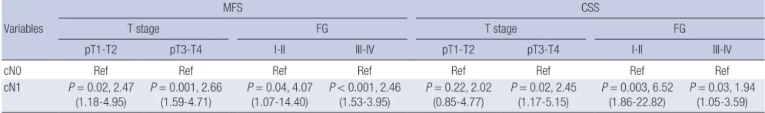
ysis of pN0, clinical lymphadenopathy was associated with poor prognosis (MFS, HR 2.27, P < 0.001; CSS, HR 2.05, P = 0.02). When the prognostic significance of cN stage was examined in stage- for-stage and grade-for-grade analyses (Table 3), patients with cN1 cancer showed significantly lower MFS and CSS rates than patients with cN0 cancer in the advanced disease category (pT3 or pT4) (P = 0.001, HR 2.66 for MFS and P = 0.02, HR 2.45 for CSS) after adjustment for all other covariates. When the prog- nostic significance of cN stage was determined by grade in sub- group analysis, cN1 was a significant predictor of MFS and CSS
for all grades relative to their pN0 counterparts (all P < 0.05). Fi- nally, the most informative independent predictors of patholo- gical LN metastasis were clinical nodal status (cN1 vs cN0, odds ratio [OR] 5.89, P < 0.001) (Table 4).
DISCUSSION
LND rates for RCC have declined in the past decade because of the rapid growth of minimally invasive techniques and a lower incidence of radiographic lymphadenopathies (16). However,
MFS
Time (months)
0 60 120 180 240
1 0.8 0.6 0.4 0.2 0
cN0cN1 cN0-censored cN1-censored
P < 0.001
MFS
Time (months)
0 60 120 180 240
1 0.8 0.6 0.4 0.2 0
pN0pN1 pN0-censored pN1-censored
P < 0.001
CSS
Time (months)
0 60 120 180 240
1 0.8 0.6 0.4 0.2 0
pN0pN1 pN0-censored pN1-censored
P < 0.001 A
CSS
Time (months)
0 60 120 180 240
1 0.8 0.6 0.4 0.2 0
cN0cN1 cN0-censored cN1-censored
P < 0.001
B
C D
Fig. 1. Survival curves by stages. (A, B) Metastasis-free survival (MFS) and Cancer-specific survival (CSS) by cN stage. (C, D) MFS and CSS by pN stage.
Fig. 2. Stratified survival curves by clinical and pathological nodal status. (A) Metastasis-free survival (MFS). (B) Cancer-specific survival (CSS).
cN0pN0 (n = 329) cN0pN1 (n = 9) cN1pN0 (n = 82) cN1pN1 (n = 20)
5 yr MFS, % (SE) 85.1 (2.2) 31.7 (18.0) 54.1 (6.3) 29.9 (10.9)
5 yr CSS, % (SE) 92.1 (1.7) 63.6 (16.9) 77.9 (5.5) 38.7 (12.8)
MFS
Time (months)
0 60 120 180 240
1 0.8 0.6 0.4 0.2 0
cN0pN0 cN0pN1 cN1pN0 cN1pN1 cN0pN0-censored cN0pN1-censored cN1pN0-censored cN1pN1-censored
P < 0.001
A
CSS
Time (months)
0 60 120 180 240
1 0.8 0.6 0.4 0.2 0
P < 0.001
B
cN0pN0 cN0pN1 cN1pN0 cN1pN1 cN0pN0-censored cN0pN1-censored cN1pN0-censored cN1pN1-censored
patients with isolated regional nodal metastases from RCC are a distinct cohort for whom resection of involved LNs might pro- vide therapeutic benefits (2, 4). Furthermore, the detrimental effects of nodal metastases on cancer-specific mortality after nephrectomy are particularly high in patients with low-stage or low-grade non-metastatic RCC (17). We similarly found that in grade-for-grade analyses, Fuhrman Grade (FG) I-II and FG III- IV patients with cN1 disease were 4.1-fold, 2.5-fold, 6.5-fold, and 1.9-fold more likely to have metastatic recurrence or cancer- specific death relative to cN0 counterparts.
The aim of our study was to investigate the diagnostic and pro- gnostic value of positive LNs on preoperative CT imaging and to identify specific subsets which might benefit from aggressive surgical resection involving LND. Predictive nomograms for LNI have been developed to prevent unnecessary LNDs and ensure adequate extension of LND templates to high-risk cases, incor- porating symptoms, radiographic lymphadenopathy, intraop- erative palpable LNs, tumor stage of ≥ pT3, presence of sarco- matoid features, nuclear grade ≥ 3 and histological necrosis (18- 21). Non-clear cell subtype and removed LNs ≥ 11, cN stage based on CT imaging was the most informative predictor of LNI in our cohort, which confirmed the findings of previous study (21).
LNI rates vary considerably regarding the presence of distant metastases and the extent of LND (7, 9). In our results, the over- all pN1 rate (7.0%) was comparable to rates reported in previ- ous studies (2.9%-6.1%) based on data from patients with M0 RCC treated with LND (8, 9, 22). We found that most patients with nodal metastases (65%) had clinically node-positive dis- ease identified on preoperative imaging. Furthermore, the pres- ence of clinical lymphadenopathy had a detrimental effect on MFS and CSS that was strongly stratified with pathological char- acteristics of pT stage, pN stage and nuclear grade. Patients with clinically positive but pathologically negative LNs (cN1pN0, 18.6%) had similar survival outcomes to patients with clinically negative but pathologically positive LNs (cN0pN1, 2.0%). How- ever, these patients had a worse prognosis than patients with clinically and pathologically negative LNs (cN0pN0, 74.8%), con- sistent with a previous report (21), implying the prophylactic ef- fects of LND via removing the means by which cancer might spread through lymphatic channels. Moreover, an inflammato- ry response to a tumor could be a sign of systemic dissemina- tion or of micro-metastases in non-sampled LNs and inadequate LND. However, comparing MFS and CSS between cN0pN1 and cN1pN1 groups indicated that the absence of evident LN me- tastasis does not preclude regional LND because of undetected LN micro-metastasis on available imaging technology.
In the present study, LN staging by CT had a sensitivity of 65%, a specificity of 80%, and an accuracy of 77%. Prior series found only a modest association between LN size and metastatic in- volvement, with 32%-43% of LNs > 1 cm containing metastatic Table 2. Multivariable Cox regression analyses predicting metastasis-free survival (MFS) and cancer-specific survival (CSS) overall and stratified according to pathological N stage amongst all non-metastatic RCC patients who under- went lymph node dissection at nephrectomy CovariatesMFSCSS OverallpN0pN1OverallpN0pN1 PHR (95% CI)PHR (95% CI)PHR (95% CI)PHR (95% CI)PHR (95% CI)PHR (95% CI) Age ≥65 yr (ref<65 yr)0.131.42 (0.90-2.25)0.521.19 (0.70-2.02)0.133.99 (0.68-23.37)<0.0013.13 (1.92-5.10)<0.0013.42 (2.02-5.82)0.145.98 (0.57-62.43) BMI<25 kg/m2 (ref≥25 kg/m2)0.411.20 (0.79-1.82)0.301.28 (0.80-2.03)0.173.90 (0.57-26.80)0.061.62 (0.99-2.65)0.481.22 (0.71-2.11)0.0123.03 (2.12-250.11) Male (ref female)0.011.81 (1.14-2.89)0.0461.72 (1.01-2.92)0.432.05 (0.35-12.13)<0.0012.98 (1.61-5.50)0.0042.92 (1.41-6.05)0.0314.96 (1.44-156.01) Grade III-IV (ref I-II)0.131.49 (0.89-2.49)0.081.62 (0.94-2.79)0.730.65 (0.06-7.54)0.671.13 (0.65-1.96)0.651.14 (0.64-2.04)0.240.18 (0.01-3.13) Tumor size≥10 cm (ref<10 cm)0.0012.09 (1.37-3.20)<0.0012.41 (1.50-3.86)0.321.99 (0.52-7.63)0.0062.05 (1.23-3.42)0.031.95 (1.07-3.58)0.571.60 (0.31-8.13) pT3-4 (ref≤pT1-2)<0.0012.60 (1.69-3.99)<0.0012.75 (1.71-4.43)0.431.88 (0.40-8.84)0.0022.33 (1.38-3.95)0.022.02 (1.12-3.63)0.174.83 (0.50-46.23) Clear cell (ref Non-clear cell)0.291.30 (0.81-2.08)0.041.85 (1.03-3.33)0.180.41 (1.11-1.50)0.101.61 (0.91-2.84)0.331.39 (0.71-2.74)0.591.66 (0.27-10.32) Sarcomatoid component (+)0.081.98 (0.91-4.30)0.092.47 (0.87-7.06)0.771.34 (0.19-9.61)0.731.20 (0.43-3.32)0.860.87 (0.18-4.16)0.831.28 (0.13-12.46) Necrosis (+)0.041.83 (1.04-3.23)0.241.56 (0.74-3.26)0.232.39 (0.57-10.00)0.781.12 (0.52-2.40)0.491.40 (0.55-3.59)0.890.87 (0.14-5.62) Capillary-lymphatic invasion (+)0.921.03 (0.56-1.91)0.841.08 (0.51-2.30)0.540.53 (0.07-3.97)0.390.72 (0.34-1.51)0.961.03 (0.41-2.55)0.080.09 (0.01-1.38) cN1 (ref cN0)<0.0012.47 (1.59-3.81)0.0012.27 (1.39-3.71)0.144.14 (0.63-27.47)0.0092.04 (1.20-3.48)0.022.05 (1.10-3.80)0.0215.40 (1.57-151.54) pN1 (ref pN0)0.012.10 (1.18-3.74)NANANANA<0.0013.70 (1.84-7.41)NANANANA HR, hazard ratio; CI, confidence interval; ref, reference.
Table 3. Multivariable Cox regression analyses predicting MFS and CSS, stratified according to pathological tumor stage and Furhman nuclear grade (FG)
Variables
MFS CSS
T stage FG T stage FG
pT1-T2 pT3-T4 I-II III-IV pT1-T2 pT3-T4 I-II III-IV
cN0 Ref Ref Ref Ref Ref Ref Ref Ref
cN1 P = 0.02, 2.47
(1.18-4.95) P = 0.001, 2.66
(1.59-4.71) P = 0.04, 4.07
(1.07-14.40) P < 0.001, 2.46
(1.53-3.95) P = 0.22, 2.02
(0.85-4.77) P = 0.02, 2.45
(1.17-5.15) P = 0.003, 6.52
(1.86-22.82) P = 0.03, 1.94 (1.05-3.59) Covariates comprised of patient age, BMI, sex, tumor size, histological subtype, sarcomatoid component, necrosis, CKI, and pN stage.
Table 4. Multivariate analysis to predict pathologic LN metastasis
Covariates Multivariate
P value OR 95% CI
Tumor size > 10 cm (vs. < 10 cm) 0.98 1.01 0.41-2.50
cN+ (vs. cN0) < 0.001 5.89 2.31-15.04
Clear cell (vs. Non-clear cell) 0.009 0.32 0.13-0.75
pT3-4 (vs. ≤ pT2) 0.80 1.13 0.44-2.88
Fuhrman grade III-IV (vs I-II) 0.06 3.68 0.93-14.54
Sarcomatoid component (+) 0.11 3.23 0.78-13.34
Necrosis (+) 0.67 1.29 0.41-4.07
Capillary-lymphatic invasion (+) 0.08 2.71 0.88-8.35 No. nodes removed ≥ 11 (vs. 1-10) 0.008 3.39 1.38-8.37 OR, odds ratio; CI, confidence interval.
disease (10, 18, 23). Nodal enlargement can be caused by reac- tive hyperplasia, which is often associated with large or exten- sive tumor necrosis or venous thrombosis, and may represent a reactive immune response (10). Furthermore, micrometastases in normal-sized nodes cannot be visualized using the above mentioned techniques (11-13). The evolution of CT technology such as multidetector CT with higher spatial resolution have improved diagnosis by reducing false-positives from reactive hyperplasia (12).
Here in, we did not use a standard template, and the extent of dissection might have been influenced by the risk of nodal metastases. In addition, most patients underwent only hilar LND, which could underestimate the true rate of LNI. Clearly, hilar node dissection alone, incorporating fatty tissue along the renal vessels to the vena cava or aorta, is insufficient. LN metastases without involvement of the renal hilum are common, with in- volvement of the interaortocaval nodes without regional hilar involvement in 35%-45% of patients (7). Improved CSS and in- creased number of LNs excised in nonmetastatic, LN-positive patients are significantly associated (24), and 15 LNs need to be removed to achieve a 90% probability of detecting at least one metastatic LN (25). These findings suggest that more extensive LND including retrocaval and/or interaortocaval dissection should be pursued in all patients with clinically node-positive RCC undergoing nephrectomy, regardless of technique (26).
In our study, selection bias was possible since the cohort was restricted to a single institution and contained mostly patients considered to be high-risk for LN metastasis who underwent LND. Surgeon preference and retrospective data collection from
patient medical records might have affected the accuracy of vari- ables related to the LND extent. Preoperative CT techniques were not standardized and radiology re-review was not performed, which could have affected the accuracy of cN staging. Finally, using the operative records, we were unable to assess factors af- fecting surgeons’ intent to perform retroperitoneal LND (RPLND) for cN0 disease such as intraoperative palpable or suspicious LNs. Additional prospective multi-institutional assessments will help evaluate our findings and further define LND as a treatment for patients with low-volume LN metastases without distant me- tastases.
DISCLOSURE
The authors declare that they have no competing conflicts of interest.
AUTHOR CONTRIBUTION
Conception and coordination of the study: Lee HW, Jeon HG, Jeong BC, Lee HM. Design of ethical issues: Lee HW, Lee HM.
Acquisition of data: Lee HW, Jeon HG, Jeong BC, Seo SI, Jeon SS, Choi HY, Lee HM. Data review: Lee HW, Jeong BC, Seo SI, Jeon SS, Choi HY, Lee HM. Statistical analysis: Lee HW. Manu- script preparation: Lee HW, Jeon HG, Lee HM. Manuscript ap- proval: all authors.
ORCID
Hye Won Lee http://orcid.org/0000-0002-0303-4658 Hwang Gyun Jeon http://orcid.org/0000-0002-5613-8389 Byong Chang Jeong http://orcid.org/0000-0002-5399-2184 Seong Il Seo http://orcid.org/0000-0002-9792-7798 Seong Soo Jeon http://orcid.org/0000-0002-3265-6261 Han Yong Choi http://orcid.org/0000-0002-1980-4899 Hyun Moo Lee http://orcid.org/0000-0003-3969-4540 REFERENCES
1. Pantuck AJ, Zisman A, Dorey F, Chao DH, Han KR, Said J, Gitlitz BJ, Fig- lin RA, Belldegrun AS. Renal cell carcinoma with retroperitoneal lymph nodes: role of lymph node dissection. J Urol 2003; 169: 2076-83.
2. Delacroix SE Jr, Chapin BF, Chen JJ, Nogueras-Gonzalez GM, Tamboli P, Matin SF, Wood CG. Can a durable disease-free survival be achieved with surgical resection in patients with pathological node positive renal cell carcinoma? J Urol 2011; 186: 1236-41.
3. Karakiewicz PI, Trinh QD, Bhojani N, Bensalah K, Salomon L, de la Taille A, Tostain J, Cindolo L, Altieri V, Ficarra V, et al. Renal cell carcino- ma with nodal metastases in the absence of distant metastatic disease:
prognostic indicators of disease-specific survival. Eur Urol 2007; 51: 1616- 24.
4. Canfield SE, Kamat AM, Sánchez-Ortiz RF, Detry M, Swanson DA, Wood CG. Renal cell carcinoma with nodal metastases in the absence of dis- tant metastatic disease (clinical stage TxN1-2M0): the impact of aggres- sive surgical resection on patient outcome. J Urol 2006; 175: 864-9.
5. Kane CJ, Mallin K, Ritchey J, Cooperberg MR, Carroll PR. Renal cell can- cer stage migration: analysis of the National Cancer Data Base. Cancer 2008; 113: 78-83.
6. Leibovich BC, Blute ML. Lymph node dissection in the management of renal cell carcinoma. Urol Clin North Am 2008; 35: 673-8; viii.
7. Crispen PL, Breau RH, Allmer C, Lohse CM, Cheville JC, Leibovich BC, Blute ML. Lymph node dissection at the time of radical nephrectomy for high-risk clear cell renal cell carcinoma: indications and recommenda- tions for surgical templates. Eur Urol 2011; 59: 18-23.
8. Capitanio U, Abdollah F, Matloob R, Suardi N, Castiglione F, Di Trapani E, Capogrosso P, Gallina A, Dell’Oglio P, Briganti A, et al. When to per- form lymph node dissection in patients with renal cell carcinoma: a nov- el approach to the preoperative assessment of risk of lymph node inva- sion at surgery and of lymph node progression during follow-up. BJU Int 2013; 112: E59-66.
9. Hutterer GC, Patard JJ, Perrotte P, Ionescu C, de La Taille A, Salomon L, Verhoest G, Tostain J, Cindolo L, Ficarra V, et al. Patients with renal cell carcinoma nodal metastases can be accurately identified: external vali- dation of a new nomogram. Int J Cancer 2007; 121: 2556-61.
10. Studer UE, Scherz S, Scheidegger J, Kraft R, Sonntag R, Ackermann D, Zingg EJ. Enlargement of regional lymph nodes in renal cell carcinoma is often not due to metastases. J Urol 1990; 144: 243-5.
11. Studer UE, Birkhäuser FD. Lymphadenectomy combined with radical nephrectomy: to do or not to do? Eur Urol 2009; 55: 35-7.
12. Türkvatan A, Akdur PO, Altinel M, Olçer T, Turhan N, Cumhur T, Akinci S, Ozkul F. Preoperative staging of renal cell carcinoma with multidetec- tor CT. Diagn Interv Radiol 2009; 15: 22-30.
13. Van Poppel H. Lymph node dissection is not obsolete in clinically node- negative renal cell carcinoma patients. Eur Urol 2011; 59: 24-5.
14. Edge SB, American Joint Committee on Cancer. AJCC cancer staging manual. 7th ed. New York, NY: Springer, 2010.
15. Eisenberg MS, Cheville JC, Thompson RH, Kaushik D, Lohse CM, Boor- jian SA, Costello BA, Leibovich BC. Association of microvascular and capillary-lymphatic invasion with outcome in patients with renal cell carcinoma. J Urol 2013; 190: 37-43.
16. Kates M, Lavery HJ, Brajtbord J, Samadi D, Palese MA. Decreasing rates of lymph node dissection during radical nephrectomy for renal cell carci- noma. Ann Surg Oncol 2012; 19: 2693-9.
17. Sun M, Bianchi M, Hansen J, Abdollah F, Trinh QD, Lughezzani G, Shar- iat SF, Montorsi F, Perrotte P, Karakiewicz PI. Nodal involvement at ne- phrectomy is associated with worse survival: a stage-for-stage and grade- for-grade analysis. Int J Urol 2013; 20: 372-80.
18. Ming X, Ningshu L, Hanzhong L, Zhongming H, Tonghua L. Value of frozen section analysis of enlarged lymph nodes during radical nephrec- tomy for renal cell carcinoma. Urology 2009; 74: 364-8.
19. Thompson RH, Raj GV, Leibovich BC, Russo P, Blute ML, Kattan MW.
Preoperative nomogram to predict positive lymph nodes during nephrec- tomy for renal cell carcinoma. J Urol 2008; 179: 212.
20. Delacroix SE Jr, Chapin BF, Wood CG. The role of lymph node dissection in renal cell carcinoma. Urol Clin North Am 2011; 38: 419-28, vi.
21. Babaian KN, Kim DY, Kenney PA, Wood CG Jr, Wong J, Sanchez C, Fang JE, Gerber JA, Didic A, Wahab A, et al. Preoperative predictors of patho- logical lymph node metastasis in patients with renal cell carcinoma un- dergoing retroperitoneal lymph node dissection. J Urol 2015; 193: 1101-7.
22. Blute ML, Leibovich BC, Cheville JC, Lohse CM, Zincke H. A protocol for performing extended lymph node dissection using primary tumor pathological features for patients treated with radical nephrectomy for clear cell renal cell carcinoma. J Urol 2004; 172: 465-9.
23. Russo P. Renal cell carcinoma: presentation, staging, and surgical treat- ment. Semin Oncol 2000; 27: 160-76.
24. Whitson JM, Harris CR, Reese AC, Meng MV. Lymphadenectomy im- proves survival of patients with renal cell carcinoma and nodal metas- tases. J Urol 2011; 185: 1615-20.
25. Capitanio U, Suardi N, Matloob R, Abdollah F, Castiglione F, Briganti A, Carenzi C, Roscigno M, Montorsi F, Bertini R. Staging lymphadenecto- my in renal cell carcinoma must be extended: a sensitivity curve analy- sis. BJU Int 2013; 111: 412-8.
26. Chapman TN, Sharma S, Zhang S, Wong MK, Kim HL. Laparoscopic lymph node dissection in clinically node-negative patients undergoing laparoscopic nephrectomy for renal carcinoma. Urology 2008; 71: 287- 91.