서 론
안정 시 심박수(resting heart rate, RHR)는 심장의 수축 기능인 심 박출량을 반영하는 인자이며, 자율신경계의 균형과 대사율을 예측 할 수 있는 유용한 측정값 중 하나이다.1) 안정 시 심박수의 상승은 관상동맥질환, 심부전, 부정맥, 급성 심장사등의 심혈관질환 뿐 아니
라 제2형 당뇨병 및 암과 관련된 사망의 증가와도 관련성이 여러 차 례 보고된 바 있다.2,3) 높은 안정 시 심박수는 심근의 산소 소모량을 증가시켜 산화스트레스의 증가와 만성 저강도 염증을 유발함으로 써 관상동맥 및 말초동맥질환의 위험도를 높일 수 있다. 더불어 안정 시 심박수 상승은 심근의 피로와 동맥벽의 탄력섬유 손상을 초래해 동맥경화성 변화 및 진행에 영향을 줄 수 있다.1)
Received July 18, 2018 Revised August 28, 2018 Accepted November 1, 2018
Corresponding author Byoungjin Park Tel: +82-31-331-8710, Fax: +82-31-331-5551 E-mail: [email protected]
ORCID: http://orcid.org/0000-0003-1733-5301
Copyright © 2019 The Korean Academy of Family Medicine
This is an open-access article distributed under the terms of the Creative Commons At- tribution Non-Commercial License (http://creativecommons.org/licenses/by-nc/4.0) which permits unrestricted noncommercial use, distribution, and reproduction in any medium, provided the original work is properly cited.
Original Article
https://doi.org/10.21215/kjfp.2019.9.2.161 eISSN 2233-9116
Korean J Fam Pract. 2019;9(2):161-166
Korean Journal of Family Practice
KJFP
건강검진 수검자에서 안정 시 심박수와 경계성 발목상완지수의 연관성
권사라, 박병진*
연세대학교 의과대학 가정의학교실
Association between Resting Heart Rate and Borderline Ankle-Brachial Index Among the Health Screen Examinees
Sa-Ra Kwon, Byoungjin Park*
Department of Family Medicine, Yonsei University College of Medicine, Seoul, Korea
Background: The ankle-brachial index (ABI) is a simple, noninvasive clinical test that is useful in the diagnosis of peripheral artery disease (PAD). The borderline ABI value is 0.91–0.99 and has also been reported in endothelial dysfunction and preclinical PAD. We investigated the relationship between resting heart rate as a surrogate marker of autonomic balance and borderline ABI in apparently healthy individuals.
Methods: We evaluated the association between resting heart rate and borderline ABI in 618 participants (348 male and 270 female) in a health examination program. The odds ratios for borderline ABI were calculated using a multivariable logistic regression analysis after adjusting for confounding variables across heart rate quartiles (Q1≤56, Q2=57–62, Q3=63–68, and Q4≥69 beats/min).
Results: The overall prevalence of borderline ABI was 13.9%. The age- and sex-adjusted resting heart rate was significantly higher in the borderline ABI group than in the control group (66.9±0.9 vs. 63.0±0.4 beats/min, P<0.001). The odds ratios (95% confidence intervals) for the borderline ABI in each heart rate quartile were 1.00, 1.04 (0.43–2.52), 1.69 (0.73–3.93), and 3.52 (1.55–7.97) after adjusting for age, sex, smoking status, alcohol intake, regular exercise, body mass index, systolic blood pressure, fasting plasma glucose level, triglyceride level, high-density lipoprotein-cholesterol level, white blood cell count, C-reactive protein level, uric acid level, γ-glutamyltransferase level, hypertension medication, diabetes medication, and dyslipidemia medication.
Conclusion: These findings indicate that a higher resting heart rate is independently associated with borderline ABI.
Keywords: Heart Rate; Ankle-Brachial Index; Peripheral Arterial Disease
Sa-Ra Kwon, Byoungjin Park. Resting Heart Rate and Borderline Ankle-Brachial Index
Korean Journal of Family Practice
KJFP
한편 발목상완지수(ankle-brachial index, ABI)는 동맥경화 및 말초 혈관질환의 정도를 예측할 수 있는 검사 방법 중 하나이며4) 심혈관 질환 및 관상동맥질환, 경동맥 질환의 발생 위험과도 연관되어 있 다.5,6) 발목상완지수가 1.4를 초과하거나 0.9 이하일 경우 말초동맥질 환을 의심할 수 있다.4) 경계성 발목상완지수(ABI, 0.9–0.99) 역시 혈 관내피세포의 기능 이상과 관련이 있고 정상 발목상완지수에 비해 심혈관질환 발생의 위험도가 높은 것으로 알려져 있다.7-9)
높은 안정 시 심박수가 비정상 발목상완지수의 독립적 위험인자 라는 보고는 이루어진 바 있으나,10) 비교적 건강한 성인에서 안정 시 심박수와 경계성 발목상완지수의 관계를 규명한 연구는 없었다. 이 에 본 연구에서는 건강검진 프로그램에 참가한 비교적 건강한 성인 들을 대상으로 안정 시 심박수와 경계성 발목상완지수의 연관성에 대해 알아보고자 하였다.
방 법
1. 연구 대상
2006년 3월부터 2007년 5월까지 질병 조기 발견을 위해 대학병원 건강증진센터를 방문하여 건강 검진을 받은 남녀 성인 641명에 대해 안정 시 심박수와 발목상완지수를 측정하였으며 발목상완지수 0.9 이하 또는 1.4 이상, C-반응 단백질(C-reactive protein, CRP) 5.0/μL 이 상, 백혈구 수치 10,000/μL 이상인 23명을 제외하고 최종적으로 618 명(남자 348명, 여자 270명)을 대상으로 하였다. 본 연구는 연세대학 교 의과대학 강남세브란스병원 연구심의위원회의 승인 심사를 완료 하였으며 각 대상자에 대해 연구 동의하에 진행되었다(IRB no.
3-2018-0064).
2. 연구 방법 1) 데이터 수집
건강검진 시 시행된 모든 검사는 숙련된 의료진이 동일한 절차로 시행하였다. 대상자들은 생활습관(흡연, 음주 포함), 신체활동(주 2 회 이상 또는 이하) 및 질병으로 치료받고 있는지 여부 등을 포함하 여 문진에 답하였으며 질병이 있는 경우 진단일 및 복용 중인 약물 에 대해서도 조사하였다.
대상자들은 흡연에 대해서는 비 흡연자, 과거 흡연자, 현재 흡연자 로 구분되었으며 음주에 대해서는 비 음주자, 금주자(알코올 섭취 주당 140 g 미만), 현재 음주자(알코올 섭취 주당 140 g 이상)로 구분되 었다. 체중과 신장은 실내복 착용 및 신발 미착용 상태에서 0.1 cm, 0.1 kg 단위로 측정하였고 신체질량지수(body mass index)는 체중을 신장의 제곱으로 나누어 구하였다(kg/m2).
12시간 이상 금식한 대상자의 상완 정맥혈을 채취하였고 백혈구 수치는 Automated blood cell counter (ADVIA 120; Bayer Co., Tarry- town, NY, USA)를 이용하여, 고밀도 지단백 콜레스테롤(high density lipoprotein-cholesterol), 중성지방(triglyceride), C-반응 단백질(CRP), 감마글루타밀전이효소(gamma-glutamyl transpeptidase) 및 요산(uric acid)은 Hitachi 7600-110 자동 화학 분석기(Hitachi, Tokyo, Japan)를 이용하여 측정하였다.
혈압은 대상자가 5분 이상의 휴식을 취한 후 앉은 자세에서 자동 혈압계(TM-2665, A&D Co., Ltd., Tokyo, Japan)를 이용하여 측정하였 으며 고혈압은 자가 보고한 과거력 또는 수축기혈압 ≥140 mmHg 또 는 이완기혈압 ≥90 mmHg인 경우로, 당뇨는 자가 보고한 과거력 또 는 공복혈장포도당 ≥126 mg/dL인 경우로 정의하였다.
2) 발목상완지수(ABI)와 안정 시 심박수(RHR)의 측정
대상자들은 측정 3시간 전부터 식사와 카페인(커피, 녹차)을 금지 하였고, 10분 이상 누운 상태로 안정을 취한 후 자동화 파형 분석기 BP-203PRE II Instrument (Omron Colin Co., Komaki, Japan)를 이용하 여 사지의 혈압과 맥박수를 측정하였다. 누운 자세의 대상자의 양 팔 과 양 발목에 혈압계를 감아 전동계식으로 동시에 사지 혈압을 측정 하였으며 상완동맥(brachial artery) 및 발등동맥(dorsalis pedis artery), 후경골동맥(posterior tibial artery)에서 각각 수축기혈압을 측정하였 다.4,6,11)
미국심장협회(American Heart Association)의 정의에 따라 발목상 완지수(ABI)는 발목 및 상완에서 측정한 수축기 혈압의 비로 나타내 었으며,11) 양측 발목동맥의 수축기 혈압을 양측 상완동맥의 수축기 혈압 중 높은 쪽으로 나누어 계산하였다.4,6) 경계성 발목상완지수 (borderline ABI)는 0.91–0.99의 값으로 정의하였다.7)
3. 통계분석
연구 대상자는 ABI에 대해 두 군으로 나누었다 : 정상 발목상완지 수(대조군, 1.0–1.40), 경계성 발목상완지수(0.91–0.99). 두 군 간의 인 구사회학적 특성 및 혈액검사상의 차이에 대한 검정을 위해 연속된 변수에 대해 일원분산분석 및 명목변수에 대해 카이제곱검정을 시 행하였다.
안정 시 심박수를 네 군으로 분류하였다: Q1≤56, Q2=57–62, Q3=63–68, Q4≥69회/분. 안정 시 심박수와 발목상완지수의 관련성에 영향을 주는 요인을 보정한 후 교차비(odds ratio)를 알아보기 위해 다변량 로지스틱 회귀분석을 시행하였다.
통계분석은 SAS Institute ver. 9.1 software (SAS Institute, Cary, NC, USA)을 이용하였으며 통계학적인 유의성은 P값이 0.05 미만인 경우
권사라, 박병진. 안정 시 심박수와 경계성 발목상완지수 Korean Journal of Family Practice
KJFP
로 정의하였다.
결 과
1. 연구 대상자의 특성
전체 대상자는 총 618명으로 연구 대상자들은 발목상완지수 0.91–0.99인 경우 경계성 발목상완지수 군(n=86, 13.9%), 1.00–1.40인 경우 정상군(n=532, 86.1%)으로 분류되었다. 연구 대상자들의 평균연 령은 경계성 발목상완지수 군에서 유의하게 낮게 나타났으며(44.2 vs. 48.5, P=0.004) 현재 흡연자의 비율은 정상군 12.8%, 경계성 발목상 완지수군 24.1%로 유의하게 높게 나타났다(P=0.020). 두 군에서 성별, 체질량지수, 과거 흡연여부, 음주, 수축기 혈압, 공복혈당, 중성지방, 고밀도지단백 콜레스테롤, 백혈구, C-반응단백질, 요산, 고혈압/당 뇨/이상지질혈증 복약 여부는 유의미한 차이가 나타나지 않았다.
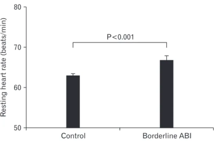
또한 안정 시 심박수의 경우 경계성 발목상완지구 군이 정상군에 비해 유의하게 높게 나타났다(67.1회/분 vs. 63회/분, P<0.001) (Table 1, Figure 1).
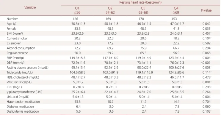
한편 연구 대상자들에 대해 안정 시 심박수에 따른 군 간의 인구 사회학적 특성 및 혈액검사상의 차이를 비교하였다. 안정 시 심박수 를 Q1≤56, Q2=57–62, Q3=63–68, Q4≥69(회/분)의 네 군으로 분류하
였으며, 4분위(≥69회/분) 군에서의 수축기 혈압(P=0.004), 이완기 혈 압(P<0.001), 공복혈당(P=0.003), 백혈구 수치(P=0.001)가 유의하게 높 게 나타났다(Table 2).
2. 안정 시 심박수에 따른 경계성 발목상완지수의 교차비 안정 시 심박수에 따른 경계성 발목상완지수의 교차비(95% 신뢰 구간)를 로지스틱 회귀분석을 통하여 조사하였다. 경계성 발목상완
Table 1. Characteristics of the study population
Variable Total (n=618) Normal (n=532) Borderline (n=86) P-value
ABI 1.00–1.40 0.91–0.99
Age (y) 47.9±11.6 48.5±11.3 44.2±12.7 0.004*
Female 43.7 53.5 42.1 0.048†
BMI (kg/m2) 23.8±2.9 23.8±2.9 23.6±2.9 0.402*
Current smoker 22.5 12.8 24.1 0.020†
Ex-smoker 20.4 20.7 18.6 0.658†
Alcohol consumption 71.0 69.8 71.2 0.779†
Regular exercise 58.4 70.9 56.4 0.011†
SBP (mmHg) 119.6±15.3 120.1±15.6 117.1±12.7 0.050*
DBP (mmHg) 73.2±11.9 73.7±12.0 69.7±11.1 0.002*
Fasting plasma glucose (mg/dL) 96.7±20.4 96.7±18.2 97.0±30.9 0.908*
Triglyceride (mg/dL) 113.0±93.1 114.5±96.1 104.2±71.8 0.245*
HDL-cholesterol (mg/dL) 47.9±12.5 47.9±12.4 48.0 ±13.0 0.912*
WBC (×103 cell/μL) 5.5±1.3 5.5±1.4 5.4 ±1.3 0.674*
CRP (mg/L) 0.7±0.9 0.7±0.7 0.5 ±0.7 0.707*
γ-glutamyltransferase (U/L) 24.4±15.8 24.9±16.1 21.1±13.6 0.039*
Uric acid (mg/dL) 5.2±1.4 5.2±1.4 4.9±1.4 0.058*
Hypertension medication 12.3 12.6 10.5 0.577†
Diabetes medication 4.7 4.9 3.5 0.569†
Dyslipidemia medication 4.7 4.1 8.1 0.103†
RHR (beats/min) 63.6±8.9 63.0±8.8 67.1±8.7 <0.001*
Values are presented as mean±standard deviation or %.
ABI, ankle-brachial index; BMI, body mass index; SBP, systolic blood pressure; DBP, diastolic blood pressure; HDL, high density lipoprotein; WBC, white blood cells; CRP, C-reactive protein; RHR, resting heart rate.
*P-value by one-way analysis of variance. †P-value by χ2 test.
Restingheartrate(beats/min)
Control Borderline ABI
P<0.001 80
70
60
50
Figure 1. The age- and sex-adjusted resting heart according to pres- ence of borderline ABI.
ABI, ankle-brachial index.
Sa-Ra Kwon, Byoungjin Park. Resting Heart Rate and Borderline Ankle-Brachial Index
Korean Journal of Family Practice
KJFP
지수의 교차비는 안정 시 심박수의 각 군에 대해 1.00, 1.23 (0.54–
3.00), 2.35 (1.06–5.20), 4.00 (1.84–8.67) (P<0.001)이었으며 나이, 성별, 흡 연, 음주, 신체활동, 체질량지수, 수축기혈압, 공복혈당, 중성지방, 고 밀도지단백 콜레스테롤, 백혈구, C-반응단백질, 요산, 감마글루타밀 전이효소, 고혈압/당뇨/이상지질혈증 복약 여부를 모두 보정하였을 때에도 안정 시 심박수의 각 군에 대한 경계성 발목상완지수의 교차 비는 1.00, 1.04 (0.43–2.52), 1.69 (0.73–3.93), 3.52 (1.55–7.97) (P=0.002)로 안정 시 심박수가 높아질수록 경계성 발목상완지수의 위험도가 유 의미하게 높아지는 것으로 나타났다(Table 3).
고 찰
본 연구는 단면연구이며 후향적 연구로 건강한 성인의 안정 시 심 박수와 경계성 발목상완지수의 관계를 분석하였다. 그 결과 말초동 맥질환 및 심혈관질환의 독립적인 위험인자인 경계성 발목상완지수 의 위험도 증가와 높은 안정 시 심박수가 유의미한 관계가 있음을 확인하였다. 이 연관성은 여러 가지 혼란변수들을 보정하였음에도 유지되었다.
경계성 발목상완지수는 발목상완지수 0.90–0.99의 값으로 정의되 었으며, 이 범위의 발목상완지수는 혈관내피세포의 기능 이상과 관 Table 2. Clinical and chemical characteristics of the study population according to heart rate quartiles
Variable
Resting heart rate (beats/min) Q1
≤56
Q2 57–62
Q3 63–68
Q4
≥69 P-value
Number 126 169 170 153
Age (y) 50.3±11.3 48.1±11.8 46.7±11.4 47.0±11.7 0.042*
Female 33.3 48.5 48.2 41.8 0.033†
BMI (kg/m2) 23.9±2.6 23.5±3.0 23.9±2.8 24.0±3.1 0.457*
Current smoker 30.2 22.5 20.6 18.3 0.104†
Ex-smoker 23.0 17.2 20.0 22.2 0.582†
Alcohol consumption 72.2 69.2 75.9 66.7 0.294†
Regular exercise 50.0 59.2 65.3 56.9 0.066†
SBP (mmHg) 119.3±15.3 117.1±16.0 119.2±14.9 123.2±14.4 0.004*
DBP (mmHg) 72.9±11.6 70.6±12.1 73.4±11.1 76.0±12.3 <0.001*
Fasting plasma glucose (mg/dL) 95.1±13.4 92.9±12.9 98.0±22.4 100.8±27.6 0.003*
Triglyceride (mg/dL) 104.6±58.5 103.0±91.9 119.1±116.9 124.3±86.6 0.114*
HDL-cholesterol (mg/dL) 48.4±12.7 48.3±13.3 48.3±12.2 46.5±11.7 0.478*
WBC (×103 cell/μL) 5.3±1.2 5.3±1.3 5.6±1.5 5.8±1.3 0.001*
CRP (mg/L) 0.7±0.8 0.7±1.0 0.7±0.9 0.8±0.9 0.296*
γ-glutamyltransferase (U/L) 25.2±16.4 22.4±14.3 24.6±17.0 25.6±15.5 0.264*
Uric acid (mg/dL) 5.4 ±1.3 5.0±1.4 5.0±1.4 5.4±1.4 0.004*
Hypertension medication 13.5 10.7 11.2 14.4 0.704†
Diabetes medication 6.4 3.0 2.4 7.8 0.060†
Dyslipidemia medication 5.6 3.6 2.4 7.8 0.103†
Values are presented as mean±standard deviation or %.
BMI, body mass index; SBP, systolic blood pressure; DBP, diastolic blood pressure; HDL, high density lipoprotein; WBC, white blood cells; CRP, C-reactive protein.
*P-value by one-way analysis of variance. †P-value by χ2 test.
Table 3. Odds ratios and 95% confidence intervals for borderline ABI according to heart rate quartiles
Model Resting heart rate (beats/min)
Q1 Q2 Q3 Q4 P for trend*
Model 1 1.00 1.23 (0.54–3.00) 2.35 (1.06–5.20) 4.00 (1.84–8.67) <0.001
Model 2 1.00 1.11 (0.46–2.64) 1.98 (0.88–4.45) 3.56 (1.63–7.78) 0.001
Model 3 1.00 1.04 (0.43–2.52) 1.69 (0.73–3.93) 3.52 (1.55–7.97) 0.002
Data are presented as odds ratio (95% confidence interval).
ABI, ankle-brachial index.
Model 1: unadjusted. Model 2: adjusted for age, sex, and body mass index. Model 3: adjusted for age, sex, smoking status, alcohol intake, regular exercise, body mass index, systolic blood pressure, fasting plasma glucose, triglyceride, high density lipoprotein-cholesterol, white blood cell count, C-reactive protein, uric acid, γ-glutamyltransferase, hypertension medication, diabetes medication, and dyslipidemia medication.
*Calculated by multivariate analysis.
권사라, 박병진. 안정 시 심박수와 경계성 발목상완지수 Korean Journal of Family Practice
KJFP
련이 있으며 죽상동맥경화 및 관상동맥질환, 말초혈관질환의 위험 도 상승과 연관이 있다.7) 혈관내피세포는 단순한 세포 한 층의 구조 이지만 항상성 유지와 관련된 수많은 물질들을 분비하고 그 기능을 관장하며12) 또한 혈관 염증이나 구조적 손상이 발생하는 지점이다.
혈관내피세포 손상으로 인한 혈관의 확장 능력 저하가 발생하면 동 맥 경직으로 이어지게 된다.7,12) 또한 선행 연구에 의하면 안정 시 심 박수 증가와 관련된 동맥벽의 구조적, 기능적 변화가 동맥경직도 상 승을 유발하며,13) 동맥경직도의 상승은 심혈관질환 위험도 상승과 연관된다.14) 따라서 경계성 발목상완지수는 정상 발목상완지수와 비교하였을 때 조기 죽상동맥경화뿐 아니라 심혈관질환 위험도를 예측하는 주요 인자로 활용할 수 있다.7)
한편 안정 시 심박수는 자율신경계 균형을 반영하는 인자로, 높 은 안정 시 심박수는 부교감신경에 비해 더 높은 교감신경의 활성을 의미하며,15) 이는 혈관벽의 저항 증가16) 및 더 많은 산소 소모와 전염 증성 사이토카인의 분비를 유발한다.17,18) TNF-α, IL-1β, IL-6 등의 전 염증성 사이토카인 분비는 일련의 과정을 통해 혈관내피세포의 기 능 이상과 혈관 이완능력의 감소를 유발하여 동맥경직을 유발한다.
안정 시 심박수의 상승이 더 많은 산소 소모 및 높은 대사율과 관 련됨을 고려할 때, 높은 안정 시 심박수는 높은 산화스트레스와 만 성 경도 염증 발생에 대한 위험을 높인다.13)
상한선 부근의 백혈구 수치는 전신적인 만성 염증상태를 반영하 는 인자 중 하나로, 본 연구에서도 안정 시 심박수가 높은 군(4분위,
≥69회/분)이 낮은 군(1분위, ≤56회/분)에 비해 백혈구 수치가 유의하 게 높게 나타남을 확인하였다.
많은 선행 연구들에서 백혈구 수치가 말초동맥질환, 경동맥 죽상 경화증, 고혈압, 당뇨와 관련이 있음이 밝혀졌으며,16,19,20) 높은 백혈구 수치가 동맥경직도와도 연관됨이 밝혀진 바,21) 본 연구에서 나타난 안정 시 심박수와 백혈구 수치의 유의한 관계성 역시 안정 시 심박수 가 산화스트레스 및 전신적 염증과 관련되고 혈관질환의 위험요소 가 될 수 있음을 시사한다.
본 연구의 제한점은 다음과 같다. 첫째, 서울에 위치한 한 종합병 원의 건강검진 수검자를 대상으로 하였기 때문에 연구 대상자들의 지역적, 사회적 특성이 제한적이며 대한민국의 건강한 성인을 대표 하기에는 한계가 있다. 둘째, 안정 시 심박수의 1회 측정을 통해 얻어 진 결과를 활용한 연구로 연구 대상자의 일상 활동 및 휴식을 반영 하지 못하였으며 측정 전후의 상황과 관련된 심박수 변화를 고려하 지 못하였고 연속적인 값인 심박수를 단면화 하였다는 단점이 있다.
그럼에도 불구하고 본 연구는 건강한 성인에서 안정 시 심박수와 경계성 발목상완지수와의 연관성을 밝힌 첫 국내 연구이며 심박수 를 혈관 질환을 예측하는 인자로서 다시 한 번 확인하는 연구라는
데에 그 의미가 있다. 본 연구 결과를 바탕으로 건강검진에서 간편하 게 확인할 수 있는 발목상완지수 및 안정 시 심박수를 통해 잠재적 인 동맥경화 및 혈관질환의 위험도를 예측하여 위험 요인의 교정을 미리 시작할 수 있을 것이며, 필요 시 추가적인 정밀검사를 통한 조 기 진단 및 치료가 가능하게 될 것이다.
CONFLICT OF INTEREST
No potential conflict of interest relevant to this article was reported.
요 약
연구배경:
발목상완지수의 측정은 말초동맥질환을 진단에 유용한 간단하고 비침습적인 검사이다. 발목상완지수 0.91–0.99의 값은 경계 성 발목상완지수로서 혈관내피세포의 기능 이상과 말초동맥질환 전단계와 관련이 있음이 보고되었다. 본 연구에서는 자율신경계 균 형의 대리표지자인 안정 시 심박수와 경계성 발목상완지수의 관련 성을 비교적 건강한 성인에서 규명하고자 한다.방법:
본 연구는 건강검진을 시행한 남자 348명, 여자 270명 총 618명 을 대상으로 경계성 발목상완지수와 안정 시 심박수의 관계를 살펴 보았다. 경계성 발목상완지수의 교차비는 안정 시 심박수와 발목상 완지수의 관련성에 영향을 주는 요인을 보정한 후 다변량 로지스틱 회귀분석을 시행하여 도출하였다.결과:
경계성 발목상완지수의 총 유병률은 13.9%였다. 나이와 성별 을 보정한 후의 안정 시 심박수는 대조군에 비해 경계성 발목상완지 수 군에서 유의하게 높게 나타났다: 66.9±0.9 및 63.0±0.4회/분 (P<0.001). 안정 시 심박수 사분위에 대한 경계성 발목상완지수의 교 차비(신뢰구간 95%)는 혼란 변수들을 보정한 후 각각 1.00, 1.04 (0.43–2.52), 1.69 (0.73–3.93), 3.52 (1.55–7.97)이었다.
결론:
이와 같은 결과는 높은 안정 시 심박수가 경계성 발목상완지 수와 독립적인 연관성이 있음을 시사한다. 따라서, 안정 시 심박수의 주기적인 측정이 말초동맥질환을 포함하여 혈관 건강을 관리하는 데 중요하다.중심단어:
심박수; 발목상완지수; 말초동맥질환 ORCID박병진, http://orcid.org/0000-0003-1733-5301
Sa-Ra Kwon, Byoungjin Park. Resting Heart Rate and Borderline Ankle-Brachial Index
Korean Journal of Family Practice
KJFP
REFERENCES
1. Perret-Guillaume C, Joly L, Benetos A. Heart rate as a risk factor for cardio- vascular disease. Prog Cardiovasc Dis 2009; 52: 6-10.
2. Aune D, Sen A, ó’Hartaigh B, Janszky I, Romundstad PR, Tonstad S, et al.
Resting heart rate and the risk of cardiovascular disease, total cancer, and all- cause mortality - a systematic review and dose-response meta-analysis of prospective studies. Nutr Metab Cardiovasc Dis 2017; 27: 504-17.
3. Aune D, Ó Hartaigh B, Vatten LJ. Resting heart rate and the risk of type 2 di- abetes: a systematic review and dose--response meta-analysis of cohort studies. Nutr Metab Cardiovasc Dis 2015; 25: 526-34.
4. WOCN Clinical Practice Wound Subcommittee, 2005. Ankle Brachial In- dex: quick reference guide for clinicians. J Wound Ostomy Continence Nurs 2012; 39(2 Suppl): S21-9.
5. Doobay AV, Anand SS. Sensitivity and specificity of the ankle-brachial index to predict future cardiovascular outcomes: a systematic review. Arterioscler Thromb Vasc Biol 2005; 25: 1463-9.
6. McDermott MM, Liu K, Criqui MH, Ruth K, Goff D, Saad MF, et al. Ankle- brachial index and subclinical cardiac and carotid disease: the multi-ethnic study of atherosclerosis. Am J Epidemiol 2005; 162: 33-41.
7. Kajikawa M, Maruhashi T, Iwamoto Y, Iwamoto A, Matsumoto T, Hidaka T, et al. Borderline ankle-brachial index value of 0.91-0.99 is associated with endothelial dysfunction. Circ J 2014; 78: 1740-5.
8. Tanaka S, Kaneko H, Kano H, Matsuno S, Suzuki S, Takai H, et al. The pre- dictive value of the borderline ankle-brachial index for long-term clinical outcomes: an observational cohort study. Atherosclerosis 2016; 250: 69-76.
9. Natsuaki C, Inoguchi T, Maeda Y, Yamada T, Sasaki S, Sonoda N, et al. Asso- ciation of borderline ankle-brachial index with mortality and the incidence of peripheral artery disease in diabetic patients. Atherosclerosis 2014; 234:
360-5.
10. Tao J, Li D, Dong Y, Wu S. [Relationship between resting heart rate and bra-
chial-ankle pulse wave velocity in healthy Chinese population]. Chin J Car- diol 2014; 42: 686-92. Chinese.
11. Aboyans V, Criqui MH, Abraham P, Allison MA, Creager MA, Diehm C, et al. Measurement and interpretation of the ankle-brachial index: a scientific statement from the American Heart Association. Circulation 2012; 126:
2890-909.
12. Syvänen K, Korhonen P, Partanen A, Aarnio P. Endothelial function in a car- diovascular risk population with borderline ankle-brachial index. Vasc Health Risk Manag 2011; 7: 97-101.
13. Park BJ, Lee HR, Shim JY, Lee JH, Jung DH, Lee YJ. Association between resting heart rate and arterial stiffness in Korean adults. Arch Cardiovasc Dis 2010; 103: 246-52.
14. Lee SH, Choi SH, Kim EK, Yang JH, Song YB, Hahn JY, et al. Borderline an- kle-brachial index is associated with poor short-term clinical outcome after coronary artery intervention. Atherosclerosis 2016; 249: 186-90.
15. Jose AD, Collison D. The normal range and determinants of the intrinsic heart rate in man. Cardiovasc Res 1970; 4: 160-7.
16. Shankar A, Klein BE, Klein R. Relationship between white blood cell count and incident hypertension. Am J Hypertens 2004; 17: 233-9.
17. Das UN. Beneficial effect(s) of n-3 fatty acids in cardiovascular diseases: but, why and how? Prostaglandins Leukot Essent Fatty Acids 2000; 63: 351-62.
18. Tracey KJ. The inflammatory reflex. Nature 2002; 420: 853-9.
19. Jee SH, Park JY, Kim HS, Lee TY, Samet JM. White blood cell count and risk for all-cause, cardiovascular, and cancer mortality in a cohort of Koreans.
Am J Epidemiol 2005; 162: 1062-9.
20. Nakanishi N, Yoshida H, Matsuo Y, Suzuki K, Tatara K. White blood-cell count and the risk of impaired fasting glucose or Type II diabetes in middle- aged Japanese men. Diabetologia 2002; 45: 42-8.
21. Lee YJ, Lee JW, Kim JK, Lee JH, Kim JH, Kwon KY, et al. Elevated white blood cell count is associated with arterial stiffness. Nutr Metab Cardiovasc Dis 2009; 19: 3-7.