INTRODUCTION
The past decade has seen a veritable explosion of research into the clinical applications of lasers in dentistry. Although lasers were once regarded as complex technology of limited use in clinical dentistry, dentists have becoming increasingly aware of the usefulness of lasers in the armamentarium of the modern dental practice, where they can be used as an adjunct or alternative to traditional approaches. Traditionally, lasers have been classified according to the physical construction of the laser (e.g., gas, liquid, solid state, or semiconductor diode), the type of medium that undergoes lasing (e.g., Erbium: Yttrium, Aluminum Garnet (Er:YAG), Erbium: Yttrium, Scandium, Gallum, Garnet (Er:YSGG), Erbium, Chromium: Yttrium, Scandium, Gallum, Garnet (Er,Cr:YSGG), and the degree of hazard to the skin or eyes following inadvertent exposure.1Currently available systems represent a high state of technical refinement with regard to performance.
Recently developed, the Er,Cr:YSGG laser uses a combination of laser energy, water, and air to ablate enamel, dentin, bone, and soft tissues.2The wavelength (2780 nm) of the Er,Cr:YSGG laser has an affinity for water and is well absorbed by water and hydroxyapatite, two of the major inorganic components of bone and dental hard tissues. Accordingly, this wavelength is playing an increasingly important role in oral maxillofacial surgery. For most patients, drills and hand pieces are inconvenient components in oral surgery, making laser osteotomy an attractive alternative.3-5The preparation of the osteotomy site demands a technique in which the local temperature does not exceed 47℃.6Eyrich compared the super-pulsed CO2laser and Er-YAG laser and the conventional drill with regard to their respective thermal effects on human bone, and found that lasers caused an even lower temperature rise than conventional drilling for osteotomies on larger bone segments compared to small bone slices.3Moreover, the laser showed acceptable efficacy, with drilling times
EFFECTS OF THE ER,CR:YSGG LASER ON BONE BED PREPARATION WITH VARIOUS LASER TIPS
Seong-Kyun Kim1, DDS, PhD, Seong-Joo Heo2, DDS, PhD, Jai-Young Koak3, DDS, PhD, Seong-Doo Hong4, DDS, PhD, Shin-Jae Lee5, DDS, PhD, Joo-Hee Lee6*, DDS, PhD
1Assistant Professor, Department of Prosthodontics and Dental Research Institute, School of Dentistry, Seoul National University, Korea
2Professor, Department of Prosthodontics and Dental Research Institute, School of Dentistry, Seoul National University, Korea
3Associate Professor, Department of Prosthodontics and Dental Research Institute, School of Dentistry, Seoul National University, Korea
4Assistant Professor, Department of Oral Pathology, School of Dentistry, Seoul National University, Korea
5Associate Professor, Department of Orthodontics, School of Dentistry, Seoul National University, Korea
6Assistant Professor, Department of Prosthodontics, Asan Medical Center, College of Medicine, University of Ulsan, Korea
Corresponding Author: Joo-Hee Lee
Department of Prosthodontics, Asan Medical Center, College of Medicine, University of Ulsan,
388-1, Pungnap-dong, Songpa-gu, Seoul, 138-736, Korea +82 2 3010 3850: e-mail, [email protected] Received April 29, 2008: Last Revision May 13, 2008: Accepted June 2, 2008.
※ This work was supported by the Korea Research Foundation Grant funded by the Korean Government (MOEHRD, Basic Research Promotion Fund) (KRF-2007-331-E00245).
comparable to those of a conventional drill. Kimura et al.
focused more on morphological changes in bovine mandibular bone after Er,Cr:YSGG laser irradiation. In the fixed position and the contact mode group, thermal damage was apparent, but it was minimal (<10 μm) in the non-fixed position and non-contact mode group.7
While the accuracy of lasers for cutting materials and tissue is generally accepted, there is no evidence-based advocacy for the use of any given laser wavelength in producing a fully-prepared osteotomy site for the placement of root-form dental implants. There are anecdotal reports about the use of erbium YAG and erbium YSGG lasers to establish a controlled incision of overlying gingival tissue and to initiate a breach of the cortical bone plate prior to the use of conventional implant drills.8-13The transduction rate of laser power depends on the laser tips, which can affect the cutting surface and efficiency of the reduction. Laser tips can be divided into parallel and tapered shapes. Tips may be made of either sapphire or zirconia. According to the manufacturer, sapphire tips can transduce 95% and zirconia tips 70% of the laser energy. As a result, different cutting surfaces and effects are expected in the clinical setting.
The mechanism of water augmentation during Er,Cr:YSGG laser ablation of hard tissue is controversial and poorly understood. One proposed mechanism, dubbed the “hydrokinetic effect”, suggests that water droplets are rapidly accelerated into the target by absorption in the laser beam.14 Hydrokinetic ablation is accomplished by hydrokinetic energy that prevents the temperature from rising. Further study is needed to examine this hypothesis.
Based on research up to now, implant bed preparation using lasers is considered a safe and reliable method. The accuracy of bone preparation using lasers has not been examined, however, and so we investigated the accuracy and effectiveness of implant bed preparation using an Er,Cr:YSGG laser.
MATERIALS AND METHODS
The upper cortical rib bone of a pig was removed and the cancellous bone was exposed. An Er,Cr:YSGG laser (Waterlase MD�; Biolase technology, Inc., San Clemente, CA) was applied to the bone. The bones and laser hand pieces were fixed with a specially produced stamp to
control against extrinsic factors such as the effect of trembling hands. The Er,Cr:YSGG laser was employed at a 5.75 W power setting, 30 Hz/sec pulse repetition, and 70 μs pulse duration with 50 % water and 60 % air spray.
Irradiation was performed in non-contact and focusing modes. According to laser tips the groups were divided as follows;
Group 1: Paralleled - shaped sapphire tip (0.6 mmФ) (MG6�; Biolase technology, Inc., Irvine, CA) Group 2: Paralleled - shaped zirconia tip (0.6 mmФ)
(MZ6�; Biolase technology, Inc., Irvine, CA) Group 3: Tapered sapphire tip (0.4 mmФ) (MT4�;
Biolase technology, Inc., Irvine, CA)
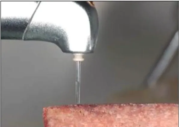
The laser tips were separated by 1 mm from the experimental bone and the laser was applied in a non- contact mode (Fig. 1). Three experiments were executed for each category group with a 15 second laser application for each experiment.
Experimental bones were sectioned and prepared for histologic measurement. Specimens were fixed in 10 % neutral buffered formalin, and decalcified in 5 % nitric acid for 24 hours. After dehydration in ascending graded alcohol, the specimens were embedded in paraffin and sectioned to 4 mm thickness. The sections were stained with hematoxylin and eosin. The histologic measurements were determined using computerized morphometry (SPOT version 4.6, Diagnostic Instruments, Inc. USA) with an Olympus IX70 microscope (Olympus, Tokyo, Japan). The length of the cut bone surface and the width of the entrance following laser application were subjected to repeat
Fig. 1. View of experimental bone and laser tip.
measurements by an oral pathologist. A higher magnification objective and zoom were used to determine whether the bone was prepared by a laser. The result was analyzed with one-way ANOVA (P<0.05) (SPSS 12.0;
SPSS, Chicago, IL, USA).
RESULTS
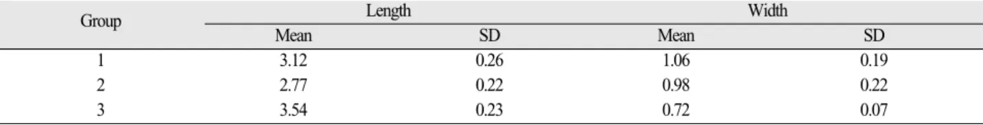
In group 1, the length of the cut was 3.12 mm and the maximum width (the entrance of the prepared bed) was 1.06 mm (Table I). In group 2, the length was 2.77 mm and the maximum width was 0.98 mm. In group 3, the length was 3.54 mm and the maximum width was 0.72 mm.
The mean length of the cut was longer in group 3 than in group 2. The width of the cut was larger than the width of the laser tip in every group.
Additional irregular bone removal was observed adjacent to the prepared edge (Fig. 2). The prepared bed was cone- shaped.
DISCUSSION
Research has revealed the advantages of erbium laser bone ablation.15Erbium laser ablation can provide clean cutting as efficiently as drilling, and the thermal damage to the surrounding area is comparable to that caused by vibration during drilling. The adequacy of bone cutting by Er,Cr:YSGG lasers, in particular, has been discussed in several studies.7,16-19 Eversole et al. found that the Er,Cr:YSGG laser was as efficient as conventional surgical bone wound healing.16 After 24 hours of irradiation, the wound cavities operated on with a Er,Cr:YSGG laser showed a clean cut margin with a thin zone of basophils characteristic of a thermal coagulative effect (40 ~ 60 μm).
Similar results were achieved in the basic histologic appearance and wound healing pattern when the Er:YAG laser was used. 11
In contrast with the popping sound of Er-based laser systems during caries removal, the one current generation Er,Cr:YSGG laser system creates a loud snapping sound even when not in contact with any structure in the mouth.1 Table I. Measurements of laser-prepared bone beds
Group Length Width
Mean SD Mean SD
1 3.12 0.26 1.06 0.19
2 2.77 0.22 0.98 0.22
3 3.54 0.23 0.72 0.07
SD: Standard deviation
Fig. 2. Photographs of cross-sections of bone prepared with laser: (A) group 1, (B) group 2, (C) group 3. Bone removal was observed at the adjacent surface.
A B C
This seeming paradox can be explained by an effect termed
‘plasma de-coupling’of the beam, in which incident laser energy heats the air and water directly in front of the laser handpiece. In the Er,Cr:YSGG laser, this heating is done intentionally in order to deliver energy onto the rear surface of atomized water molecules, with the goal of accelerating them to a higher speed (so-called hydrokinetic cutting).20
In this study, we observed bone removal adjacent to the areas that received direct laser application. We speculate that this was due to a hydrokinetic effect on the surrounding bone. Lasers, by their nature, focus their energy directly forward in a straight beam, but the sprayed water affected by the laser does not necessarily follow the same direction.
As a result, excited water molecules with new vectors may impact the adjacent bone, especially if the bone is weak. To compensate, it is necessary to use less laser implant bed preparation compared with drill bed preparation in clinical applications. Special care will be needed in patients with weak bone quality. Additionally, vertical control of the laser handpiece would be useful, with minimum control to the way of lateral wall. In clinical applications, the laser should not be applied continuously to the same area, and the time of application should be reduced to when the laser tip reaches the final depth for implant installation.
A tapered tip with a 400-micron diameter channels the laser with a high power density, making it good for operations that demand precision and short duration. On the contrary, a parallel-shaped tip with a 600-micron diameter can be used for a wide range of applications. Based on this preliminary research, there were performance differences between the tips. The MZ6� tip is less effective in transforming the laser energy, and so the severed length of the tissue was small. The MT4� tip produced longer cuts because it is characterized by high power density. The performance of the MG6�tip was, on average, between the other two. All of the laser tips removed a larger width of bone compared with the original diameter, irrespective of their straight-forward character. If an original implant surgery protocol using drills is adopted using lasers without alteration, the size of the bone bed prepared by the laser alone may be larger than expected.
Currently, lasers are used not only by physicians (e.g. for incision of soft tissue, excision and coagulation) but also for periodontal uses such as application into periodontal
pockets, removal of calculus and melanin pigments, root canal treatment, tooth preparation, and removal of carious dentin. Laser technology has also been adopted to implant therapy in many cases. Though the protocol for implant surgery is designed for de facto drill usage, many practitioners transfer it blindly to laser implant surgery. New protocols for lasers must be developed and introduced in order to optimize their treatment potential. Research on the best way to utilize laser technology will allow broader usage of lasers and enhance safety and efficacy.
REFERENCES
1. Walsh LJ. The current status of laser applications in dentistry. Aust Dent J 2003;48:146-55.
2. van As G. Erbium lasers in dentistry. Dent Clin N Am 2004;48:1017-59.
3. Eyrich G. Hard-tissue drilling and cutting with a 9.6 um CO2laser. Med Habililationsschrift, Zurich. 2004.
4. Ivanenko M, Sader R, Afilal S, Werner M, Hartstock M, von Hanisch C, Milz S, Erhardt W, Zeilhofer H-F, Hering P. In vivo animal trials with a scanning CO2
laser osteotome. Lasers Surg Med 2005;37:144-8.
5. Aoki A, Sasaki KM, Watanabe H, Ishikawa I. Lasers in nonsurgical periodontal therapy. Periodontol 2000 2004;36:59-97.
6. Eriksson A, Albrektsson T. Temperature threshold lev- els for heat-induced bone tissue injury. A vital micro- scoping study in the rabbit. J Prosthet Dent 1983;
50:101-7.
7. Kimura Y, Yu DG, Fujita A, Yamashita A, Murakami Y, Matsumoto K. Effects of erbium, chromium:YSGG laser irradiation on canine mandibular bone. J Periodontol 2001;72:1178-82.
8. Sasaki KM, Aoki A, Ichinose S, Yoshino T, Yamada S, Ishikawa I. Scanning electron microscopy and Fourier transformed infrared spectroscopy analysis of bone re- moval using Er:YAG and CO2lasers. J Periodontol 2002;73:643-52.
9. Sasaki KM, Aoki A, Ichinose S, Ishikawa I.
Ultrastructural analysis of bone tissue irradiated by Er:YAG laser. Lasers Surg Med 2002;31:322-32.
10. Salina S, Maiorana C, Iezzi G, Colombo A, Fontana F, Piattelli A. Histological evaluation, in rabbit tibiae, of osseointegration of mini-implants in sites prepared with Er:YAG laser versus sites prepared with traditional burs. J Long Term Eff Med Implants 2006;16:145-56.
11. Schwarz F, Olivier W, Herten M, Sager M, Chaker A, Becker J. Influence of implant bed preparation using an Er:YAG laser on the osseointegration of titanium im-
plants: a histomorphometrical study in dogs. J Oral Rehabil 2007;34:273-81.
12. Kesler G, Romanos G, Koren R. Use of Er:YAG laser to improve osseointegration of titanium alloy implant-a comparison of bone healing. Int J Oral Maxillofac Implants 2006;21:375-9.
13. el-Montaser M, Devlin H, Dickinson MR, Sloan P, Lloyd RE. Osseointegration of titanium implant in er- bium-YAG laser-prepared bone. Implant Dent 1999;8:79-85.
14. Rizoiu IM, DeShazer LG. New laser-matter interaction concept to enhance hard tissue cutting efficiency.
Laser-Tissue Interaction 1994;Vol.2134A:309-17.
15. Hossain M, Nakamura Y, Yamada Y, Kimura Y, Matsumoto, N, Masumoto, K. Effects of Er,Cr:YSGG laser irradiation in human enamel and dentin: ablation and morphological studies. Journal of Clinical Laser Medicine and Surgery, 1999b;17:155.
16. Eversole LR, Rizoiu IM. Preliminary investigations on
the utility of an erbium, chromium:YSGG laser. J Calif Dent Assoc 1995;23:41-7.
17. Aoki A, Yoshino T, Akiyama F, Miura M, Kinoshita A, Oda S, Watanabe H, Ishikawa I. Comparative study of Er:YAG laser and rotating bur for bone ablation: SEM and longterm histological examinations. Lasers in Dentistry 2003;389-91.
18. Wang X, Ishizaki NT, Suzuki N, Kimura Y, Matsumoto K. Morpholgoical changes of bovine mandibular bone irradiated by Er,Cr:YSGG laser: an in vitro study. J Clin Laser Med Surg 2002;20:245-50.
19. Wang X, Zhang C, Matsumoto K. In vivo study of the healing processes that occur in the jaws of rabbits fol- lowing perforation by an Er,Cr:YSGG laser. Lasers Med Sci 2005;20:21-7.
20. Riziou I, Kimmel A. Atomized fluid particles for elec- tromagnertically induced cutting. US patent 5,741,247.
1998.
EFFECTS OF THE ER,CR:YSGG LASER ON BONE BED PREPARATION WITH VARIOUS LASER TIPS
Seong-Kyun Kim1, DDS, PhD, Seong-Joo Heo2, DDS, PhD, Jai-Young Koak3, DDS, PhD, Seong-Doo Hong4, DDS, PhD, Shin-Jae Lee5, DDS, PhD, Joo-Hee Lee6*, DDS, PhD
1Assistant Professor, Department of Prosthodontics and Dental Research Institute, School of Dentistry, Seoul National University, Korea
2Professor, Department of Prosthodontics and Dental Research Institute, School of Dentistry, Seoul National University, Korea
3Associate Professor, Department of Prosthodontics and Dental Research Institute, School of Dentistry, Seoul National University, Korea
4Assistant Professor, Department of Oral Pathology, School of Dentistry, Seoul National University, Korea
5Associate Professor, Department of Orthodontics, School of Dentistry, Seoul National University, Korea
6Assistant Professor, Department of Prosthodontics, Asan Medical Center, College of Medicine, University of Ulsan, Korea
STATEMENT OF PROBLEM: Preparation of implant beds with lasers is considered a safe and reliable method, but the accuracy of this technique has not been examined. PURPOSE: The purpose of this study was to evaluate the accuracy and effectiveness of implant bed preparation using an Er,Cr:YSGG laser. MATERIAL AND METHODS: An Er,Cr:YSGG laser was applied to pig rib bone. The laser was employed at a 5.75 W power setting, 30 Hz/sec pulse repetition, and 70 μs pulse duration with 50 % water and 60% air spray. According to laser tips the groups were divided as follows; Group 1: paralleled - shaped sapphire tip (0.6 mmФ), Group 2: paralleled - shaped zirconia tip (0.6 mmФ), Group 3: tapered sapphire tip (0.4 mmФ). The Er,Cr:YSGG laser tip was separated by 1 mm from the bone and applied for 15 seconds in a non-contact mode. After the application, the bone was sectioned for specimens. Histologic measurements were determined by computerized morphometry. The length of the prepared bone surface was measured and the width of the entrance was measured. The re- sults were analyzed with one-way ANOVA (P<0.05). RESULTS: The prepared length of group 3 was longer than that of group 2. The prepared bone width was larger than the width of the laser tip in every group. Additional bone removal was observed adjacent to the pre- pared area and displayed an irregular surface. CONCLUSION & DISCUSSION: Different cutting effects were observed according to the laser tip, emphasizing the importance of proper tip selection in the clinical setting. This preliminary study supported the existence of hydro- kinetic effects.
KEY WORDS: Er.Cr:YSGG laser, Laser tip, Bone cutting length, Bone cutting width, Shape of bone cutting
Corresponding Author: Joo-Hee Lee
Department of Prosthodontics, Asan Medical Center, College of Medicine, University of Ulsan, 388-1, Pungnap-dong, Songpa-gu, Seoul, 138-736, Korea +82 2 3010: e-mail, [email protected] Received April 29, 2008: Last Revision May 13, 2008: Accepted June 2, 2008.