Boiled - Water Extract from Hizikia fusiformis Showing Antioxidant Effects
Mi-Soon Jang, Jiyoung Kim, Chiwon Lim, Yeon-Kye Kim
andHee-Yeon Park*
Biotechonology Research Center, National Fisheries Research & Development Institute, Busan 619-902, Korea Received October 25, 2005; Accepted December 12, 2005
Boiled-water extract (BWE) of effects of BWE, 1,1-dephenyl-2-picrylhydrazyl (DPPH) and thiobarbituric acid (TBA) methods wereHizikia fusiformis has antioxidant effects. To investigate antioxidant used. BWE exhibited strong radical-scavenging activity (87.3% at final conc. 2000 µg BWE/ml in an assay mixture) on DPPH and good inhibitory activity (64% at final conc. 1000 µg BWE/ml in an assay mixture) on lipid peroxidation. Raw 264.7 cells treated with lipopolysaccharide (LPS) showed 26.7-fold increase in nitric oxide (NO) (48.9 µM) compared with control group (1.83 µM). When treated with BWE and LPS, NO production was inhibited by BWE dose-dependently.
Keywords: Hizikia fusiformis, DPPH, TBA, NO, antioxidant Oxidative stress is related to physiological and pathological
conditions such as inflammation, diabetes, shock, arthritis, carcinogenesis, and aging.1-3) Antioxidants can markedly defer or inhibit the oxidation of molecules by inhibiting the initiation or multiplication of oxidative chain reactions.4,5) Synthetic antioxidants, such as butylated hydroxy anisole (BHA) and butylated hydroxy toluene (BHT), are very effective but they may possess mutagenic activity.6) For this reason, many studies have been carried out and some antioxidative substance have been found from natural sources.7-9) Long studies on the natural antioxidants using a variety of seaweeds and plants as dietary and medicinal sources revealed they have antioxidant, antimutagenic, antitumor, and anticoagulant activities and play very important roles in lipid metabolism.10) In particular, Hizikia fusiformis, distributed in the southern and western coastal areas of Korea including Jeju Island, has been extensively studied as a resource of dietary fiber.10-14) Organic solvent extracts of H. fusiformis have been reported to possess strong antioxidant and antitumor activities.12) In addition, its hot water extract have strong anticoagulant and antitumor activities.13,14)
Nitric oxide (NO) is a multifunctional biomolecule involved in many physiological and pathological processes. In the immune system, NO is generated by inducible NO synthase (iNOS) in response to inflammatory stimuli such as lipopoly-
saccharide (LPS) and interferon-γ (IFN-γ) in macrophages15,16) and acts as a cytotoxic agent against invading microorganisms and tumor cells; however, excess NO is related to inflammation, septic shock, rheumatoid arthritis, and autoimmune diseases.17)
In this study, antioxidant effects of boiled-water extract of Hizikia fusiformis (BWE), which have not been fully investigated were examined.
Materials and Methods
Materials.L-Ascorbic acid, BHA, DPPH, Griess reagent, linoleic acid, 3-[4,5-dimetylthiazol-2-yl]-2,5-diphenyltetrazolium bromide (MTT), and TBA were purchased from Sigma Chemicals Co. (St. Louis, MO, USA). Trichloroacetic acid (TCA) was obtained from Lancaster (Eastgate, UK). Raw 264.7, a mouse macrophage cell line, was purchased from Korean Cell Line Bank. Dulbecco’s Modified Eagle Medium (DMEM) and fetal bovine serum (FBS) were obtained from WelGENE Inc. (Korea). H. fusiformis was collected from Wando, Chonnam.
PreparationofH. fusiformis extract.H. fusiformis was rinsed carefully in freshwater and steam-heated to 120oC at 2.5 kg/cm2 for 50 min. One liter of the obtained layer was mixed with 3l of 100% ethanol. After centrifugation at 5,000 rpm for 20 min, the supernatant was separated, dissolved with 75% ethanol, and filtered through Whatman No 2. filter paper.
The filtrate was dried on a rotary evaporator (EYELA, Japan) under reduced pressure at 40oC. The residue was re-extracted with 75% ethanol evaporated in vacuo freeze-dried to give powered extract, and stored in a desiccator at room temperature until use.
DPPH free radical-scavenging method. DPPH free radical-scavenging assay measures the ability of antioxidants to scavenging free radicals. This assay was performed according to the method of Blois18) with some modification.
Namely, 100µl of various concentrations (final conc. 0.1~
*Corresponding author
Phone: 82-51-720-2457; Fax: 82-51-720-2456 E-mail: [email protected]
Abbreviations: BWE, boiled-water extract from Hizikia fusiformis; DPPH, 1,1-dephenyl-2-picrylhydrazyl; TBA, thiobarbituric acid; LPS, lipopolysaccharide; NO, nitric oxide; BHA, butylated hydroxy anisole;
BHT, butylated hydroxy toluene; iNOS, inducible nitric oxide synthase;
IFN-γ, interferon-γ; DMEM, Dulbecco’s Modified Eagle Medium; FBS, fetal bovine serum, TCA, trichloroacetic acid; MTT, 3-[4,5-dimetylthia- zol-2-yl]-2,5-diphenyltetrazolium bromide.
2000µg each sample/ml in an assay mixture) ofsample were added to 900µl of DPPH solution. DPPH solution was prepared as 39µg · ml−1 in ethanol. The reaction mixture was shaken vigorously for 20 sec and left at room temperature for 10 min. Subsequently, the amount of DPPH remaining was determined using a UV-visible spectrophotometer (Mecasy, Korea) at 518 nm. DPPH free radical-scavenging activity (electron-donating activity) was calculated using the following formula:
Electron donating ability (EDA) (%)= ×100 TBAmethod.The level of lipid peroxidation, in terms of thiobarbituric acid reactive substances (TBARS), was estimated by TBA reaction. A mixture consisting of 80 mg sample in 5 ml ethanol or distilled water, 5 ml of 0.2 M phosphate buffer (pH 7.0), and 10 m of 20% linoleic acid in ethanol was incubated in a vial at 40oC for 96 h. The mixture (2 ml) was combined with 1 m of 35% TCA and 1.5 ml of 0.75% TBA, adjusted with distilled water to a final volume of 5 ml, and heated to 95oC for 40 min. After cooling to room temperature, 1 ml acetic acid and 2 ml chloroform were added to each sample, and the mixture (final conc. 1000µg each sample/ml
in a mixture) was shaken. After centrifugation at 3,000 rpm for 5 min, the supernatant was isolated, and the absorbance was measured spectrophotometrically at 532 nm. Lipid peroxidation inhibitry activity was calculated using the following formula:
Inhibitory activity (%) = ×100
Cellculture.Raw 264.7 cells were cultured in DMEM with 10% FBS at 37oC in a CO2 incubator (SANYO, Japan). BWE was dissolved in the culture medium. LPS was dissolved in distilled water and diluted in the culture medium.
Cellviabilityassay.Cell viability was determined by the MTT method. The cells were plated at 1×10⁵ cells/well in 96- well plates and incubated for 24 h. One hour after BWE (final conc. 1~100µg BWE/ml in a well) treatment, the cells were treated with LPS (final conc. 1µg LPS/ml in a well) and incubated for 24 h. Subsequently, 10µl MTT stock solution (5 mg · ml−1 in PBS) was added to the cells, which were then incubated at 37oC and 5% CO2 for 4 h. The culture supernatant was removed, and the formazan crystals, formed from MTT by NADH-generating dehydrogenases in metabolically active cells, were dissolved in 150µl DMSO : EtOH (1 : 1). Cell viability was evaluated in comparison to the control group (taken as 100%) by measuring the intensity of the blue color using a microplate reader (Bio-Tek, USA).
Measurement of NO production. Nitrite as the end product of NO was measured by the Griess reagent (1%
sulfanilamine, 0.1% naphthylethylenediamine dihydrochyloride in 2.5% phosphoric acid), as an indirect assay procedure for NO production.19) The cells were seeded at 1×10⁵ cells/well in 96-well plates treated with LPS (final conc. 1µg LPS/ml in
a well) for 24 h, and added with various concentrations of BWE (final conc. 1~100µg BWE/ml in a well) 1 h before LPS treatment. The cells were then placed in a growth medium lacking phenol red and 50µl culture medium was mixed with an equal volume of the Griess reagent and incubated at room temperature for 10 min. Nitrite concentration was determined by measuring the absorbance at 560 nm in a microplate reader (Bio-Tek, USA). The calibration curve was prepared using sodium nitrite (Sigma Chemicals Co.) as a standard.
Statistics. All experiments were repeated at least three times. Statistical analysis was performed using a one-way ANOVA, followed by LSD in SPSS version 10 (SPSS Inc., USA). p-Values less than 0.05 were considered significant.
Results and Discussion
EffectofBWEonDPPHfreeradical-scavengingactivity.
DPPH, a free radical, was used to investigate the effect of BWE on the radical-scavenging activity. DPPH directly abstracted hydrogen atoms from the phenolic compounds at a rapid rate. This reaction, which can be observed directly though the change in color from violet to yellow, can also be measured by the decrease in absorbance at 518 nm.20)
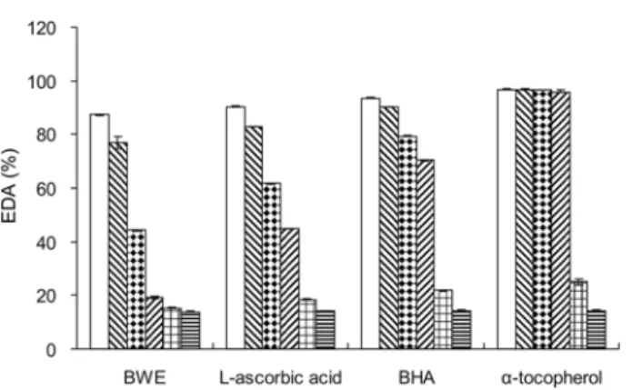
Changes in DPPH free radical-scavenging activity with varying contents of BWE added were compared against the reference samples added with BHA, L-ascorbic acid, and α- tocopherol (Fig. 1). BWE caused a dose-dependant DPPH free radical-scavenging activity exhibiting the strongest scavenging activity (87.3%) at final conc. 2000µg BWE/ml
in an assay mixture, and 77.07 and 44.23% at final conc. 1000 and 100µg/ml in an assay mixture, respectively. BHA and a- tocopherol showed stronger activity than BWE. Kang et al.21) reported that the hot water extract of Angelica gigas showed 66.3% at final conc. 1000µg · ml−1 and 27.1% inhibition at Acontrol–Asample
Acontrol
---
Acontrol–Asample
Acontrol ---
Fig. 1. Effects of BWE on DPPH free radical-scavenging activity.
Values indicate means±S.E.M. (n = 3). □, with final conc.
2000µg each sample/mlin an assay mixture; ▨, with final conc. 1000µg each sample/ml in an assay mixture; ▒, with final conc. 100µg each sample/mlin an assay mixture; ▧, with final conc. 10µg each sample/mlin an assay mixture; ▦, with final conc. 1µg each sample/ml in an assay mixture; ▤, with final conc. 0.1µg each sample/ml in an assay mixture.
final conc. 100µg · ml−1 in the same assay system. These results demonstrate that even though BWE was lower than BHA or α-tocopherol in radical-scavenging activity, BWE also have strong DPPH free radical-scavenging activity.
Effect of BWE on lipid peroxidation. Effect of BWE on lipid peroxidation during incubation at 40oC are shown in Fig.
2. The inhibitory activity of BWE on lipid peroxidation increased time-dependently, 64% at final conc. 1000µg/ml in a mixture after 72 h incubation. BHA and α-tocopherol also significantly inhibited lipid peroxidation, 81.66 and 77.54% at final conc.
1000µg/ml in a mixture, respectively. However, L-ascorbic acid, a known antioxidant, showed higher lipid peroxidation than that of the positive control. Similar results were obtained by Franker et al.22) and Jang et al.23), who reported that hydrophilic antioxidants such as L-ascorbic acid and catechin were more effective in bulk oil than in oil-in-water emulsion system. In the oil-in-water system, the hydrophilic antioxidants are dissolved and become diluted during the water phase.22) These results suggest that, although the inhibitory activities on lipid peroxidation of BWE were lower than those of BHA or
α-tocopherol, BWE nevertheless significantly inhibited lipid peroxidation.
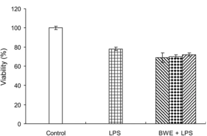
Effect of BWE on cell viability. To confirm the effect of BWE on cell viability, the cells were initially treated with BWE (final conc. 1~100µg BWE/ml in a well) followed by stimulation with LPS (final conc. 1µg LPS/ml in a well).
After 24 h incubation, cell viability was quantified. The control group showed 100% cell viability, whereas that of LPS-treated group was 78%. BWE and LPS-treated group showed 72%, 70%, and 69% cell viability at final conc. 1, 10, and 100µg BWE/ml in a well,respectively (Fig. 3). Results of this experiment revealed that BWE had a considerable effect on cytotoxic effects. BWE is still crude extract therefore, further research will be necessary for the identification of the effective compound.
Effect of BWE on LPS-induced NO production. Excess NO reacts with superoxide to form spontaneously the potent oxidant peroxynitrite. Thus, inhibitory effects of NO production are related to antioxidant effects. To clarify whether BWE could inhibit LPS-induced NO production, BWE (final conc.
1~100µg BWE/ml in a well) was added to the cells 1 h prior to treatment with LPS (final conc. 1µg LPS/ml in a well) for 24 h and nitrite concentration was analyzed (Fig. 4). LPS treatment significantly increased NO concentration by
Fig. 2. Effects of BWE on inhibition lipid peroxidation dur- ing incubation at 40oC. Values indicate means±S.E.M. (n = 3).
○, BWE; ●, L-ascorbic acid; □, BHA; ■, α-tocopherol. All samples were measured at final conc. 1000 µg/ml in a mixture.
Fig. 3. Effects of BWE on cell viability in LPS-treated Raw 264.7 cells. Cell viability was determined by MTT method. The cells (1×10⁵ cells/well) were plated on 96-well plates for 24 h, treated with LPS (final conc. 1µg LPS/ml in a well) for 24 h.
BWE (final conc. 1~100µg BWE/ml in a well) was added 1 h before LPS treatment. Values indicate means±S.E.M. (n = 3).
control (□), without BWE and LPS; LPS (▦), with final conc.
1µg LPS/ml in a well; BWE + LPS (▨), with final conc. 100
µg BWE and 1µg LPS/ml in a well; HWE + LPS (▒), with final conc. 10µg BWE and 1µg LPS/ml in a well; HWE + LPS (▧), with final conc. 1µg BWE and 1µg LPS/ml in a well.
Fig. 4. Effects of BWE on NO production in LPS-treated Raw 264.7 cells. NO production was assessed using Griess reagent. The cells (1×10⁵cells/well) were plated and treated with LPS (final conc. 1µg LPS/ml in a well) for 24 h. BWE (final conc. 1~100µg BWE/ml in a well) was added 1 h before LPS treatment. Values indicate means±S.E.M. (n = 3). control (□), without BWE and LPS; LPS (▦), with final conc. 1µg LPS/ml
in a well; BWE + LPS (▨), with final conc. 100µg BWE and 1µg LPS/ml in a well; HWE + LPS (▒), with final conc.
10µg BWE and 1µg LPS/ml in a well; HWE + LPS (▧), with final conc. 1µg BWE and 1µg LPS/ml in a well. Significant differences from the control are p< 0.05 (*).
approximately 26.7-fold compared with the control group (1.83µM). The cells treated with BWE showed a reduction in LPS-induced NO production in a concentration manner. In particular, the cells treated with BWE (containing final conc.
100µg BWE/ml in a well) showed significant inhibitory effects. These results might indicate that BWE could have anti-inflammatory effect by reducing NO production. However, as NO production is regulated by iNOS in various cell types including macrophage, further investigation is needed to confirm whether the decreased NO production is correlated with anti-inflammatory effect. In addition to NO, many proinflammatory mediators such as tumor necrosis factor-α, interleukins, and prostaglandin E2 have been suggested to be involved in various pathophysiological processes of inflammation.24-26) It is known that the anti-inflammatory action of drugs is achieved is by inhibiting other pro- inflammatory mediators as well as NO.27) In addition, further study is needed to determine anti-inflammatory effects in vivo, and to examine whether BWE could suppress pro- inflammatory mediators.
This study examined whether BWE can scavenge free radical, inhibit lipid peroxidation, and suppress NO production in LPS-treated macrophages. It was demonstrated that BWE has antioxidant effects by showing the activities of free radical-scavenging and lipid peroxidation inhibiting. It is also likely that BWE can exert anti-inflammatory effect through the suppression of NO production.
Acknowledgment
This work is funded by a grant from the National Fisheries Research and Development Institute (RP-2005-BT-012).
References
1. Chae, S. W., Kim, J. S., Kang, K. A., Bu, H. D., Lee, Y.
K., Hyun, J. W. and Kang, S. S. (2004) Antioxidant Activ- ity of Jionoside D from Clerodendron trichotomum. Biol.
Pharm. Bull. 27, 1504-1508.
2. Kang, K. A., Lee, K. H., Chae, S. W., Zhang, R., Jung, M.
S., Kim, S. Y., Kim, H. S., Kim, D. H. and Hyun. J. W.
(2005) Cytoprotective effect of tectorigenin, a metabolite formed by transformation of tectoridin by intestinal microf- lora, on oxidative stress induced by hydrogen peroxide. Eur.
J. Pharmacol. 519, 16-23.
3. Saha, K., Lajis, N. H., Israf, D. A., Hamzah, A. S., Khozirah, S., Khamis, S. and Syahida, A. (2004) Evalua- tion of antioxidant and nitric oxide inhibitory activities of selected Malaysian medicinal plants. J. Ethnopharmacol.
92, 263-267.
4. Gülçin, ., Büyükokuro lu M. E., Oktay, M. and Küfre- vioglu Ö. . (2003) Antioxidant and analgesic activities of turpentine of Pinus nigra Arn. subsp. pallsiana (Lamb.) Holmboe. J. Ethnopharmacol. 86,51-58.
5. Javanmardi, J., Stushnoff, C., Locke, E. and Vivanco, J. M.
(2003) Antioxidant activity and total phenolic content of Iranian Ocimum accessions. Food Chem. 83, 547-550.
6. Namiki, M. (1990) antioxidant, antimutagens in food. Crit.
Res. Food Sci. Nutr. 29, 273-300.
7. Ramarathnam, N., Osawa, T., Ochi, H. and Kawakishi, S.
(1995) The contribution of plant food antioxidants to human health. Trends Foor Sci. Technol. 6, 75-82.
8. Hu, C. and Kitts DD. (2000) Studies on the antioxidant activity of Echinacea root extract. J. Agric. Food Chem. 48, 1466-1472.
9. Zhang, K. Q., Bao, Y., u, P., Rosen, R. T. and Ho, C. T.
(1990) Antioxidative components of Tanshen (Salvia miltio- rrhiza Bung). J. Agric. Food Chem. 38, 1194-1197.
10. Jung, B. M., Ahn, C. B., Kang, S. J., Park, J. H. and Chung, D. H. (2001) Effects of Hijikia fusiformis extracts on lipid metabolism and liver antioxidative enzyme activi- ties in triton-induced hyperlipidemic rats. J. Korean Soc.
Food Sci. Nutr. 30, 1184-1189.
11. Kim, S. A., Kim, J., Woo, M. K., Kwak, C. S. and Lee, M.
S. (2005) Antimutagenic and cytotoxic effects of ethanol extracts from five kinds of seaweeds. J. Korean Soc. Food Sci. Nutr. 34, 451-459.
12. Yan, X., Chuda, Y., Suzuki, M. and Nagata, T. (1999) Fucoxanthin as the major antioxidant in Hijikia fusiformis, a common edible seaweed. Biosci. Biotechnol. Biochem. 63, 605-607.
13. Kim, K. I., Seo, H. D., Lee, H. S., Jo, H. Y. and Yang, H.
C. (1998) Studies on the blood anticoagulant polysaccaride isolated from hot water extracts of Hizikia fusiformis. J.
Korean. Soc. Food Sci. Nutr. 27,1204-1210.
14. Ryu, B. H., Kim, D. S., Cho, K. and Sin, D. B. (1989) Antitumor activity of seaweeds agarne sarcoma-180. Kor. J.
Food. Sci. Technol. 21, 595-600.
15. Morikawa, A., Koide, N., Sugiyama, T., Mu, M. M., Has- san, F., Islam, S., Ito, H., Mori, I., Yoshida, T. and Yoko- chi, T. (2004) The enhancing action of D-galactosamine on lipopolysaccharide-induced nitric oxide production in RAW 264.7 macrophage cells. FEMS Immunol. Med. Microbiol.
41, 211-218.
16. Yoo, H. Y., Lim, Y. J., Park, S. E., Kim, J. M. and Park, Y.
C. (2004) Overexpression of redox factor-1 negatively regu- lates NO synthesis and apoptosis in LPS-stimulated RAW 264.7 macrophages. FEBS Lett. 556, 39-42.
17. Kim, Y. H., Moon, J. S., Lee, K. S., Park, S. Y., Cheong, J.
H., Kang, H. S., Lee, H. Y. and Kim, H. D. (2004) Ca2+/ calmodulin-dependent protein phosphatase calcineurin medi- ates the expression of iNOS through IKK and NF-κB activ- ity in LPS-stimulated mouse peritoneal macrophages and RAW 264.7 cells. Biochem. Biophys. Res. Comm. 314, 695- 18. Blois, M. S. (1958) Antioxidant determination by the use of703.
a stable free radical. Nature26, 1199-1200.
19. Tunçtan, B., Altug, S., Uludag, O., Demirkay, B. and Aba- cioglu, N. (2004) Effects of cyclooxygenase inhibitors on nitric oxide production and survival in a mice model sep- sis. Pharmacol. Res. 48, 37-48.
I· I·
20. Ancerewicz, J., Migliavacca, E., Carrupt, P. A., Testa, B., Brée, F., Zini, R., Tillement, J. P., Labidalle, S., Guyot, D., Chauvet-Monges, A. M., Crevat, A. and Ridant, A. L.
(1998) Structure-property relationships of trimetazidine derivatives and model compounds as potential antioxidants.
Free Rad. Biol. Med. 25, 113-120.
21. Kang, S. A., Han, J. A., Jang, K. H. and Choue, R. W.
(2004) DPPH radical scavenger activity and antioxidant effects of Cham-Dang-Gui (Angelica gigas). J. Korean Soc.
Food Sci. Nutr. 33, 1112-1118.
22. Frankel, E. N., Huang, S. W., Kanner, J., and German J. B.
(1994) Interfacial pehnomena in the evaluation of antioxi- dants: bulk oils vs emulsions. J. Agric. Food Chem. 42, 1054-1059.
23. Jang, M. S., Eun J. B., Ushio H., and Ohshima T. (2004) Antioxidative properties of mushroom Flammulina veluti- pes crude extract on the oxidation of cod liver oil in emul- sion. Food Sci. Biotechnol. 13, 215-218.
24. Ilieva, I., Ohgami, K., Shiratori, K., Koyama, Y., Yoshida, K., Kase, S., Kitamei, H., Takemoto, Y., Yazawa, K. and
Ohno, S. (2004) The effects of Ginkgo biloba extract on lipopolysaccharide-induced inflammation in vitro and in vivo. Exp. Eye. Res. 79, 181-187.
25. Lee, E. S., Ju, H. K., Moon, T. C., Lee, E. K., Jahng, Y. D., Lee, S. H., Son, J. K., Baek, S. H. and Chang, H. W.
(2004) Inhibition of nitric oxide and tumor necrosis factor-α
(TNF-α) production by propenone compound through blockade of nuclear factor (NF)-κB activation in cultured murine macrophages. Biol. Pharm. Bull. 27, 617-620.
26. Xaus, J., Comalada, M., Valledor, A. F., Lloberas, J., Lopez-Soriano, F., Argilies, J. M., Bogdan, C. and Celada, A. (2000) LPS induces apoptosis in macrophages mostly through the autocrine production of TNF-α. Blood 95, 3823-3831.
27. Kim, R. G., Shin, K. M., Kim, Y. K., Jeong, H. J., Ha, J.
H., Choi, J. W., Park, H. J. and Lee, K. T. (2003) Inhibi- tion of methanol effect from aerial parts of Saururus chin- ensis on lipopolysaccharide-induced nitric oxide and prostaglandin E2 production from murine macrophage Raw 264.7 cells. Biol. Pharm. Bull. 24,481-486.