ISSN 2288-1069 (Online)
http://dx.doi.org/10.12925/jkocs.2020.37.6.1545
Effect of Aucklandia lappa Decne Extract on Antioxidant
Hea-Jin Lee
*․Hyun-Ji Lim․Mi-Hye Lim
✝Department of Beauty Healthcare, Daejeon University
(Received November 28, 2020; Revised December 9, 2020; Accepted December 11, 2020)
Abstract : In the present study, we investigated the antioxidant activity of Aucklandia lappa Decne (AL). Cell viability was measured in an MTT assay. Antioxidant effects were evaluated based on total polyphenol/flavonoid contents, ABTS radical scavenging activity, DPPH radical scavenging activity, SOD activity, and ROS content. AL was found not to be toxic at concentrations of 1 μg/mL, 10 μ g/mL, and 100 μg/mL, respectively. The phenolic content was higher in AL-D than in AL-E, while the flavonoid content was higher in AL-E than in AL-D. AL-E exhibited higher ABTS radical scavenging activity than AL-D, and the EC
50values for BHA were 217.1 μg/mL in AL-D and 180.5 μg/mL in AL-E. AL-E also showed the highest DPPH radical scavenging activity. EC
50values for BHA were 114.2 μg/mL in AL-D and 95.8 μg/mL in AL-E. The SOD-like activity of AL-E was higher than that of AL-D. The EC
50values for ascorbic acid were 48.5 μg/mL in AL-D and 72.9 μ g/mL in AL-E, indicating that both AL extracts have a SOD activity higher than that of ascorbic acid. AL-E reduced relatively more ROS than AL-D. With 100 μg/mL AL-E, the reduction level was almost similar to that of dexamethasone. Our results demonstrate that AL have antioxidant effects, and we believe that they could be very valuable as raw materials for anti-aging products, based on their antioxidant activity.
Keywords : Aucklandia lappa Decne, Antioxidant, raw material, Medicinal herb, Natural products analysis
1. Introduction
Free radicals are fundamental to any biochemical process and represent an essential part of aerobic life and metabolism. Free radicals have a effect on growth and differentiation of cell in the processes of respiration and metabolism. And it defenses body from antigen. The free radicals produced
✝
Corresponding author (E-mail: [email protected])
in the body is decomposed into water and CO
2on normal procedure to finally discharge.
However, free radicals of about 2-4% remain harmful free radicals which have strong reactivity[1]. Free radical such as H
2O
2, O
2-, hydroxyl radical and ·OH cause oxidative stress[1-3].
Oxidative stress is caused by oxidative damage occurring from an imbalance between pro-oxidants and antioxidants in the cell.
Oxidative stress can cause impaired bio-
membrane ion-transport and protein
degradation[4], serious abnormalities of cellular
metabolism including lipid peroxidation[5], liver cirrhosis, fatty liver, arteriosclerosis, cardiovascular diseases[6], cancer[7], aging[8]
and DNA damage[9]. Oxidative stress also triggers the release of inflammatory factors[10].
To cope with oxidative stress, the body generates antioxidants enzymes such as SOD, catalase, glutathion peroxidase, NAD(P)H:
Quinone reductase and the thioredoxin system which are called detoxication enzymes.
However, when excess amounts of reactive oxygen are produced and these enzymes are limited, supplementary antioxidants have to be provided to prevent or remediate oxidative stress[11-13]. Synthetic antioxidants such as BHA and BHT have been developed as food supplements, cosmetics, or drugs to prevent oxidation. With regard to food additives, there are strict regulations on the target food and the amount of antioxidant that can be included because of safety disputes[14-16].
Synthetic antioxidants that act directly on the skin can cause a variety of side effects such as cancer, chromosomal abnomality, up-regulation of cholesterol and symptom of calcium- scarcity[17-18]. Thus, the quality and safety of this natural antioxidant is very important [17-18]. The various functional cosmetics are developed on natural materials with antioxidant effects[8,10,14,17-18].
Aucklandia lappa Decne (AL) is included in the “National Standard of Traditional Medicinal (Herbal and Botanical) Materials” as it is used to treat various diseases including indigestion, abdominal inflation, diarrhea and dysentery[19]. Morphologically, AL is characterized by its conical shape, with a length of 10∼30 cm and a diameter of 1∼3 cm. It is dark brown/brown in color, has many wrinkles, and has soft/tough texture[20].
AL grows abundantly in Yunnan and Gamsuk of China. AL contain sesquiterpene and sesquiterpene lacton. In addition to polyene alcohols, triterpene, lignams, amino acid-sesquiterpene, alkaloids, tannins and other componants have also been reported[21].
Costunolide and dehydrocostus lactone are best known for therapeutic components of AL.
These compounds are effective against gastrointestinal hyperkinesis[22] and have antiulcer and cholagogue activities[23]. In oriental medicine, AL are used for pain relief, to treat weakened spleens, to aid in food digestion, to treat acute diarrhea, or to help loss of appetite[24]. A number of studies investigated the component/pattern analysis of AL[25], their toxicity on A549 cells[26], lipid induction[27], anti-cancer activities[28], prostate cancer cell growth inhibition[29], insulin resistance[24] and hepatocellular protection[30]. In this study, we investigated the effect of AL on antioxidant activities to evaluate its possible use in the care of skin.
2. Materials and Methods
2.1. Aucklandia lappa Decne extract Aucklandia lappa Decne was purchased from an Omniherb (Hallym Farm, Daegu, Korea).
Aucklandia lappa Decne was used in manner of the reflux extraction in 80% ethanol (C
2H
5OH) and DW at 80℃ for 3 hours, followed by filteration over paper (No. 2, Whatman, USA). The filtrate was evaporated by rotary vacuum evaporator. After the remains was freeze-dried in freeze dryer. Dried extract was dissolved in DW at a concentration of 20 mg/mL and then stored at -70℃ until use. The dried extract yields are each 16.80% in AL-E (80% ethanol extraction) and 16.40% in AL-D (AL water extraction).
2.2. HPLC pattern analysis
We carried out HPLC (Shimadzu, Japan)
pattern analysis on the AL formulation to
evaluate consistency. The AL extracts were
analyzed by HPLC under the conditions listed
in Table 1.
Table 1. HPLC conditions
Column : Agilent Eclipse C18 (250×4.6) mm, 5 μm UV : 215 nm
Flow : 1.0 mL/min Inj. Vol. : 30 μL
Temp. : 40°C Eluent : A (DW), B (Acetonitrile)
Time (min) 0 20 25 40 50
A (%) 80 10 10 0 0
B (%) 20 90 90 100 100
2.3. Cell culture
Mouse origin Raw 264.7 cells (Korea Cell Line Bank, Seoul, Korea) were grown at 37℃
in a humidified 5% CO
2incubator (Forma scientific, USA) in Dulbecco’s modified eagle's medium (DMEM; Gibco BRL Co., USA), containing 10% FBS (Gibco BRL Co., USA) and 1% antibiotic-antimycotic (A/A; Gibco BRL Co., USA) until 85% confluence.
2.4. MTT Assay
Raw 264.7 cell viability was assessed in an MTT assay. Cells were seeded in a 96 well plate (2×10
4cells/well) and grown at 37℃ in a humidified 5% CO
2incubator in DMEM containing 10% FBS and 1% A/A for 24h.
Cells were incubated with AL at 1 μg/mL, 10 μg/mL, or 100 μg/mL. After 1 day, 10 μL of MTT solution (Daeil Lab service, Korea) was added to each well. After incubating for 30 min, absorbance was measured at 450 nm using a microplate reader (Molecular Devices, USA).
2.5. Total polyphenol content analysis Total phenolics were determined using FCR as described by Velioglu et al. (1998)[31] with slight modifications. Briefly, AL extracts dissolved in DW (10 mg/mL) was mixed with 500 μL of Foiln-Ciocalteu's phenol reagent, ethanol (FCR; Sigma Co., USA) and allowed to sit at room temperature for 3 min. Then, 1 mL of Na
2CO
3solution and 7.5 mL of DW were added to the mixture. After 40 min, absorbance was measured at 760 nm. Results were expressed as gallic acid (GAE; Sigma
Co., USA) per gram.
2.6. Total flavonoid content analysis The flavonoid content was determined according to the method described by Kumaran and Karunakaran (2006)[32] with slight modifications. Briefly, 500 μL of extract (10 mg/mL) in ethanol (diluted 10-fold) was mixed with 100 μL of 10% aluminum nitrate (Sigma Co., USA), 100 μL of 1 M potassium acetate (Sigma Co., USA) and 4.3 mL of 80%
ethanol. Absorbance was measured at 415 nm after 40 min. The amount of flavonoids in the extract in AL was expressed in terms of quercetin (Sigma Co., USA) (μg/mL).
2.7. ABTS radical scavenging assay The scavenging activity of the extract on ABTS (Sigma Co., USA) free radicals was assessed according to the method reported by Arnao et al. (2001)[33] with slight modifications. Briefly, a 5 μL solution of the extract at different concentrations (1 μg/mL, 10 μg/mL, or 100 μg/mL) was mixed with 95 μL of 7.4 mM ABTS in PBS. The mixture was shaken vigorously and allowed to stand at room temperature in the dark for 10 min.
Blank solutions were prepared for each test
sample solution (5 μL) and 95 μL of PBS
while the negative control was 95 μL of 7.4
mM ABTS solution plus 5 μL of DW. BHA
(Sigma Co., USA) was used as the positive
control. The absorbance (optical density, or
OD) of the assay mixtures was measured at
732 nm. ABTS radical inhibition was
calculated using the equation: ABTS radical
scavenging activity (%) = (Blank OD-Sample OD)/Blank OD×100
2.8. DPPH radical scavenging assay The scavenging activity of the extract on DPPH (Sigma Co., USA) free radicals was assessed according to the method reported by Gyamfi et al. (1999)[34] with slight modifications. Briefly, a 100 μL solution of the extract at different concentrations (1 μ g/mL, 10 μg/mL, or 100 μg/mL) was mixed with 150 μL of 0.5 mM DPPH in ethanol.
The mixture was shaken vigorously and allowed to stand at 37℃ in the dark for 30 min. Blank solutions were prepared with each test sample solution (100 μL) and 150 μL of ethanol while the negative control was 150 μ L of 0.5 mM DPPH solution plus 100 μL of DW. BHA was used as the positive control.
The absorbance of the assay mixture was measured at 517 nm. DPPH radical inhibition was calculated using the equation: DPPH radical scavenging activity (%) = (Blank OD-Sample OD)/Blank OD×100
2.9. SOD-like activity assay
The SOD (Sigma Co., USA) activity of the extract was assessed according to the method reported by Martin et al. (1987)[35] with slight modifications. Briefly, a 200 μL solution of the extract at different concentrations (1 μ g/mL, 10 μg/mL, or 100 μg/mL) was mixed with 2.6 mL of 50 mM Tris-HCl buffer (Sigma Co., USA) and 0.2 mL of pyrogallol (Sigma Co., USA). The mixture was shaken vigorously and allowed to stand at 37℃ in the dark for 10 min. Subsequently, 100 μL of 1 N HCl was added to the mixture and the absorbance of the assay mixture was measured at 420 nm. L-ascorbic acid (vitamin C) (Sigma Co., USA) was used as the positive control. SOD activity was calculated using the equation: SOD activity (%) = (Blank OD- Sample OD)/Blank OD×100
2.10. ROS assay
ROS reduction by the extract was assessed according to the method reported by Lee et al.
(2014)[10] with slight modifications. Raw 264.7 cells were seeded in a 12 well plate (2×10
5cells/well) and grown at 37℃ in a humidified 5% CO
2incubator in DMEM containing 10% FBS and 1% antibiotic- antimycotic for 24 h. Cells were incubated with extract at a concentration of 1 μg/mL, 10 μg/mL, or 100 μg/mL and LPS (Sigma Co., USA) at a concentration of 1 μg/mL.
After 1 day, 100 μL of 10 μM DCF-DA (Sigma Co., USA) was added to the pellet and the mixture was incubated at 37℃ in the dark for 15 min. ROS levels were measured by flow cytometery (Becton Dickinson, USA).
ROS inhibition was calculated using the equation: ROS inhibition (%) = Sample FI/
Control FI×100
2.11. Analysis
Results are expressed as mean±standard deviation. Statistical significance was evaluated with a one-way analysis of variance using SPSS ver. 18.0 (Chicago, IL, USA).
3. Results and Discussion
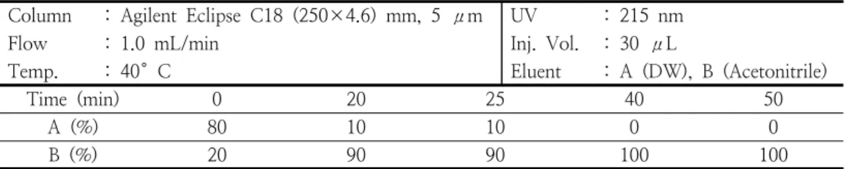
3.1. HPLC pattern analysis of AL extracts Analysis result of AL extract were able to identify that costunolide was eluted at 22.62 min. Dehydrocotus lactone was eluted at 21.11 min (Fig. 1-A, B). Also, pattern of AL extracts shows a difference in the content of the two representative compounds (costunolide and dehydrocostus lactone) according to the solvent (Fig. 1-C, D). The content of costunolide and dehydrocostus lactone was each 28.256 mg/L or 21.169 mg/L in AL-E (Fig. 1-C). AL-D was each 1.449 mg/L or 3.009 mg/L (Fig. 1-D). The result was higher in AL-E than in AL-D (Fig. 1-C, D).
Costunolide and dehydrocostus lactone are the
Fig. 1. HPLC pattern analysis of AL extracts. Costunolide (A), dehydrocostus lactone (B). AL 80% ethanol extraction (C) and AL water extraction (D).
Fig. 2. Cell viability of Raw 264.7 cells after AL extracts treatment. Cell viability was measured in an MTT assay. The results are expressed as the mean ± standard deviation of three independent experiments.
representative components of AL. The compound analysis by HPLC corroborates previous reports on AL[25,36]. Costunolide and dehydrocostus lactone have been suggested to possess various biological activities, including anti-tumor, anti-ulcer, antioxidant and anti-inflammatory effects[37-39]. Their levels were significantly higher in ethanol extracts than in water extracts. In addition, the levels of costunolide were higher than those of dehydrocostus lactone. It is thought that the difference of content in ALE and ALD be able to affect the anti-oxidation.
3.2. Cell viability
Treatment with AL extracts at a concentration of 1 μg/mL, 10 μg/mL, or 100 μg/mL caused no cytotoxicity in Raw 264.7 cells (Fig. 2). Therefore, we concluded that the extracts can be used for cellular treatments at the indicated concentrations in our
experiments.
3.3. Total polyphenol and flavonoid contents The total polyphenol contents of AL extracts were measured using gallic acid as reference material. The results are shown in Table 2.
The total flavonoid content of AL extracts were measured using gallic acid as a reference material. Results are shown in Table 2.
Phenolic component which reacts during the oxidation-reduction as a substrate has the hydroxy (-OH) group in the structure[40-41].
Hydroxy Group has a nature that combine with other compounds easily[42]. Phenolic component is reported to have the antioxidant and anti-microbial effects because of its propensity to combine with protein[40].
Flavonoid is reported that flavonoid degrade
production of ROS[43]. Polyphenol and
flavonoid are reported effect of antivirus, anti-
inflammation and anti-cancer[43]. Especially,
Table 2. Total phenolic or flavonoid content of AL extracts
Sample Total phenolic contents (GAE μg/mL) Total flavonoid contents (quercetin μg/mL)
AL-D
1342.2±9.5 78.4±3.5
AL-E
2301.3±6.1 111.6±5.0
1
AL-D, AL water extraction
2
AL-E, AL 80% ethanol extraction.
(A) (B)
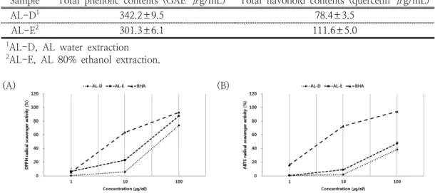
Fig. 3. DPPH (A) and ABTS (B) free radical scavenging activity of AL extracts. The results are expressed as the mean ± standard deviation of three independent experiments.
flavonoid is reported that antioxidant properties of flavonoids are effected mainly via scavenging of superoxide anions[44]. So polyphenol and flavonoid-based materials have been generally used to prevent or improve chronic diseases caused by oxidative stress[10].
Our results demonstrate that the phenolic content in AL-D was higher than in AL-E, while the flavonoid content was higher in AL-E than in AL-D. The difference in contents of polyphenol and flavonoid between the two extracts was assumed to have an effect on the antioxidant, effects.
3.4. ABTS and DPPH radical scavenging activity
The ABTS radical scavenging activities of AL extracts at a concentration of 1 µg/mL, 10 µg/mL and 100 µg/mL were measured. AL-D extracts exhibited 0.6±1.5%, 2.1±0.8% and 38.5±3.4% ABTS radical scavenging activity at the respective concentrations (Fig. 3-A). For AL-E the respective activities were 0.7±0.5%, 9.1±1.1% and 47.7±2.8% (Fig. 3-A). The DPPH radical scavenging activities of AL extracts at a concentration of 1 µg/mL, 10 µg/mL and 100 µg/mL were measured. AL-D
extracts exhibited 0.4±1.0%, 5.9±1.5% and 74.0±1.1% DPPH radical scavenging activity at the respective concentrations (Fig. 3-B). For AL-E, these values were 6.6±1.7%, 23.0±
1.9% and 87.5±1.0% activity, respectively (Fig. 3-B). Both DPPH radical and ABTS radical play a role as a stabilizer of the unstable and harmful free radicals originated from the internal body by supplying proton ion and stabilizing the unstability of free radical[45]. Therefore, a unknown specific material is used as an index that evaluate the antioxidant function which eliminates the hydroxyl radical or superoxide radical produced by physical or antioxidant function of the body[45]. AL-E showed a relatively higher outcome in terms of DPPH radical scavenging activity, and the EC
50(y=
19.013ln(x)+10.305, R²=0.964) values for
each BHA were 114.2 μg/mL in AL-D and
95.8 μg/mL in AL-E. In addition, the ABTS
radical scavenging activity was higher for
AL-E than for AL-D, and the EC
50(y=16.918ln(x)+22.092, R²=0.9361) values for
each BHA were 225.8 μg/mL in AL-D and
188.2 μg/mL in AL-E. Specifically from the
result of the radical scavenging activity, AL-E
Fig. 4. SOD-like activity of AL extracts.
Activities were determined by absorbance measurement at 420 nm.
The results are expressed as the mean
± standard deviation of three independent experiments.
Fig. 5. Effect of AL extracts on ROS production in Raw 264.7 cells. ROS was measured using FACS. The results are expressed as the mean ± standard deviation of three independent experiments (
*p < 0.05,
**p < 0.01,
***