www.e-arms.org 15 Any burn injury that involves more than the deep dermis
often requires reconstructive treatment. Skin graft is the paradigm of burn reconstruction for coverage, and prevention of wound contraction by relatively simple methodology. Local flap advancement also provides an excellent option with the benefit of stable coverage.1 V-Y fasciocutaneous advancement flap in the gluteal region is frequently used to cover the defect.
However, in case of large burn wounds, this kind of flap cannot provide adequate coverage due to the lack of normal surrounding tissues. We presented a case of full-thickness burn in the gluteal region. The defect size was too wide to be covered by general local flap advancement, but we noticed intact surrounding fat tissue available after the removal of dry eschar at first debridement. We devised a plan to apply the vacuum-
assisted closure (VAC) device to induce healthy granulation tissues, and confirmed the intactness of vascular network of subcutaneous fat tissue by a repeat preoperative Doppler.
CASE REPORT
A 30-year-old man made a suicide attempt with an ignition material which resulted in third-degree burn covered by dry eschar over his buttock. He had a depressive disorder and CO intoxication caused by the ignition material led to brain damage. He was in rigid posture and unable to move by himself.
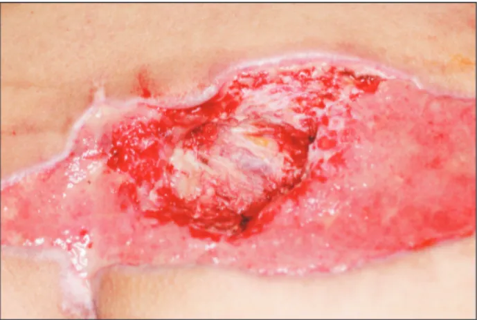
His wound was covered with dry and wet eschar, hence it was difficult to determine the exact degree of the burn (Fig. 1).
After escharectomy and debridement under general
The Adipofascial V-Y Advancement Flap with Skin Graft for Coverage of the Full-Thickness Burns of the Gluteal Region
Yoo Jung Lee, Myong Chul Park, Dong Ha Park, Il Jae Lee*
Department of Plastic and Reconstructive Surgery, Ajou University Hospital, Suwon, Korea
CC This is an open-access article distributed under the terms of the Creative Commons Attribution Non-Commercial License (http://creativecommons.org/licenses/by-nc/4.0) which permits unrestricted noncommercial use, distribution, and reproduction in any medium, provided the original work is properly cited.
Copyright © 2016 by the Korean Society for Microsurgery. All Rights Reserved.
Received April 24, 2016 Revised May 3, 2016 Accepted May 3, 2016
*Correspondence to: Il Jae Lee
Department of Plastic and Reconstructive Surgery, Ajou University Hospital, 164 WorldCup-ro, Yeongtong-gu, Suwon 16499, Korea
Tel: +82-31-219-5614 Fax: +82-31-219-5610 E-mail: [email protected] Financial support: None.
Conflict of interest: None.
Any types of burn injury that involve more than deep dermis often require reconstructive treatment. In gluteal region, V-Y fasciocutaneous advancement flap is frequently used to cover the defect. However, in case of large burn wounds, this kind of flap cannot provide adequate coverage because of the lack of normal surrounding tissues. We suggest V-Y adipofascial flap using the surrounding superficially damaged tissue. We present the case of a patient who was referred for full-thickness burn on gluteal region. We performed serial debridement and applied vacuum-assisted closure device to defective area as wound preparation for coverage. When healthy granulation tissue grew adequately, we covered the defect with surrounding V-Y adipofascial flap and the raw surface of the flap was then covered with split-thickness skin graft. We think the use of subcutaneous fat as an adipofascial flap to cover the deeper defect adjacent to the flap is an excellent alternative especially in huge defect with uneven depth varying from subcutaneous fat to bone exposure in terms of minimal donor site morbidity and reliability of the flap. Even if the flap was not intact, it was reuse of the adjacent tissue of the injured area, so it is relatively safe and applicable.
Key Words: Buttocks, Burns, Third degree burn, Adipofascial flap, V-Y advancement flap
ARMS
Archieves of Reconstructive MicrosurgeryCase Report
pISSN 2383-5257 eISSN 2288-6184 Arch Reconstr Microsurg 2016;25(1):15-18 http://dx.doi.org/10.15596/ARMS.2016.25.1.15
Arch Reconstr Microsurg Vol. 25. No. 1. May 2016
16
anesthesia, necrotic area was found deep within the muscle layer and the defect had exposure of muscle and bone over his buttock (Fig. 2). Total size of burn wound was about 18×7 cm, (depth was deep to subcutaneous fat layer to bone) and there was 3.5×3.5 cm sized necrotic area with exposure of sacrum and its periosteum in the middle of the wound. The defect size was too wide to be covered by general local flap advancement, but we noticed there was intact surrounding fat tissue looking available after removal of dry eschar at first debridement. We performed serial debridement and applied the VAC device to the defective area as wound preparation for coverage (Fig. 3).
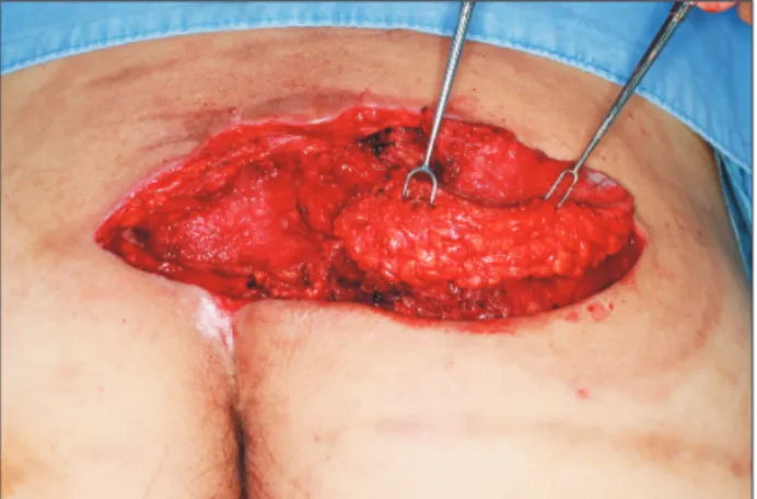
Once healthy granulation tissue grew adequately, we covered the defect with a surrounding V-Y adipofascial flap. Adipofascial flap was elevated over the right lateral aspect of the defect, measuring 8×6 cm (Fig. 4). The flap was advanced leftward to cover the defect and the raw surface of the flap was then covered with split-thickness skin graft taken from the posterior thigh at the same time. The donor site of the V-Y adipofascial flap was repaired primarily (Fig. 5). Split-thickness skin graft over the flap was secured with tie-over dressing. Grafted skin was well accepted and there was no postoperative complication (Fig. 6).
DISCUSSION
The principle of the V-Y advancement flap was first des- cribed by Blasius for reconstruction of smaller defects.2 V-Y advancement flap is widely used because of its simplicity and minimal donor site morbidity.3 Unlike conventional V-Y advancement flap, we used the adipofascial V-Y advancement
flap that lacks epidermal and dermal layer to cover the full- thickness burn exposing bone on the gluteal area.
Heymans et al.4 used adipofascial flap for full-thickness burns exposing the tibial crest. They advocated that the exposed bone secondary to a recent burn does not require muscular coverage because it is not initially infectious, hence adipofascial pedicled flaps would be excellent for reconstruction of full-thickness burns exposing bone.
A case of adipofascial flap on the gluteal region was previously reported. Yazar et al.5 used bilateral transpositional adipofascial flaps to cover the pilonidal sinus defect. They reported that it was technically easy, reliable, cosmetically acceptable with minimal incisions, and had decreased morbidity compared to the alternatives. But in this case, we applied damaged adipofascial flap after full thickness burn injury. Even though depth of burn in jury was deep to bone, after debridement and application of VAC device, we found that remained fat tissue was good enough to use as an adipofascial V-Y advancement flap.
The depth of burn injury was various from skin and subcutaneous tissue to muscle and bone according to the location because the buttock was exposed to thermal injury for long time–about 1 day. The damage was especially deep to gluteal muscles in the middle of the lesion. We debrided necrotic tissues serially and applied the VAC device for wound preparation. These procedures helped the formation of new connective tissue and capillaries, and granulation tissue on the surface of a wound.6,7 Granulation tissue formation is promoted by increasing tissue perfusion, by inactivating capillary
Fig. 1. Third-degree burn covered by dry eschar on gluteal area. Fig. 2. After escharectomy, the defect had exposure of muscle and bone over his buttock.
Yoo Jung Lee, et al. Adipofascial V-Y Advancement Flap for the Full-Thickness Burns of the Gluteal Region
www.e-arms.org 17
autoregulation and by switching off the mitotic process.8 We presumed that once healthy granulation tissue appeared, the vascular supply of the underlying fat layer was intact enough to use as an adipofascial flap with skin grafting. We confirmed the intactness of vascular supply by the use of Doppler.
Although the subdermal plexus was absent, our flap was reliable owing to the subcutaneous vascular network. Extensive subcutaneous vascular network exists between the dense
and loose adipose tissue, which affects the hemodynamic characteristics of a flap.9
The use of subcutaneous fat tissue as an adipofascial flap to cover the deeper defect adjacent to the flap is an excellent alternative especially in a large defect with uneven depth varying from subcutaneous fat to bone exposure. The adipofascial flap has minimal donor site morbidity and high reliability. It is relatively safe and applicable as the reuse of tissue adjacent to
Skin graft
A B
C D
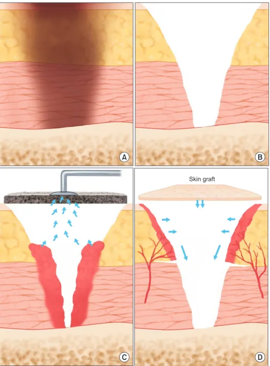
Fig. 3. Operative findings and procedure. (A) Initial wound was third degree burn covered by dry eschar. (B) After escharectomy, defect was deep to bone exposure. (C) Vacuum-assisted closure device was applied to defective area as wound preparation for coverage. (D) Adipofascial flap was elevated and advanced and the raw surface was covered with split-thickness skin graft.
Arch Reconstr Microsurg Vol. 25. No. 1. May 2016
18
the injured area, even if the flap is not intact.
REFERENCES
1. Orgill DP, Ogawa R. Current methods of burn reconstruction.
Plast Reconstr Surg 2013;131:827e-36e.
2. Hauben DJ. Ernst Blasius's contributions to plastic surgery. Plast Reconstr Surg 1984;74:561-70.
3. Venkataramakrishnan V, Mohan D, Villafane O. Perforator based V-Y advancement flaps in the leg. Br J Plast Surg 1998;51:431-5.
4. Heymans O, Verhelle N, Peters S, Nélissen X, Oelbrandt B. Use of the medial adipofascial flap of the leg for coverage of full- thickness burns exposing the tibial crest. Burns 2002;28:674-8.
5. Yazar M, Kurt Yazar S, Celet Ozden B, Guven E, Basaran K, Alyanak A, et al. Cosmetic closure of pilonidal sinus defects with bilateral transpositional adipofascial flaps. J Plast Surg Hand Surg 2013;47:292-6.
6. Argenta LC, Morykwas MJ. Vacuum-assisted closure: a new method for wound control and treatment: clinical experience.
Ann Plast Surg 1997;38:563-76; discussion 77.
7. Morykwas MJ, Argenta LC, Shelton-Brown EI, McGuirt W.
Vacuum-assisted closure: a new method for wound control and treatment: animal studies and basic foundation. Ann Plast Surg 1997;38:553-62.
8. Philbeck TE Jr, Whittington KT, Millsap MH, Briones RB, Wight DG, Schroeder WJ. The clinical and cost effectiveness of externally applied negative pressure wound therapy in the treatment of wounds in home healthcare Medicare patients.
Ostomy Wound Manage 1999;45:41-50.
9. De Lorenzi F, Vaienti L. Subcutaneous tissue flaps: theoretical principles and clinical applications. Eur J Plast Surg 2000;23:267- 71.
Fig. 4. Adipofascial flap was elevated over the right lateral aspect of the defect, measuring 8×6 cm.
Fig. 5. The adipofascial flap was advanced leftward and the raw surface of the flap was covered with split-thickness skin graft at the same time.
The donor site and remained defect were repaired primarily.
Fig. 6. Postoperative follow-up: after 2 months.