ISSN 0378-6471 (Print)⋅ISSN 2092-9374 (Online)
http://dx.doi.org/10.3341/jkos.2016.57.1.106
Original Article
산화스트레스가 유도된 망막색소상피세포에서 폴리페놀 화합물이 보이는 세포보호 효과
Cytoprotective Effect of Polyphenolic Compounds against Oxidative Stress in Cultured Retinal Pigment Epithelial Cells
서경훈⋅유승영⋅곽형우
Kyung Hoon Seo, MD, Seung Young Yu, MD, PhD, Hyung Woo Kwak, MD, PhD
경희대학교 의학전문대학원 경희의료원 안과학교실
Department of Ophthalmology, Kyung Hee University Medical Center, Kyung Hee University School of Medicine, Seoul, Korea
Purpose: Grape seed-derived polyphenols (GSPs) provide a concentrated source of polyphenols having antioxidant capacity. In this study we investigated the cytoprotective effect of GSP against oxidative stress-induced cell damage in cultured human reti- nal pigment epithelial (RPE) cells.
Methods: Cultured adult retinal pigment epithelium (ARPE)-19 cells were incubated with GSP from Vitis vinifera (0.1, 0.5, 1, 5 or 10 μg/mL) for 24 hours and treated with hydrogen peroxide (H2O2, 0.4 mM) for 24 hours to induce oxidative stress. Cell viability was measured using 3-(4, 5-dimethylthiazol-2-yl)-2, 5-diphenyltetrazolium bromide (MTT) assay. Intracellular reactive oxygen species (ROS) was quantified using 2′,7′-dichlorofluorescein diacetate (DCF-DA) fluorescence.
Results: The percentage of viable RPE cells was significantly lower in cultures treated with H2O2 0.4 mM than in control cultures.
GSP significantly reduced H2O2-induced cell death in a dose dependent manner. GSP at 0.1, 0.5, 1, 5 and 10 μg/mL significantly reduced cell mortality due to the treatment with H2O2. Intracellular ROS production increased significantly in cultures treated with H2O2 0.4 mM compared with control. There was a significant dose-dependent decrease in intracellular ROS levels after treat- ment of RPE with GSP.
Conclusions: GSP, a natural polyphenolic compound, can protect RPE cells from H2O2-induced oxidative stress and reduce in- tracellular ROS production by scavenging free radicals. This suggests potential effects of polyphenolic compounds against reti- nal diseases associated with oxidative stress.
J Korean Ophthalmol Soc 2016;57(1):106-112
Key Words: Grape seed extract, Oxidative stress, Polyphenols, Proanthocyanidins, Retinal pigment epithelium
■Received: 2015. 6. 19. ■ Revised: 2015. 7. 23.
■Accepted: 2015. 11. 20.
■Address reprint requests to Hyung Woo Kwak, MD, PhD Department of Ophthalmology, Kyung Hee University Medical Center, #23 Kyungheedae-ro, Dongdaemun-gu, Seoul 02447, Korea
Tel: 82-2-958-8455, Fax: 82-2-966-7340 E-mail: [email protected]
* This study was presented as a narration at the 113th Annual Meeting of the Korean Ophthalmological Society 2015.
* This study was supported by a research agreement between Hanlim Pharm. Co. and Kyung Hee University.
ⓒ2016 The Korean Ophthalmological Society
This is an Open Access article distributed under the terms of the Creative Commons Attribution Non-Commercial License (http://creativecommons.org/licenses/by-nc/3.0/) which permits unrestricted non-commercial use, distribution, and reproduction in any medium, provided the original work is properly cited.
식물성 영양분이 풍부한 식품의 경우 심혈관 질환으로 인한 사망률을 낮추고 종양성 질환이나 노화를 예방하는 데 효과적임이 여러 역학연구를 통해 보고되었다.1-3 식물성 영양분이 풍부한 식품은 섬유소나 비타민, 식물 스테롤, 황 화합물, 카로테노이드, 유기산 등이 작용함으로써 유익한 효과를 나타내는 것으로 알려져 있는데, 그 중에서도 폴리 페놀이 지니는 여러 가지 보호 효과들이 점점 더 주목 받고 있다.
폴리페놀은 화학구조에 따라 다양한 화합물의 형태로 존
재하는데, 식품에 따라 그 함유량이 다르다.4 폴리페놀과 인체에서 생성되는 폴리페놀의 대사물질은 여러 다른 기전 을 통하여 보호작용을 나타내고 현재 활발히 연구 중인 분 야이다. 예를 들어 안토시아닌(anthocyanin)이 풍부한 들쭉 추출물이나 프로안토시아니딘(proanthocyanidin)과 같이 포 도에서 추출한 폴리페놀 화합물 등이 동물모델뿐만 아니라 당뇨병과 알츠하이머병 환자에서 연구되고 있다.5-7
산소의 단계적 환원의 과정에서 생성되는 활성산소는 정 상적인 대사과정에서도 나타날 수 있지만 과량이 존재할 경우 미토콘드리아에 심각한 손상을 유발할 수도 있고 이 런 활성산소가 세포의 항산화 능력을 넘어서서 존재하게 되어 세포에 지속적인 위협을 가하게 되는 상태를 산화스 트레스라고 한다. 이런 산화스트레스는 노화와 관련된 퇴 행성 질환에 중요한 역할을 한다. 망막은 신체 내에서 대사 작용이 가장 활발한 조직 중의 하나로 산소요구량이 높으 며 지속적으로 빛에 노출되고 고농도의 불포화지방산을 포 함하고 있어 이러한 요소들이 산화적 손상의 발생에 관여 하는 것으로 알려져 있다. 망막색소상피세포는 망막과 맥 락막모세혈관 사이에 단일층을 이루고 있는 세포로서 생리 적으로 광수용체의 영양공급과 대사물질의 배출 및 세포탈 락물의 탐식 등 시력의 유지에 중요한 역할을 하며 산소 소 모량이 많아 산화스트레스에 노출되어 있다.
따라서 많은 연구들이 산화스트레스로부터 망막색소상 피를 보호하는 방법 및 물질에 대해 관심을 기울이고 있으 며, 특히 꾸준히 섭취할 수 있는 식품에서 유래한 생화합물 에 대한 연구가 활발히 이루어지고 있다.8-11 Vitis vinifera 에서 얻은 포도씨 유래 폴리페놀 화합물은 프로안토시아니 딘이 풍부하게 함유되어 있어 만성 질환에서의 유익한 효 과로 건강보조식품으로 널리 이용되고 있고, 일부에서는 강한 항산화 효과로 산화스트레스 모델에서 치료적인 효과 를 얻기도 했다.12,13
이에 저자들은 포도씨에서 유래한 폴리페놀 화합물이 산 화스트레스가 유도된 인간 망막색소상피세포에서 어떠한 세포보호 효과를 보이는지 확인해 보고자 하였다.
대상과 방법
세포 배양
인체망막색소상피세포주(ARPE-19; ATCC No. CRL-2302) 를 95%의 공기와 5%의 이산화탄소 환경의 습윤 배양기에 서 37℃의 온도로 배양하였다. 배양액은 basal Dulbecco’s modified Eagle’s medium과 Ham’s F12 medium의 1:1 혼 합배양액(DMEM/F-12)으로 fetal bovine serum 10%와 pen- icillin (100 U/mL), streptomycin (100 μg/mL)을 함유하고
있다(penicillin-streptomycin, Gibco BRL, Grand Island, NY, USA). 3일마다 배양액을 교환하였다. 배양된 세포주의 5번 째에서 6번째 계대배양된 세포를 실험에 사용하였다.
폴리페놀 화합물
폴리페놀 화합물의 세포보호 효과를 확인하기 위해 배양 된 망막색소상피세포에 포도씨 유래 폴리페놀(Grape seed- derived polyphenols, GSP)을 24시간 동안 전처리하였다.
GSP는 유럽종 포도인 Vitis vinifera의 건조된 씨를 분쇄한 다음 에탄올을 이용하여 가온 추출한 후 감압여과 및 농축 하여 분말상태로 얻었다.14 실험 전 정확한 농도에 맞게 1 ng/mL에서 100 μg/mL까지 10배씩 농도를 증가시키면서 증류수에 녹여서 준비하였다.
산화스트레스 유도
상피세포주를 1×10⁵ cell/mL 수준으로 배양 후 산화스트 레스를 유도하기 위하여 H2O2 (Sigma, St Louis, MO, USA) 200, 400, 600 μM 농도로 24시간 동안 처리하였다.
세포활성도 검사
산화스트레스를 유도하기 전, 폴리페놀 화합물 자체의 세포독성을 확인하기 위해 GSP로 24시간 동안 처리한 망 막상피세포들을 Pieters et al15의 방법을 사용하여 세포활성 도를 측정하였다. 망막색소상피세포를 0.05% trypsin EDTA (Gibco, Grand Island, NY, USA)로 떼어 96 well microplate (Falcon, Lincoln Park, NJ, USA)에 0.5×10⁴ cell/mL로 분주 하였다. 그 후 18시간 동안 배양한 뒤에 GSP를 농도별로 10 μL씩 넣고 이때 약물 대신 PBS (Gibco, Grand Island, NY, USA)를 넣어 세포의 대조군으로 삼고, 세포 대신 배양액 만을 넣어 대조군으로 삼았다. 잘 흔든 후 24시간 동안 CO2 배 양기에서 배양한 후 모든 well에 3-(4,5-dimethylthiazol-2-yl)-2, 5-diphenyl tetrazoliumbromide (MTT) 용액(5 mg/mL, Sigma, St Louis, MO, USA) 10 μL를 가해주고 다시 37℃, 5%
CO2에서 4-5시간 더 배양하여 MTT가 환원되도록 하였다.
각 well에서 80 μL씩 버린 다음 150 μL DMSO (Sigma, St Louis, MO, USA)를 넣고 10분 동안 흔들어서 생성된 formazan 결정 을 잘 녹여서 microplate reader (Bio-Tek, Cambridge, MA, USA)를 이용하여 540 nm에서 흡광도를 측정하였다.
산화스트레스 유도와 폴리페놀 화합물의 세포보호 효과 관찰
본 연구에서는 H2O2 (Sigma, St Louis, MO, USA)를 200, 400, 600 μM 농도로 배양액에 넣고 24시간 동안 세포를 배 양하는 방법으로 산화스트레스를 유도하였다. 폴리페놀 화
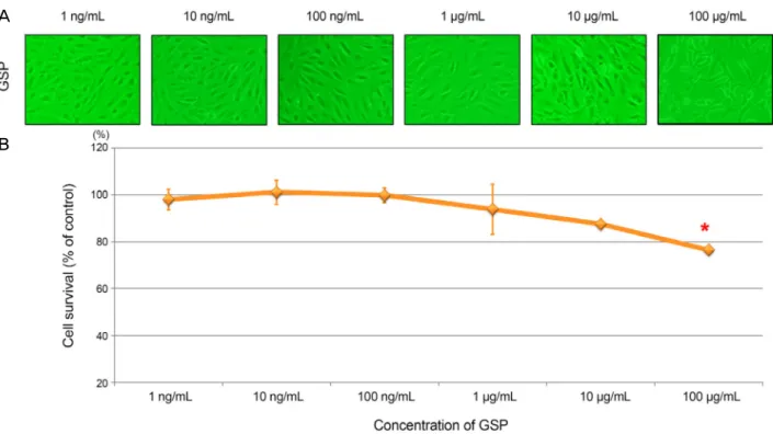
Figure 1. Cytotoxic effect of grape seed-derived polyphenols (GSP). The ARPE-19 cells were incubated with different concen-
tration of GSP for 24 hours. (A) GSP showed cytotoxicity at 100 μg/mL. (B) The asterisk (*) indicates p < 0.05 versus untreated control. ARPE = adult retinal pigment epithelium.합물의 보호 효과를 확인하기 위해 산화스트레스를 유도하기 전 앞선 실험에서 세포독성이 없는 것으로 확인된 10 μg/mL 이하의 (100 ng/mL, 500 ng/mL, 1 μg/mL, 5 μg/mL, 10 μg/mL) GSP를 24시간 동안 망막색소상피세포와 배양하였다. 이후 세포를 0.05% trypsin EDTA (Gibco)로 떼어 새로운 plate 에 옮겨 각각의 H2O2 농도 (200, 400, 600 μM)로 24시간 동안 배양하여 산화스트레스를 유도하였다. 이때 H2O2 대 신 PBS (Gibco)를 넣어 세포의 대조군으로 삼았다. 다양한 농도의 GSP의 세포보호 효과는 앞서 세포활성도 검사에서 기술하였던 것과 같이 MTT assay로 확인하였다.
세포 내 반응산소종(reactive oxygen species, ROS)의 측정
산화스트레스로 인한 세포 내 반응산소종의 생성을 비형광 색소인 2′,7′-dichlorofluorescein diacetate (DCF-DA, Invitrogen, Carlsbad, CA, USA)를 이용하여 측정하였다. 각각의 농도 의 GSP가 전처리된 망막상피세포에 200, 400, 600 μM 농 도의 H2O2를 각각 1 μM의 DCF-DA를 함께 투여 후 37℃
에서 4시간 동안 5% CO2 배양기에서 배양하였다. 그 후 excitation 파장 485 nm와 emission 파장 530 nm에서 형광 강도를 측정하였다. 폴리페놀 화합물 대신 PBS를 넣은 세 포를 대조군으로 삼았다.
통계분석
실험 결과는 3회의 독립된 실험 후 평균 ± 표준편차의 형태로 표시하였다. 통계분석은 SPSS version 18.0 (SPSS Inc., Chicago, IL, USA)을 이용하였으며, Wilcoxon signed rank test를 이용하여 대조군과 실험군 간의 차이를 비교하 였다. p<0.05인 경우를 통계적으로 유의한 것으로 간주하 였다.
결 과
우선 여러 농도의 GSP를 망막색소상피세포에 노출시켜 배양하면서 이들 농도에 따른 세포의 활성도를 조사하였다.
10 μg/mL 이하에서 세포의 활성도가 85% 이상이었으나 100 μg/mL에서는 세포활성도가 76.5 ± 1.5%로 대조군에 비해 유의하게 감소한 것으로 확인되었다(p=0.013, Fig. 1).
따라서 본 실험에서는 10 μg/mL 이하의 GSP (100 ng/mL, 500 ng/mL, 1 μg/mL, 5 μg/mL, 10 μg/mL)를 실험에 적합 한 농도로 판단하고 사용하였다.
또한 GSP 처리를 하지 않은 망막색소상피세포에 농도를 달리하여 H2O2 (200, 400, 600 μM)를 노출시킨 후 24시간 배양한 뒤 세포의 활성도를 조사하였다. H2O2에 노출되기 전에 비하여 H2O2에 노출되었을 경우에 세포활성도가 유 의하게 감소하는 것을 확인할 수 있었으며, 이는 H2O2 농
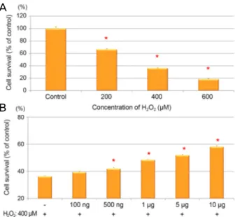
A
B
Figure 2. Grape seed-derived polyphenols (GSP) protected
against H2O2 cell death in ARPE-19 cells. (A) The ARPE-19 cells were treated with H2O2 at dose of 200, 400, and 600 μM for 24 hours. (B) The cells were pretreated with GSP for 24 hours then exposed to H2O2 (400 μM) for 24 hours. Data was expressed as a percentage of the untreated control. The aster- isk (*) indicates p < 0.05 versus untreated control (A), or H2O2-induced cells without GSP pretreatment (B). ARPE = adult retinal pigment epithelium.Figure 3. Grape seed-derived polyphenols (GSP) reduced
H2O2-induced intracellular reactive oxygen species (ROS) generation in ARPE-19 cells. (A) The ARPE-19 cells were treated with H2O2 at dose of 200, 400, and 600 μM for 24 hours. (B) The cell were pretreated with GSP for 24 hours then treated with 400 μM for 4 hours. The intracellular ROS was examined by a fluorescence spectrophotometer using DCF- DA. Data was expressed as a percentage of the untreated control. The asterisk (*) indicates p < 0.05 versus untreated control (A), or H2O2-induced cells without GSP pretreatment (B). ARPE = adult retinal pigment epithelium; DCF-DA = 2′,7′-dichlorofluorescein diacetate.
도가 높아질수록 증가하는 경향을 보였다(p<0.05, re- spectively, Fig. 2A). 특히 400 μM에서의 세포활성도는 대 조군의 절반 정도인 36.3%로 GSP의 보호 효과를 확인하기 위한 적합한 농도로 판단하였다.
24시간 동안 GSP를 농도를 달리하여(100 ng/mL, 500 ng/mL, 1 μg/mL, 5 μg/mL, 10 μg/mL) 전처리한 망막색소 상피세포에 400 μM의 H2O2를 노출시킨 후 각각 24시간 동 안 배양한 뒤 세포활성도를 확인하였다. GSP를 추가한 경 우에는 H2O2만 처리하였을 때보다 세포활성도가 상대적으 로 증가하였으며 GSP의 농도가 높아질수록 세포활성도가 더 증가하였다(Fig. 2B).
GSP의 세포보호 효과의 기전을 알아보기 위해 산화스트 레스로 인해 발생하는 반응산소종의 양을 측정하였다.
H2O2의 농도를 달리하여(200, 400, 600 μM) DCF-DA와 함 께 4시간 동안 배양한 후 세포 내 반응산소종이 대조군과 비 교하여 유의하게 증가함을 확인하였다(Fig. 3A). 세포활성도 검사와 마찬가지로 24시간 동안 GSP를 농도를 달리하여 (100 ng/mL, 500 ng/mL, 1 μg/mL, 5 μg/mL, 10 μg/mL) 전 처리한 망막색소상피세포에 400 μM의 H2O2를 노출시킨 후 각각 4시간 동안 배양한 뒤 세포 내 반응산소종의 축적 을 비교하였다. GSP를 추가한 경우에는 H2O2만 처리하였 을 때보다 세포 내 반응산소종의 축적이 농도에 비례하여
저하되었다(Fig. 3B, Fig. 4).
고 찰
노화와 퇴행성 질환의 원인이 되는 산화스트레스에 대항 하는 신체의 방어기제에는 여러 가지가 있고, 이를 강화시 키기 위한 가장 손쉬운 방법은 항산화 효과를 지닌 식품을 섭취하는 것이다.16 이들 식품들에서 항산화 효과를 보이는 성분들은 화학구조에 따라 비타민과 미네랄, 식물 화합물 (phytochemical) 등으로 나눌 수 있다.
식품으로서의 항산화제의 섭취는 여러 역학연구에서 그 효용성과 안정성이 보고되었으나, 항상 기대 이상의 효과 를 보여주는 것은 아니다.17 이는 항산화제 성분이 많이 포 함된 식품의 경우 식품 내의 다른 성분이나, 다른 식이요인 에 의해 영향을 받을 수 있기 때문이다. 따라서 생체이용률 (bioavailability)을 높이기 위해 추출물 형태가 널리 이용되 고 있다. 식물 화합물의 한 형태인 폴리페놀은 화학구조에 따라 다양한 항산화 효과가 보고되었고, 그중에서도 본 연 구에서 사용한 포도씨 추출물의 경우 높은 농도의 프로안
A
B
A
B
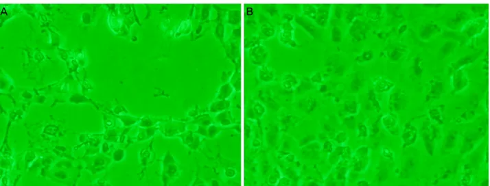
Figure 4. Morphology of ARPE-19 cells exposed to H
2O2 (400 μM) with/without grape seed-derived polyphenols (GSP). (A) 400 μM H2O2 without GSP. After 24-hour exposure, (B) 400 μM H2O2 with GSP. ARPE = adult retinal pigment epithelium.토시아니딘을 함유하고 있다.4
우리 체내에서 이용하는 전체 산소 소모량의 1-5%가 반 응산소종으로 전환되는데 이는 산화스트레스의 주요 원인 이 된다. 사람의 망막은 산소소모량이 많으며 특히 망막색 소상피세포는 시세포외절을 탐식하기 때문에 반응산소종 의 생성량이 많다. 또한 미토콘드리아의 전자 전달계를 통 한 세포 내 반응산소종이 생성된다.18-20 이러한 반응성 산 소의 생성과 소거의 평형유지는 산화스트레스로 인한 세포 손상에 대한 생체의 항상성 유지에 대단히 중요한 요소로 서 망막색소상피세포에서 생성된 유리기를 제거하는 항산 화계는 superoxide dismutase (SOD)와 catalase 및 gluta- thione peroxidase (GSHPx) 등 항산화 효소계와 gluta- thione, uric acid, vitamin C, vitamin E, melanin 등 비효소 계 항산화 물질로 구성되어 있다.
산화스트레스가 유도된 망막색소상피세포는 세포자멸사 가 유도되거나, 미토콘드리아 DNA의 손상, 혈관내피세포 성장인자(vascular endothelial growth factor, VEGF)의 증 가, 항산화 효소의 감소, 염증반응의 증가와 같은 변화를 보인다. 항산화 물질은 이러한 스트레스 반응 중 특정 경로 에 작용하여 세포보호 효과를 나타낸다. 녹차나 양파에 함 유되어 있는 quercetin의 경우 caspase-3의 활성을 억제하여 세포자멸사 경로를 차단하며, 브로콜리 추출물인 sulfor- aphane은 항산화 효소인 Nrf2-thioredoxin 시스템을 활성화 시켜 항산화 효과를 증대시킨다.21-24 포도 껍질에 많은 re- sveratrol과 turmeric 향신료에 많은 curcumin은 각각 NF-κ B 경로를 억제하여 염증반응을 억제한다.25,26
본 연구에 사용된 포도씨 유래 폴리페놀 화합물은 유럽종 포도인 Vitis vinefera에서 추출하였으며 (+)-catechin, (-)-epi- catechin, (-)-epicatechin 3-O-gallate를 포함하는 Proanthocyanidins
가 주요 성분으로 되어있다.14 Proanthocyanidins의 경우 기존 의 연구들에서 직접적으로 반응산소종을 제거하고 세포막 에 존재하는 polyunsaturated fatty acids (PUFAs)의 지질과 산화(lipid peroxidation)를 약화시켜 산화스트레스로 인한 퇴행성 손상을 억제할 수 있는 것으로 보고되었다.27,28
본 연구에서도 100 μg/mL의 고농도의 GSP를 전처리하 였을 경우 세포활성도가 감소하였으나(Fig. 1), 10 μg/mL 이하의 농도에서는 농도에 비례하여 산화스트레스로부터 망막상피세포를 보호하는 효과를 보였으며(Fig. 2), 반응산 소종의 생성이 억제됨을 관찰하였다(Fig. 3). 고농도의 GSP 가 세포손상을 유발하는 기전에 대해서는 본 연구를 통해 알 수는 없었지만, 고농도의 식물화합물의 경우 역설적으 로 미토콘드리아 내의 superoxide radical (O2-
)의 생성을 증 가시키거나 세포 DNA를 손상시키고 세포자멸사를 유발할 수 있음이 보고되었다.29-31 하지만 10 μg/mL 이하의 농도에 서 관찰되는 세포보호 효과는 프로안토시아니딘의 항산화 효과로 생각할 수 있다.
Age-Related Eye Disease Study research group (AREDS) 의 연구에 따르면 항산화 작용을 하는 비타민을 장기적으로 복용할 경우 나이관련 황반변성의 진행을 늦출 수 있는 것 으로 확인되었다.32,33 AREDS 연구를 통해 항산화 물질을 규칙적으로 섭취하는 것이 나이관련 황반변성의 진행을 억 제하는 데 효과가 있음을 확인할 수 있었으며, 본 연구 결과 와 연관하여 고려해 보면 포도씨 유래 폴리페놀 화합물도 나이관련 황반변성의 진행을 억제하기 위한 좋은 후보군이 될 수 있음을 보여준다고 할 수 있다. 향후 포도씨에서 유래 한 폴리페놀 화합물에서 위와 같은 효과를 강화시킬 수 있 도록 생체이용률을 높이고 동물모델이나 실제 환자에서 그 결과를 확인하는 추가적인 연구가 필요하리라 판단된다.
A B
REFERENCES
1) Devore EE, Kang JH, Breteler MM, Grodstein F. Dietary intakes of berries and flavonoids in relation to cognitive decline. Ann Neurol 2012;72:135-43.
2) McCullough ML, Peterson JJ, Patel R, et al. Flavonoid intake and cardiovascular disease mortality in a prospective cohort of US adults. Am J Clin Nutr 2012;95:454-64.
3) Wang Y, Stevens VL, Shah R, et al. Dietary flavonoid and proan- thocyanidin intakes and prostate cancer risk in a prospective cohort of US men. Am J Epidemiol 2014;179:974-86.
4) Manach C, Scalbert A, Morand C, et al. Polyphenols: food sources and bioavailability. Am J Clin Nutr 2004;79:727-47.
5) Takikawa M, Inoue S, Horio F, Tsuda T. Dietary anthocyanin-rich bilberry extract ameliorates hyperglycemia and insulin sensitivity via activation of AMP-activated protein kinase in diabetic mice. J Nutr 2010;140:527-33.
6) Vepsäläinen S, Koivisto H, Pekkarinen E, et al. Anthocyanin-en- riched bilberry and blackcurrant extracts modulate amyloid pre- cursor protein processing and alleviate behavioral abnormalities in the APP/PS1 mouse model of Alzheimer's disease. J Nutr Biochem 2013;24:360-70.
7) Wang J, Ferruzzi MG, Ho L, et al. Brain-targeted proanthocyanidin metabolites for Alzheimer's disease treatment. J Neurosci 2012;
32:5144-50.
8) Dong X, Li Z, Wang W, et al. Protective effect of canolol from oxi- dative stress-induced cell damage in ARPE-19 cells via an ERK mediated antioxidative pathway. Mol Vis 2011;17:2040-8.
9) Li Z, Dong X, Liu H, et al. Astaxanthin protects ARPE-19 cells from oxidative stress via upregulation of Nrf2-regulated phase II enzymes through activation of PI3K/Akt. Mol Vis 2013;19:1656-66.
10) Hanneken A, Lin FF, Johnson J, Maher P. Flavonoids protect hu- man retinal pigment epithelial cells from oxidative-stress-induced death. Invest Ophthalmol Vis Sci 2006;47:3164-77.
11) Kil HK, Song YM, Chun K. The efficacy of vaccinium uliginosum for early age-related macula degeneration. J Korean Ophthalmol Soc 2013;54:1255-60.
12) Moini H, Rimbach G, Packer L. Molecular aspects of procyanidin biological activity: disease preventative and therapeutic potentials.
Drug Metabol Drug Interact 2000;17:237-59.
13) Vigna GB, Costantini F, Aldini G, et al. Effect of a standardized grape seed extract on low-density lipoprotein susceptibility to oxi- dation in heavy smokers. Metabolism 2003;52:1250-7.
14) Gabetta B, Fuzzati N, Griffini A, et al. Characterization of proan- thocyanidins from grape seeds. Fitoterapia 2000;71:162-75.
15) Pieters R, Huismans DR, Leyva A, Veerman AJ. Adaptation of the rapid automated tetrazolium dye based (MTT) assay for chemo- sensitivity testing in childhood leukemia. Cancer Lett 1988;41:
323-32.
16) Uddin S, Ahmad S. Dietary antioxidants protection against oxida- tive stress. Biochem Educ 1995;23:2-7.
17) Jerome-Morais A, Diamond AM, Wright ME. Dietary supple- ments and human health: for better or for worse? Mol Nutr Food Res 2011;55:122-35.
18) Schraermeyer U, Heimann K. Current understanding on the role of
retinal pigment epithelium and its pigmentation. Pigment Cell Res 1999;12:219-36.
19) Cai J, Nelson KC, Wu M, et al. Oxidative damage and protection of the RPE. Prog Retin Eye Res 2000;19:205-21.
20) Lee JH, Kim JW. Effect of glucose on the production of reactive oxygen species in retinal pigment epithelial cells. J Korean Ophthalmol Soc 2010;51:276-81.
21) Murota K, Mitsukuni Y, Ichikawa M, et al. Quercetin-4'-glucoside is more potent than quercetin-3-glucoside in protection of rat in- testinal mucosa homogenates against iron ion-induced lipid peroxidation. J Agric Food Chem 2004;52:1907-12.
22) Kook D, Wolf AH, Yu AL, et al. The protective effect of quercetin against oxidative stress in the human RPE in vitro. Invest Ophthalmol Vis Sci 2008;49:1712-20.
23) Nguyen T, Sherratt PJ, Pickett CB. Regulatory mechanisms con- trolling gene expression mediated by the antioxidant response element. Annu Rev Pharmacol Toxicol 2003;43:233-60.
24) Tanito M, Masutani H, Kim YC, et al. Sulforaphane induces thio- redoxin through the antioxidant-responsive element and attenuates retinal light damage in mice. Invest Ophthalmol Vis Sci 2005;46:979-87.
25) King RE, Kent KD, Bomser JA. Resveratrol reduces oxidation and proliferation of human retinal pigment epithelial cells via ex- tracellular signal-regulated kinase inhibition. Chem Biol Interact 2005;151:143-9.
26) Singh S, Aggarwal BB. Activation of transcription factor NF-kap- pa B is suppressed by curcumin (diferuloylmethane) [corrected]. J Biol Chem 1995;270:24995-5000.
27) Li MH, Jang JH, Sun B, Surh YJ. Protective effects of oligomers of grape seed polyphenols against beta-amyloid-induced oxidative cell death. Ann N Y Acad Sci 2004;1030:317-29.
28) Zhang B, Osborne NN. Oxidative-induced retinal degeneration is attenuated by epigallocatechin gallate. Brain Res 2006;1124:
176-87.
29) De Marchi U, Biasutto L, Garbisa S, et al. Quercetin can act either as an inhibitor or an inducer of the mitochondrial permeability transition pore: a demonstration of the ambivalent redox character of polyphenols. Biochim Biophys Acta 2009;1787:1425-32.
30) Wätjen W, Michels G, Steffan B, et al. Low concentrations of fla- vonoids are protective in rat H4IIE cells whereas high concen- trations cause DNA damage and apoptosis. J Nutr 2005;135:
525-31.
31) Bouayed J, Bohn T. Exogenous antioxidants - double-edged swords in cellular redox state: Health beneficial effects at physio- logic doses versus deleterious effects at high doses. Oxid Med Cell Longev 2010;3:228-37.
32) Age-Related Eye Disease Study Research Group. A randomized, placebo-controlled, clinical trial of high-dose supplementation with vitamins C and E, beta carotene, and zinc for age-related mac- ular degeneration and vision loss: AREDS report no. 8. Arch Ophthalmol 2001;119:1417-36.
33) Age-Related Eye Disease Study 2 Research Group. Lutein + zeax- anthin and omega-3 fatty acids for age-related macular degener- ation: the Age-Related Eye Disease Study 2 (AREDS2) random- ized clinical trial. JAMA 2013;309:2005-15.
= 국문초록 =
산화스트레스가 유도된 망막색소상피세포에서 폴리페놀 화합물이 보이는 세포보호 효과
목적: 포도씨 유래 폴리페놀(Grape seed-derived polyphenols, GSP)의 경우 폴리페놀 함유량이 높은 생화합물로서 본 연구에서는 배양된 인간 망막색소상피세포에서 산화스트레스를 유도한 후 GSP가 보이는 세포보호 효과에 대하여 알아보고자 하였다.
대상과 방법: 배양된 인간 망막색소상피세포인 adult retinal pigment epithelium (ARPE)-19를 GSP에 24시간 동안 다양한 농도로 전처치(0.1, 0.5, 1, 5 and 10 μg/mL, respectively)한 후, 산화스트레스 유도를 위해 과산화수소(H2O2, 0.4 mM)로 24시간 동안 처리하 였다. 3-(4, 5-dimethylthiazol-2- yl)-2, 5-diphenyltetrazolium bromide (MTT) assay를 통해 세포의 생존성을 측정하였고, 2′,7′
-dichlorofluorescein diacetate (DCF-DA) assay로 세포 내 반응성산소종의 생성을 측정하였다.
결과: 과산화수소로 산화스트레스가 유도된 망막색소상피세포의 경우, 세포의 생존율이 산화스트레스를 받지 않은 대조군에 비해 유 의하게 낮았다. GSP의 전처치 농도에 비례하여 산화스트레스가 유도된 망막색소상피세포의 생존율이 증가하였는데, 0.1, 0.5, 1, 5 and 10 μg/mL 농도에서 생존율이 유의하게 증가하였다. 세포 내 반응성산소종의 발생은 과산화수소로 산화스트레스가 유도된 망막 색소상피세포에서 유의하게 증가하였고, GSP로 전처치하였을 경우 농도에 비례하여 유의하게 감소하였다.
결론: 천연 폴리페놀 화합물인 GSP는 산화스트레스가 유도된 망막색소상피세포에서 세포 내 반응성산소종의 발생을 감소시키고, 세 포를 보호하는 효과를 보여주었다. 따라서 산화스트레스와 연관이 있는 망막질환을 치료하는 데 폴리페놀 화합물이 도움을 줄 수 있을 것이라 생각된다.
<대한안과학회지 2016;57(1):106-112>