DOI : 10.3341/jkos.2009.50.8.1247
= 증례보고 = 접수번호 : 50-08-12-20
인체망막색소상피세포에서 헴산화효소-1의 발현에 대한 산화스트레스 및 항산화물질의 영향
김영준⋅진희승 인하대학교 의과대학 안과학교실
목적: 인체망막색소상피세포에 산화스트레스 및 항산화물질이 헴산화효소-1 (heme oxygenase-1, HO-1)의 발현에 미치는 영향을 알아 보고자 하였다.
대상과 방법: 배양한 망막색소상피세포를 과산화수소(hydrogen peroxide, H2O2) 농도를 달리하여 각각 24시간, 48시간 동안 노출시킨 후 역전사중합효소반응을 통해 HO-1의 발현 정도를 관찰하였다. 또한 망막색소상피세포에 항산화제인 N-acetylcystein (NAC)을 30분 동안 전 처치한 뒤 과산화수소에 노출시킨 후 HO-1의 발현 정도를 비교하였다.
결과: 과산화수소에 노출된 망막색소상피세포에서 HO-1의 발현이 증가하였으며, 이는 과산화수소의 농도와 비례하여 증가하였다 (p<0.05). NAC와 함께 H2O2에 노출된 경우에는 H2O2 단독으로 노출시킨 경우보다 HO-1의 발현이 감소되었다(p<0.05).
결론: 망막색소상피세포에서 HO-1은 산화스트레스 정도에 비례하여 증가하고 항산화제에 의해 감소한다. HO-1은 연령관련황반변성 같은 산화스트레스로 유발되는 망막질환을 예방하는데 도움이 될 것으로 기대된다.
<대한안과학회지 2009:50(8):1247-1253>
■ 접 수 일: 2008년 12월 29일 ■ 심사통과일: 2009년 5월 19일
■ 책 임 저 자: 진 희 승
인천시 중구 신흥동 3가 인하대학교병원 안과
Tel: 032-890-2408, Fax: 032-890-2403 E-mail: [email protected]
* 본 논문의 요지는 2008년 대한안과학회 제99회 춘계학술대회에서 구연으로 발표되었음.
* 본 논문은 인하대학교의 지원을 받아 연구되었음.
서양에서 연령관련황반변성은 65세 이상 인구의 주요 실명 원인으로 알려진 노인성 질환으로 평균 수명의 증가와 함 께 연령관련황반변성의 이환율도 높아지고 있다.1-3연령관 련황반변성의 시력저하는 황반부의 광수용체 파괴 및 망막 색소상피와 브루크막의 변성과 관련이 있다.4망막색소상피 세포는 망막과 맥락막모세혈관 사이에 단일층을 이루고 있 는 세포로서 혈액-망막 장벽 역할을 한다.5망막은 산소 소 모가 많은 조직으로 망막색소상피세포는 항상 고농도의 산 소 하에서 기능을 하며 산화스트레스에 노출되어 있다. 만 성적인 산화스트레스는 망막색소상피세포를 손상시키거나 죽게 하므로, 연령관련황반변성의 중요한 위험인자로 여겨 지고 있다.6,7
그러나 아직까지 연령관련황반변성은 뚜렷한 치료법이 없는 상태다. Verteporfin을 이용한 광역학레이저 치료나 bevacizumab, ranibizumab과 같은 anti-vascular endothelial growth factor (VEGF) 제제를 이용한 약물치료 등이 시도 되고 있으나, 성공적인 신생혈관의 소실이 있다 하더라도
이미 소실된 시력을 호전시키는 데에는 한계가 있다. 또한, 이러한 치료는 일회성으로 끝나는 것이 아니라 반복적으로 시행되어야 하는 경우가 많고 고가의 치료비용 때문에 연령 관련황반변성의 예방법의 개발이 필요한 실정이다.
유해한 자극에 대하여 스트레스 단백질이 활성화되는 것은 세포들의 공통된 반응이다.8 망막색소상피세포에서도 산화 스트레스에 대항하여 몇 가지 방어작용을 하는 효소를 분 비하는 것으로 알려져 있다.9그 중 최근 많은 연구가 진행 되고 있는 물질이 헴산화효소(Heme oxygenase, HO)이다.
HO은 헴의 이화작용에서 속도를 제어하는 효소로 헴 한 분 자를 일산화탄소, 이가철 이온, 그리고 빌리버딘으로 분해 한다.10 빌리버딘은 빌리버딘 환원효소에 의해 빌리루빈으 로 전환되는데, 빌리루빈이 항산화작용을 하는 것으로 밝혀
졌다.11,12이가철 이온은 페리틴의 생성을 유도하여 펜톤반
응에 의해 유발되는 산화 손상을 감소시킨다.13 일산화탄소는 혈관확장, 혈소판 응집 억제, 항 세포사작용, 항 염증작용 등을 유발한다고 알려져 있다.14-17
HO에는 세 가지 isoform이 발견되었다.18-20이중 HO-1 은 인체 장기 전반에 걸쳐 분포되어 있으며, 산화스트레스 를 포함한 다양한 자극에 반응하여 생성이 증가된다고 알 려져 있다.21 인체망막색소상피세포에서는 transforming growth factor-beta (TGF-β), 저산소증, prostaglandin D2, 과산화수소(Hydrogen peroxide, H2O2)에 의해 HO-1 의 발현이 증가된다는 연구결과들이 있다.22-25HO-2 역시 인체 장기 전반에 고루 분포되어 있으며 특히 뇌와 고환에
Table 1. Primer sequences of HO-1 and GAPDH used for RT-PCR
Primer Strand Sequence Length
HO-1 Forward 5’-GTCCGCAACCCGACAGCA-3’ 183 bp
HO-1 Reverse 5’-TCCTCCAGGGCCACATAGATG-3’
GAPDH Forward 5’-CCACTGGCGTCTTCACCAC-3’ 481 bp
GAPDH Reverse 5’-CCTGCTTCACCACCTTCTTG-3’
Table 2. PCR conditions of HO-1 and GAPDH
Gene Start Denaturation Annealing Polymerization Cycle
HO-1 94℃, 7 min 94℃, 45 sec 64℃, 45 sec 72℃, 45 sec 40 cycle
GAPDH 94℃, 7 min 94℃, 45 sec 64℃, 45 sec 72℃, 45 sec 26 cycle
많이 존재한다고 알려져 있다. 그러나 HO-2는 HO-1을 유도하는 어떤 외부 자극에도 반응하지 않는 특징이 있다.18,26 세 번째 isoform인 HO-3는 McCoubrey 등이 규명한 효소로 촉매작용이 거의 없는 것으로 알려져 있다.20그러나 Hayashi 등이 최근 연구에서 HO-3는 HO-2의 전사물에서 나온 가유 전자일 것이라는 보고를 하였다.27
그러나 아직까지 망막색소상피세포에서는 HO로 유발되 는 이러한 변화에 대한 연구가 충분히 이루어지지 않은 상 태이다. 이에 저자들은 본 연구를 통해 인체망막색소상피세 포에 산화스트레스 및 항산화물질이 HO-1의 발현에 미치 는 영향에 대하여 알아보고자 하였다.
대상과 방법
세포주 배양
인체망막색소상피세포주(ARPE-19;ATCC No. CRL- 2302)를 37℃, 95% air, 5% CO2가 공급되는 습윤화된 배양기에서 배양하였다. 배양액은 56℃에서 30분간 열처리한 FBS (Gibco BRL, NY, USA) 10%와 penicillin (100 U/ml), streptomycin (100 μg/ml)과 amphotericin B (1 μg/ml)를 포함한 항생제(antibiotic-antimycotic; Gibco BRL)를 함 유한 DMEM:F-12 (1:1) 배지(Gibco BRL)를 사용하였다.
실험에는 4번째에서 6번째 계대배양한 세포들이 사용되었 고, 배양액은 3일마다 새로 교환하였다.
세포활성도 검사
망막색소상피세포에 산화스트레스를 유발할 H2O2의 적 정 농도를 알아보기 위하여 농도반응검사를 통해 세포활성 도를 알아보았다. 96 well microplate (Falcon, NJ, USA)에 망막색소상피세포를 5×10³/ml로 희석시키고 세포부유액 90 μl씩을 각각 넣었다. 그리고 18시간 배양 후에 측정하고 자 하는 H2O2를 농도별로 (0, 50, 100, 250, 500, 750,
1000 µM) 10 μl씩 넣고 대조군으로 phosphate buffered saline (PBS; Gibco BRL) 10 μl를 넣고 2일간 37℃, 5%
CO2 배양기에서 배양하였다. 모든 well에 3-(4,5-dime- thylthiazol-2-yl)-2,5-diphenyl tetrazolium bromide (MTT) 용액 (5 mg/ml PBS, Sigma-Aldrich, St Louis, USA) 10 μl를 가해주고 다시 37℃, 5% CO2에서 4시간 더 배양하여 MTT가 환원되도록 하였다. 각 well에서 media를 버린 다음 dimethyl sulfoxide (DMSO; Sigma-Aldrich)를 150 μl씩 넣고 10분 동안 흔들어서 생성된 MTT formazan 결정을 녹여서 microplate reader (Bio-Tek, MA, USA)를 이용하여 540 nm에서 흡광도를 측정하였다. 세포활성도의 정도는 실험군과 대조군의 흡광도의 차이를 %로 나타내었다.
세포활성도 검사는 3회 반복하여 실험하였다.
산화스트레스 유도 및 HO-1 유전자 발현
망막색소상피세포를 6 well plate (Falcon)에 1×10⁵ cell/ml씩 넣고 24시간 배양 후, PBS로 2번 세척한 다음 serum free media로 바꿔 주었다. Serum free media를 최소한 4시간 이상 처리하고 새로운 serum free media로 바꿔 준 후 H2O2를 농도별로 처리하였다(0, 100, 250, 500 µM). H2O2처리 후 두 그룹으로 나누어 각각 24시간, 48시 간 동안 배양한 후 PBS로 2번 세척하고 나서Total RNA Purification kit (Invitrogen, California, USA)를 이용하여 RNA를 분리하였다. 분리한 RNA를 정량하여 RT Premix (Bioneer, USA)에 total RNA 1 μg과 oligo dT 100 pmole을 넣어 20 μl의 반응액을 합성하였다. PCR Premix(Bioneer) 에 합성한 cDNA와 HO-1, glycerol-3-phosphate dehy- drogenase (GAPDH) primer를 각각 넣고 PCR을 시행하였다.
PCR 조건과 primer는 Table 1, 2와 같다. 합성된 PCR 산물 10 μl씩을 1.5% agarose gel에서 전기영동 한 후 KODAK image station 4000R (Kodak, USA)을 이용하여 gel 사진 을 찍었다. 본 실험은 5회 반복하여 실행하였다.
Figure 1.Human retinal pigment epithelial (RPE) cells were cultured for 24 hours at various concentrations of H2O2. RPE cell survival showed progressive decrease with increasing concentrations of H2O2. The cell viability was determined using MTT assay. The experiment was performed three times independently.
A B
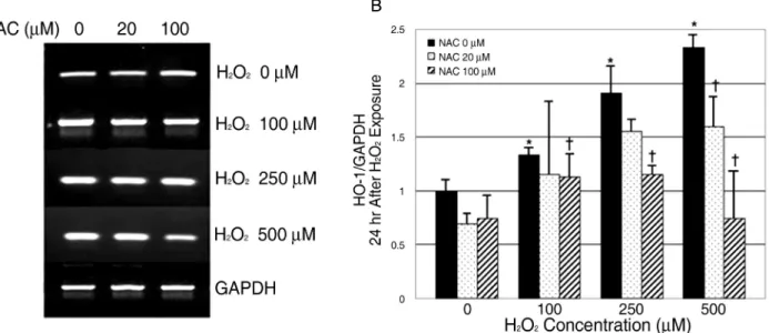
Figure 2. RT-PCR results of HO-1 mRNA expression in 24 hours culture. Human retinal pigment epithelial cells were cultured at various concentrations of H2O2 with or without NAC for 24 hours. HO-1 mRNA expression increased with H2O2 concentration increase. And after NAC treatment, HO-1 mRNA expression decreased in comparison to the cells without NAC treatment. The mRNA expression of HO-1 was normalized to GAPDH (house keeping gene). Data are shown as mean±SD of five experiments. *p<0.05 versus control (H2O20 μM, NAC 0 μM).
†p<0.05 versus the cells treated with H2O2 without NAC.
항산화제처리
산화스트레스 유도 방법과 동일하게 망막색소상피세포 를 처리한 후 H2O2를 농도별로 (0, 100, 250, 500 µM) 가 한 뒤 30분 후 항산화제로 N-acetylcystein (NAC)을 농 도별로(0, 20, 100 µM) 처리하였다. 약물 처리한 세포를 두 그룹으로 나누어 각각 24시간, 48시간 동안 배양한 후 총 RNA를 분리하여 RT-PCR을 시행하였다. 본 실험은 5회
반복하여 시행하였다.
통계분석
실험 결과는 5회의 독립된 실험 후 평균±표준편차의 형 태로 표시하였다. 통계분석은 SPSS version 12.0 (SPSS Inc., IL, USA)을 이용하였으며 Wilcoxon signed rank test를 이용하여 대조군과 실험군 간의 차이를 비교하였다. p<0.05인 경우 통계적으로 유의한 것으로 간주하였다.
결 과
세포활성도
MTT assay를 통하여 세포를 죽이지 않으면서 세포 변 형을 일으킬 수 있는 적정 H2O2의 농도를 조사하였다. H2O2
농도 50 μM 에 이틀간 배양하였을 때 세포생존율이 PBS를 넣은 대조군에 비해 98.3±6.8% 수준을 보였으며, 100 μM 에서는 97.3±6.3%를 보였다. H2O2250 μM에서는 83±5.5%
였으며 이후 점차적으로 감소하는 양상을 보였다(Fig. 1).
산화스트레스 유도 및 HO-1 유전자 발현
망막색소상피세포를 산화제인 H2O2에 농도를 달리하여
A B
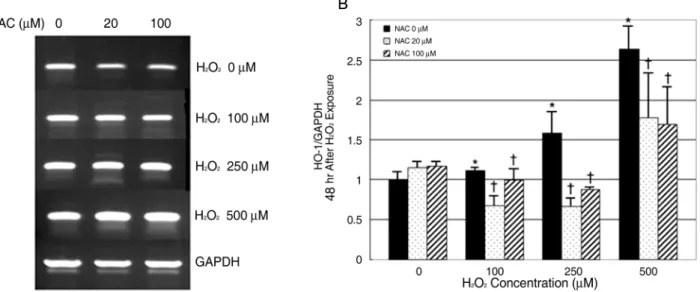
Figure 3. RT-PCR results of HO-1 mRNA expression in 48 hours culture. Human retinal pigment epithelial cells were cultured at various concentrations of H2O2 with or without NAC for 48 hours. HO-1 mRNA expression increased with H2O2 concentration increase. And after NAC treatment, HO-1 mRNA expression decreased in comparison to the cells without NAC treatment. The mRNA expression of HO-1 was normalized to GAPDH (house keeping gene). Data are shown as mean±SD of five experiments. *p<0.05 versus control (H2O20 μM, NAC 0 μM).
†p<0.05 versus the cells treated with H2O2 without NAC.
(0, 100, 250, 500 µM) 노출시킨 후 24시간 배양한 뒤 RT- PCR을 통해 HO-1 유전자 발현을 측정하였다. H2O2에 노 출되기 전에 비하여 H2O2에 노출되었을 경우에 HO-1 유 전자 발현이 유의하게 증가하는 것을 확인할 수 있었으며 이는 H2O2 농도가 높아질수록 증가하는 경향을 보였다 (p<0.05, Fig. 2A, B).
H2O2에 48시간 동안 노출시킨 후 시행한 검사에서도 24 시간 배양 결과와 마찬가지로 HO-1 유전자 발현이 H2O2
농도가 높아질수록 유의하게 증가하였다(p<0.05, Fig. 3A, B).
항산화제에 의한 HO-1 유전자 발현의 변화
30분간 항산화제인 NAC를 농도를 달리하여(0, 20, 100 µM) 전처리한 망막색소상피세포에 H2O2 농도를 달리하여 (0, 100, 250, 500 µM) 노출시킨 후 각각 24시간 동안 배 양한 뒤 RT-PCR을 시행하였다. NAC를 추가한 경우에는 H2O2만 처리하였을 때 보다 HO-1 유전자 발현이 상대적 으로 감소하였으며 NAC 농도가 높은 군에서 상대적으로 HO-1 유전자 발현이 더 적었다(p<0.05, Fig. 2A, B).
상기와 같은 방법으로 약물 처리한 후 48시간 배양한 뒤 시행한 RT-PCR에서도 24시간 배양 후 시행한 검사와 마 찬가지로 NAC를 추가한 경우에는 H2O2만 처리한 경우보 다 HO-1 유전자 발현이 유의하게 감소하였다(p<0.05).
그러나 24시간 배양했을 때와는 달리 NAC 농도에 따른 일 관된 차이는 보이지 않았다(Fig. 3A, B).
고 찰
본 논문은 연령관련황반변성의 치료 및 예방에 대해 알 아보기 위해 인체망막색소상피세포에서 산화스트레스 및 항산화제가 HO-1의 발현에 미치는 영향을 살펴보았다.
연령관령환반변성은 만성적인 산화스트레스에 대한 노출 과 나이가 들면서 항산화 방어 기제의 약화로 유발된다.6,7적 극적인 치료에도 불구하고 이미 손상된 시력의 회복에는 한계가 있기 때문에 연령관련황반변성에 대한 예방이 중요시 되고 있다. Age related eye disease study research group (AREDS)의 보고에 의하면 항산화 효과가 있는 비타민과 아연을 지속적으로 섭취할 경우 연령관련황반변성의 진행을 유의하게 감소시킨다고 한다.28 이는 연령관련황반변성이 어느 정도 예방될 수 있다는 것을 시사하는 보고로 그 의의가 있다고 할 수 있다.
HO에는 세 가지 isoform이 존재하며 이 중 HO-1이 유도 가능한 형태로 본 실험에서도 HO-1을 사용하였다. HO-1 은 인체의 여러 장기에서 외부 자극에 의해 유도되는 효소 로 항산화작용 뿐 아니라 항염증작용, 항세포사작용, 혈관 확장작용, 면역억제작용, 혈소판응집억제작용 등을 통해 이 식거부반응, 혈전형성, 급성경색에 의한 손상 등을 감소시
키는 효과가 있다고 알려져 있다.29-31상기와 같은 연구들 이 아직까지 망막색소상피세포에서는 이루어지지 않고 있 으나 연령관련황반변성의 또 다른 위험요소로 알려져 있는 드루젠의 생성에 염증반응과 면역반응이 관련이 되어 있다는 보고가 있다.32따라서 HO-1의 발현으로 유도되는 일련의 반응들은, 망막색소상피세포에서 연령관련황반변성의 병태 생리를 개선하는 방향으로 작용한다고 생각할 수 있다.
Hanneken et al은 항산화작용이 있는 flavonoid를 인체 망막색소상피세포에 가하였을 경우에 HO-1의 발현이 증 가되었으며, HO-1이 증가된 세포에서 산화스트레스에 의 한 세포사가 감소했다고 보고한 바 있다.33 Frank et al은 삼출성 연령관련황반변성이 있는 망막색소상피세포의 세포 질에서 HO-1과 catalase의 활동성이 증가되어 있으며 나 이가 들수록 활동성이 떨어진다고 하였다.34이는 망막색소 상피세포가 만성적으로 산화스트레스에 노출될 경우 그 방 어기전으로 HO-1이 증가되며, 나이가 들수록 HO-1의 활 동성이 감소되기 때문에 연령관련황반변성의 발병 위험이 높아지는 것으로 해석된다. 또한, Milbury et al은 bilberry (Vaccinium myrtillus) 추출물을 망막색소상피세포에 처리 하였을 경우 H2O2를 가하였을 때 생성되는 반응성 산소종 이 감소하였으며 HO-1의 발현이 증가하였다고 하였다.35 본 연구에서는 인체망막색소상피세포에 H2O2를 농도를 달리하여 가한 뒤 24시간, 48시간 배양 후 HO-1의 발현을 비교하고, 항산화제로 잘 알려져 있는 NAC를 H2O2와 함께 처리한 뒤 HO-1의 발현을 비교해 보았다. 적정농도의 H2O2를 구하기 위해 MTT assay를 시행한 결과 100 μM 까지는 97.3±6.3%의 높은 세포생존율을 보였으나 250 μM 에서는 83±5.5%였으며 이후에는 계속적으로 감소하였다 (Fig. 1). 그러나 24시간, 48시간 배양한 망막색소상피세포 에서는 모두 H2O2의 농도가 증가할수록 HO-1의 발현이 증가하는 경향을 보였다(Fig. 2, 3). 이는 망막색소상피세 포가 산화스트레스에 대항하여 HO-1의 생성을 증가시킨 다고 생각할 수 있을 것이며, 본 연구에서는 H2O2농도 500 μM 까지만 확인을 하였으나, 세포의 생존이 일정수준 이상 유지되는 경우에서는 H2O2농도에 비례하여 HO-1의 발현이 증가할 것이라고 추측할 수 있겠다. 이러한 결과로 미루어 볼 때 HO-1은 망막색소상피세포에서 산화스트레스 정도에 대한 표지자로 활용될 여지가 있다고 생각할 수 있을 것이다.
망막색소상피세포에서 항산화제에 의한 HO-1의 발현을 알아보기 위하여 본 실험에서는 항산화제로 NAC를 이용하 였다. 24시간, 48시간 배양실험에서 모두 NAC를 30분간 전처리 한 후 H2O2를 가한 경우에서는 H2O2만 단독으로 처 리한 경우보다 HO-1의 발현이 감소하였다. 이는 항산화제 가 망막색소상피세포에 가해지는 산화스트레스를 감소시키
기 때문에 상대적으로 HO-1의 발현이 감소된 것이라고 생 각할 수 있을 것이다. 24시간 배양한 망막색소상피세포에 서는 NAC 농도가 높았던 경우에서 HO-1의 발현이 더감 소하는 양상을 보였으나 48시간 배양한 망막색소상피세포 에서는 일관된 반응을 보이지 않았다(Fig. 2, 3).
연령관련황반변성은 다양한 기전으로 일어날 수 있다.
그러나 장기적인 산화스트레스가 그 주요 기전으로 알려져 있다. 따라서 평상시에 산화스트레스를 감소시킬 수 있다면 연령관련황반변성을 예방하는 효과를 가져 올 수 있을 것 이다. 본 실험을 통해 인체망막색소상피세포에서 산화스트 레스와 항산화제가 HO-1의 발현에 미치는 영향을 확인할 수 있었다. 인체망막색소상피세포에서 HO-1의 작용기전에 대한 연구가 더 진행되고, 생체 내에서의 연구가 이루어진 다면 HO-1을 활용하여 연령관련황반변성을 예방하는데 많은 도움이 될 것이다.
참고문헌
1) Friedman DS, O'Colmain BJ, Muñoz B, et al. Eye Diseases Prevalence Research Group. Prevalence of Age-Related Macular Degeneration in the United States. Arch Ophthalmol 2004;122:
564-72.
2) Klein R, Wang Q, Klein BE, et al. The relationship of age related maculopathy, cataract, and glaucoma to visual acuity. Invest Ophthalmol Vis Sci 1995;36:182-91.
3) Williams RA, Brody BL, Thomas RG, et al. The psychosocial impact of macular degeneration. Arch Ophthalmol 1998;116:
514-20.
4) Young RW. Pathophysiology of age-related macular degeneration.
Surv Ophthalmol 1987;31:291-306.
5) Strauss O. The retinal pigment epithelium in visual function.
Physiol Rev 2005;85:845-81.
6) Beatty S, Koh H, Phil M, et al. The role of oxidative stress in the pathogenesis of age-related macular degeneration. Surv Ophthal- mol 2000;45:115-34.
7) Winkler BS, Boulton ME, Gottsch JD, Sternberg P. Oxidative damage and age-related macular degeneration. Mol Vis 1999;5:32.
8) Jaattela M. Heat shock proteins as cellular lifeguards. Ann Med 1999;31:261–71.
9) David JT, Michael VM, David AN. Phagocytosis and H2O2
induce catalase and metallothionein gene expression in human retinal pigment epithelial cells. Invest Ophthalmol Vis Sci 1995;
36:1271-9.
10) Tenhunen R, Marver HS, Schmid R. The enzymatic conversion of heme to bilirubin by microsomal heme oxygenase. Proc Natl Acad Sci 1968;61:748-55.
11) Clark JE, Foresti R, Green CJ, Motterlini R. Dynamics of haem oxygenase-1 expression and bilirubin production in cellular protection against oxidative stress. Biochem J 2000;348:615-9.
12) Stocker R, Yamamoto Y, McDonagh AF, et al. Bilirubin is an antioxidant of possible physiological importance. Science 1987;
235:1043-6.
13) Baranano DE, Wolosker H, Bae BI, et al. A mammalian iron ATPase induced by iron. J Biol Chem 2000;275:15166-73.
14) Peyton KJ, Reyna SV, Chapman GB, et al. Heme oxygenase-1- derived carbon monoxide is an autocrine inhibitor of vascular smooth muscle cell growth. Blood 2002;99:4443-8.
15) Otterbein LE, Bach FH, Alam J, et al. Carbon monoxide has anti-inflammatory effects involving the mitogen-activated protein kinase pathway. Nat Med 2000;6:422-8.
16) Otterbein LE, Zuckerbraun BS, Haga M, et al. Carbon monoxide suppresses arteriosclerotic lesions associated with chronic graft rejection and with balloon injury. Nat Med 2003;9:183-90.
17) Ryter SW, Otterbein LE. Carbon monoxide in biology and medicine. Bioessays 2004;26:270-80.
18) Maines MD, Trakshel GM, Kutty RK. Characterization of two constitutive forms of rat liver microsomal heme oxygenase: only one molecular species of the enzyme is inducible. J Biol Chem 1986;261:411-9.
19) Maines MD. Heme oxygenase: function, multiplicity, regulatory mechanisms, and clinical applications. FASEB J 1988;2:2557-68.
20) McCoubrey WK Jr, Huang TJ, Maines MD. Isolation and charac- terization of a cDNA from the rat brain that encodes hemoprotein heme oxygenase- 3. Eur J Biochem 1997;247:725-32.
21) Ryter SW, Alam J, Choi AM. Heme oxygenase-1/carbon monoxide: From basic science to therapeutic applications. Physiol Rev 2006;86:583-650.
22) Kutty RK, Nagineni CN, Kutty G, et al. Increased expression of heme oxygenase-1 in human retinal pigment epithelial cells by transforming growth factor-beta. J Cell Physiol 1994;159:371-8.
23) Kuesap J, Li B, Satarug S, et al. Prostaglandin D2 induces heme oxygenase-1 in human retinal pigment epithelial cells. Biochem Biophys Res Commun 2008;367:413-9.
24) Udono-Fujimori R, Takahashi K, Takeda K, et al. Expression of heme oxygenase-1 is repressed by interferon-gamma and induced by hypoxia in human retinal pigment epithelial cells. Eur J Biochem 2004;271:3076-84.
25) Alizadeh M, Wada M, Gelfman CM, et al. Downregulation of Differentiation Specific Gene Expression by Oxidative Stress in ARPE-19 Cells. Invest Ophthalmol Vis Sci 2001;42:2706-713.
26) McCoubrey WK Jr, Maines MD. The structure, organization and differential expression of the gene encoding rat heme oxygenase-2.
Gene 1994;139:155-61.
27) Hayashi S, Omata Y, Sakamoto H, et al. Characterization of rat heme oxygenase-3 gene: Implication of processed pseudogenes derived from heme oxygenase-2 gene. Gene 2004;336:241-50.
28) Age-Related Eye Disease Study Research Group. A randomized, placebo-controlled, clinical trial of high-dose supplementation with vitamins C and E, beta carotene, and zinc for age-related macular degeneration and vision loss: AREDS report No. 8. Arch Ophthalmol 2001;119:1417-36.
29) Morita T. Heme oxygenase and atherosclerosis. Arterioscler Thromb Vasc Biol 2005;25;1786-95.
30) Hoekstra KA, Godin DV, Cheng KM. Protective role of heme oxygenase in the blood vessel wall during atherogenesis. Biochem Cell Biol 2004;82:351-9.
31) Nath KA. Heme oxygenase-1: A provenance for cytoprotective pathways in the kidney and other tissues. Kidney Int 200670:
432-43.
32) Donoso LA, Kim D, Frost A, et al. The Role of Inflammation in the Pathogenesis of Age-related Macular Degeneration. Surv Ophthalmol 2006;51:137-52.
33) Hanneken A, Lin FF, Johnson J, Maher P. Flavonoids Protect Human Retinal Pigment Epithelial Cells from Oxidative-Stress–
Induced Death. Invest Ophthalmol Vis Sci 2006;47:3164-77.
34) Frank RN, Amin RH, Puklin JE. Antioxidant enzymes in the macular retinal pigment epithelium of eyes with neovascular age-related macular degeneration. Am J Ophthalmol 1999;127:
694-709.
35) Milbury PE, Graf B, Curran-Celentano JM, Blumberg JB.
Bilberry (Vaccinium myrtillus) anthocyanins modulate heme oxygenase-1 and glutathione S-transferase-pi expression in ARPE- 19 cells. Invest Ophthamol Vis Sci 2007;48:2343-9.
=ABSTRACT=
Effects of Oxidative Stress and Antioxidant on the Expression of Heme Oxygenase-1 in Human RPE
Young Jun Kim, MD, Hee Seung Chin, MD, PhD
Department of Ophthalmology, Inha University School of Medicine, Incheon, Korea
Purpose: To evaluate the effects of oxidative stress and antioxidantson heme oxygenase-1 (HO-1) in cultured human retinal pigment epithelial (RPE) cells.
Methods: Cultured RPE cells were challenged with different concentrations of hydrogen peroxide (H2O2), and the HO-1 mRNA level was determined by RT-PCR after 24 hours and 48 hours of incubation independently. Additionally, the HO-1 mRNA level was measured after preincubating RPE cells with N-acetylcystein (NAC) as the antioxidant for 30 minutes and then challenging the cells with H2O2.
Results: The expression of the HO-1 mRNA level increased after H2O2 exposure, and this level was proportional to the increased H2O2 concentration (p<0.05). Expression of the HO-1 mRNA level decreased when the RPE cells were preincubated with NAC (p<0.05).
Conclusions: In human RPE cells, the HO-1 level increased in proportion to the degree of oxidative stress and decreased with exposure to antioxidants. Expression of HO-1 may be helpful in preventing retinal disease, which occurs due to oxidative stresses such as age-related macular degeneration.
J Korean Ophthalmol Soc 2009;50(8):1247-1253
Key Words: Age related macular degeneration, Antioxidant, Heme oxygenase-1, Oxidative stress, Retinal pigment epithelium
Address reprint requests to Hee Seung Chin, MD, PhD Department of Ophthalmology, Inha University School of Medicine
#7-206 Shinheung-dong, Jung-gu, Incheon 400-711, Korea
Tel: 82-32-890-2408, Fax: 82-32-890-2303, E-mail: [email protected]