919 대한안과학회지 2009년 제 50 권 제 6 호 J Korean Ophthalmol Soc 2009;50(6):919-922 DOI : 10.3341/jkos.2009.50.6.919
www.ophthalmology.org
= 증례보고 = 접수번호 : 50-06-12-10
일산화질소가 R28 세포의 생존에 미치는 영향
이정일⋅김재우
대구가톨릭대학교 의과대학 안과학교실
목적: 일산화질소(Nitric oxide, NO)가 R28 세포의 생존에 미치는 영향을 알아보고자 하였다.
대상과 방법: R28 세포주를 계대배양한 후 24-well plate에 옮긴 다음 SNAP에 1, 10, 100 μM의 농도로 노출시켰으며 L-NAME에도 동시에 노출시켰다. 혈청의 영향을 알기 위해 무혈청배지와 10% 우태아혈청을 포함한 DMEM 배지를 이용하였으며 1일과 3일에 세포의 생존을 MTT assay를 이용하여 조사하였다.
결과: 노출된 NO의 농도에 비례하여 유의하게 R28 세포의 생존이 저하되었으며(p<0.05), L-NAME는 이러한 NO에 의한 R28 세포에 대한 생존억제효과를 상쇄시켰다(p>0.05). 이러한 결과는 혈청의 포함 유무에 관계없이 1일째와 3일째에 유사하게 나타났다.
결론: NO는 망막전구세포주인 R28 세포에 대해 세포독성을 나타내어 세포의 생존을 저하시킴을 알 수 있었다. 망막신경세포의 손상 기전을 연구하기 위해서는 R28 세포의 세포독성의 손상 기전에 대한 자세한 연구가 필요할 것으로 생각된다.
<대한안과학회지 2009:50(6):919-922>
■ 접 수 일: 2008년 12월 11일 ■ 심사통과일: 2009년 3월 10일
■ 통 신 저 자: 김 재 우
대구시 남구 대명4동 3056-6 대구가톨릭대학교 병원 안과
Tel: 053-650-4728, Fax: 053-627-0133 E-mail: [email protected]
일산화질소(Nitric oxide, NO)는 자유 유리기로써 생체막을 투과하여 내피세포에서의 이완작용, 신경계에서의 조절작용, 면역계에서의 면역매개물질 등 생체에서 중요한 역할을 하는 것으로 알려져 있으며1,2세포의 생존에 대한 NO의 효과는 세 포의 종류에 따라 세포의 증식을 유발하거나 반대로 세포의 고 사를 유발하는 경우도 있는 것으로 알려져 있는데3,4안구에서 도 NO 합성효소가 다양하게 발현되는 것으로 알려져 있고,5-8 녹내장의 경우 글루탐산 등에 의해 신경절세포의 고사가 유발 되며9,10이는 NO와 관련되어 있는 것으로 알려져 있다.11,12
망막신경세포 손상의 연구를 위해서 망막신경세포의 일차 적인 세포배양은 매우 힘들어 최근 여러 가지 세포배양모델 이 개발되어 사용되고 있다.13그 중 하나가 망막전구세포인 R28 세포인데14본 연구에서는 R28 세포를 이용하여 NO가 세포의 생존에 미치는 영향을 알아보고 시신경 보호에 관한 세 포배양모델로서의 유용성에 대해 고찰해 보고자하였다.
대상과 방법
세포배양과 면역염색
R28 세포주(immortalized with the 12S, nontumorigenic
portion of the adenovirus E1A gene from P6 rat retinal tissue)를 수립한 Dr. Seigel에게서 공여받은 R28를 분주하 여 배양한 후 항체에 대한 면역염색을 각각 시행하였다. 코 팅한 커버글라스 위에 세포를 배양한 후 5분간 고정하고 나 서 GFAP, vimemtin, S-100, neurofilament에 대한 일차 항체(Code No. M0581, DAKO, Denmark)에 각각 반응시 킨 후 Streptavidin-Biotin 표지 2차 항체(Ultratech HRP, Code No. 2391, Immunotech, France)에 반응시키고 DAB (Ultratech AEC, Code No. 2393, Immunotech, France)로 발색시켜 봉입한 다음 광학현미경으로 관찰하였다. 이때 사 용한 배지는 고농도의 포도당이 포함된 DMEM (Dulbecco’s modified Eagles Medium) 배지에 10% 우태아혈청, 비필 수아미노산과 비타민, sodium bicarbonate, L-glutamine, Gentamicin를 첨가하였으며 배양에 사용된 배지와 첨가물은 모두 미국 Gibco사의 제품을 사용하였다.
약물처리
R28 세포를 배양접시에서 계대배양한 후 24-well plate에 옮긴 다음 NO 공여자인 SNAP (S-nitroso-N-acetyl-D, L-penicillamine, Sigma, USA)에 1, 10, 100 μM의 농도로 노출시켰으며 NOS 저해제인 L-NAME (Nω-Nitro-L- arginine methyl ester, Sigma, USA)에도 동시에 노출시켰다.
혈청이 미치는 영향을 알아보기 위해 무혈청배지와 10%
우태아혈청을 포함한 배지를 이용하였으며 각각 1일과 3일에 세포의 생존과 NO의 생성을 조사하였다.
920
- 대한안과학회지 2009년 제 50 권 제 6 호 -
www.ophthalmology.org
Figure 1. Phase-contrast photograph of R28 cells in monolayer culture (×100).
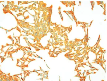
Figure 2.R28 cells immunostained with vimentin (×100).
Figure 3.Survival of R28 cells after exposed to SNAP for 1 day in serum-free and 10% serum-containing media.
Cellular survival was decreased significantly with expo- sure to NO in a dose-dependent manner (*p<0.05).
MTT와 Griess assay
세포의 생존을 알아보기 위해 MTT assay를, NO의 생성 을 알아보기 위해 Griess assay를 각각 시행하였다. MTT assay는 약물처리한 세포의 배지에 MTT (Sigma, USA)를 각 well당 100 μL씩 투여한 후 4시간 동안 정치배양한 다음 염류용액으로 씻어낸 후 DMSO (Sigma, USA)를 각 well당 0.5 mL씩 넣어 10분 이상 흔든 다음 96-well plate에 200 μL 씩옮겨 Spectrophotometer (SIRIO-S, Italy)로 570 nm 에서 측정하였다. 이때 세포의 생존 정도는 실험군의 값을 약물처리를 하지 않은 대조군의 비로 나누어 백분율로 나타 내었다. Griess assay15는 약물 처리한 세포의 배지에동량의 Griess 반응액(Sigma, USA)를 섞은 후 96-well plate에 옮겨 Spectrophotometer로 540 nm에서 아질산염의양을 측정하였다. 이때 표준치를 구하기 위해 sodium nitrite (Sigma, USA)를 단계적으로 희석하여 사용하였다.
통계적 처리
모든 실험은 공여받은 R28 세포를 배양한 후 4계대에서 5 계대 사이의 세포를 이용하였고 3회 이상 반복하여 시행하였 다. 모든 실험에서 대조군은 약물처리를 하지 않은 군으로 하였으며 실험군과 대조군의 비교는 두 군 간에는 unpaired t-test를 이용하였으며 유의 수준은 0.05% 이하로정하였다.
결 과
세포배양과 면역염색
R28 세포는 녹여서 분주한 후 빠르게 증식하여 3~4일 내에 배양접시에 충만하게 증식하였으며(Fig. 1), GFAP과 vimentin에 대해서는 강하게 면역염색 되었으나 S-100과 neurofilament에 대해서는 비교적 약한 반응을 보였다(Fig. 2).
NO가 R28 세포의 생존에 미치는 영향
NO 공여자인 SNAP에 하루 동안 노출시켰을 때 10 μM 의 농도부터 노출된 NO의 농도에 비례하여 유의하게 R28 세포의 생존이 저하되었으며(p<0.05), L-NAME를 투여 했을 경우에는 약물처리를 하지 않은 대조군에 비해 유의 한 생존율의 차이를 보이지 않았다(Fig. 3). 이러한 결과는 배지에 포함된 혈청의 유무에 관계없이 3일째도 유사하게 나타났다(Fig. 4).
NO 공여자에 의한 NO의 생성
NO 공여자인 SNAP에 하루 동안 노출시켰을 때 배지에 서의 nitrite 양은 공여된 NO의 농도의 증가에 따라 10 μM 의 농도부터 유의하게 증가하였으며 L-NAME에 의해 NO 의 생성이 유의하게 억제되었다(p<0.05)(Fig. 5). 이러한
921 - 이정일 외 : 일산화질소와 R28 세포 -
www.ophthalmology.org
Figure 5.NO production of R28 cells after exposed to SNAP for 1 day. SNAP increased NO production sig- nificantly (*p<0.05).
Figure 4.Survival of R28 cells after exposed to SNAP for 3 days in serum-free and 10% serum-containing media.
Cellular survival was decreased significantly with expo- sure to NO in a dose-dependent manner (*p<0.05).
결과는 배지에 포함된 혈청의 유무에 관계없이 1일째와 3일 째에 유사하였다.
고 찰
NO는 생리적인 조절인자로 작용할 뿐만 아니라 병리적인 매개체로도 작용한다.1,2NO는 저농도에서는 인체에서 중요한 생리적인 조절인자로 작용하지만 고농도에서는 반응성 산화 물질로 전환되어 세포에 병적인 손상을 유발하기도 한다. 그 중 NO가 세포의 생존에 미치는 영향은 세포의 종류에 따라 다르게 나타나는데3 녹내장이 있는 경우에는 NO와 NO를 합성하는 조직의 이상이 나타난다고 알려져 있다.16,17
녹내장에서 망막의 신경절세포가 글루탐산 등에 의한 세 포독성에 의한 세포고사에 의한다는 것이 알려진 이후 시 신경보호를 위한 많은 연구가 이루어지고 있는데 세포학적 수준에서의 연구의 재료로서 사람의 신경절세포의 일차배 양이 보고되기도 하였으나18 실제 망막신경절 세포를 분리 하여 유지하고 증식시키기는 대단히 힘들다.19따라서 이의 대안으로 세포에 유전학적 기법을 도입하여 조작한 세포주 를 이용한 연구가 행해지고 있다.13그 중 본 연구에서 사용 한 R28 세포는 쥐의 망막세포에 아데노바이러스 E1A 유전 자의 비종양형성부위인 12S를 이용하여 만든 세포주로서 망막의 구조와 기능적 분화의 연구에 많이 이용되고 있다.
R28 세포는 망막의 각종 구성세포의 형질을 발현하며 망막에 이식해도 생존하는 것으로 알려져 있다.20또한 저산소증이 나 혈청의 결핍에 대해 세포고사를 나타낼 뿐만 아니라21-23 녹내장에서 망막신경절세포의 세포고사에 관여하는 것으로 알려진 글루탐산이나 GABA 수용체를 발현하므로 이와 관 련된 세포고사와 세포독성에 관한 연구에 유용하게 사용될 가능성이 있다.24흥분성 글루탐산에 의해 망막세포의 세포 고사가 유발되고 NO는 글루탐산 등에 의한 흥분성 세포독 성과 관련이 있으므로11,12,25본 연구에서 R28 세포를 이용
하여 NO에 의한 생존효과를 조사한 결과 NO는 R28 세포 에 대해 생존저하효과를 나타내었다. 비록 본 연구에서는 세포사의 기전까지는 자세히 밝혀내지 못하였으나 신경세 포독성이 망막신경전구세포인 R28 세포에서 NO에 의해 세 포독성이 유발된다는 것을 알 수 있었다. 또한 R28 세포는 배양조건에 따라 망막전구세포에서 망막신경절세포로도 분 화할 수 있으므로22망막의 전반적인 세포학적 연구뿐만 아 니라 순수한 망막신경절세포의 연구에 사용될 가능성이 있을 것이다.
결론적으로 본 연구에서 NO는 망막전구세포인 R28 세 포에 대해 세포독성을 나타내어 세포의 생존을 저하시킴을 알 수 있었다. 망막의 신경세포와 망막신경절세포의 손상 기전과 시신경보호 연구를 위해 향후 R28 세포의 세포독성의 기전에 대한 자세한 연구가 필요할 것으로 생각된다.
참고문헌
1) Moncada S, Palmer RM, Higgs EA. Nitric oxide: physiology, pathophysiology, and pharmacology. Pharmacol Rev 1991;43:
109-42.
2) Bredt DS, Snyder SH. Nitric oxide: a physiologic messenger molecule. Annu Rev Biochem 1994;63:175-95.
3) Brüne B, Knethen A, Sandau KB. Nitric oxide and its role in apoptosis. Eur J Pharmacol 1998;351:261-72.
4) Beck KF, Eberhardt W, Frank S, et al. Inducible NO synthase:
Role in cell signaling. J Exp Biol 1999;202:645-53.
5) Becquet F, Courtois Y, Goureau O. Nitric oxide in the eye:
multifaceted roles and diverse outcomes. Surv Ophthalmol 1997;
42:71-82.
6) Meyer P, Champion C, Schlotzer-Schrehardt U, et al. Localization of nitric oxide synthase isoforms in porcine ocular tissues. Curr Eye Res 1999;18:375-80.
7) Geyer O, Podos SM, Mittag T. Nitric oxide synthase activity in tissues of the bovine eyes. Graefes Arch Clin Exp Ophthalmol 1997;235:786-93.
8) Nathanson JA, McKee M. Identification of an extensive system of nitric oxide-producing cells in the ciliary muscle and outflow pathway. Invest Ophthalmol Vis Sci 1995;36:1765-73.
9) Quigley HA, Nickells RW, Kerrigan LA, et al. Retinal ganglion
922
=ABSTRACT=
Effect of Nitric Oxide on the Survival of R28 Cells
Jeong Il Lee, MD, Jae Woo Kim, MD, PhD
Department of Ophthalmology, Catholic University of Daegu College of Medicine, Daegu, Korea
Purpose: To evaluate the effect of nitric oxide (NO) on the survival of R28 cells.
Methods: After immunostaining for GFAP, vimentin, S-100, and neurofilament, R28 cells were exposed to S-nitroso-N-acetyl-D, L-penicillamine (SNAP) at various concentrations, with and without the NO inhibitor, Nω-Nitro-L-arginine methyl ester (L-NAME) for 1 and 3 days. Cellular survival of R28 cells and the production of NO were quantified by rapid colorimetric assays using the MTT and Griess assay, respectively. To evaluate the effect of serum, 10% serum or serum-free media were used separately.
Results: R28 cells showed strong immunoreactivity to GFAP and vimentin compared to S-100 or neurofilament. SNAP inhibited the survival of R28 cells in a dose-dependent manner, and this effect of NO on the cellular survival was abolished by L-NAME.
These results were similar after exposure for 1 and 3 days, regardless of the presence of serum in the media.
Conclusions: The current results suggest that NO decreased the survival of R28 cells. Further studies are necessary to evaluate the mechanism of cytotoxicity of the R28 cells.
J Korean Ophthalmol Soc 2009;50(6):919-922 Key Words: Neurotoxicity, Nitric oxide, R28 cells
Address reprint requests to Jae Woo Kim, MD, PhD
Department of Opthalmology, Catholic University of Daegu College of Medicine
#3056-6 Daemyeung 4-dong, Nam-gu, Daegu 705-718, Korea Tel: 82-53-650-4728, Fax: 82-53-627-0133, E-mail: [email protected]
- 대한안과학회지 2009년 제 50 권 제 6 호 -
www.ophthalmology.org cell death in experimental glaucoma and after axotomy occurs by
apoptosis. Invest Ophthalmol Vis Sci 1995;36:774-86.
10) Otori Y, Wei J-Y, Barnstable CJ. Neurotoxic effects of low doses of glutamate on purified rat retinal ganglioncells. Invest Ophthal- mol Vis Sci 1998;39:972-81.
11) Morgan J, Caprioli J, Koseki Y. Nitric oxide mediates excitotoxic and anoxic damage in rat retinal ganglion cells cocultured with astroglia. Arch Ophthalmol 1999;117:1524-9.
12) Vorwerk CK, Hyman BT, Miller JW, et al. The role of neuronal and endothelial nitric oxide synthase in retinal excitotoxicity.
Invest Ophthalmol Vis Sci 1997;38:2038-44.
13) Seigel GM. The golden age of retinal cell culture. Mol Vis 1999;5:4 14) Seigel GM, Mutchler AL, Imperato EL. Expression of glial markers
in retinal precusor cell line. Mol Vis 1996;2:2
15) Green LC, Wagner DA, Glogoski J, et al. Analysis of nitrate, nitrite and [15N]nitrate in biologic fluids. Anal Biochem 1982;
126:131-8.
16) Nathanson JA, McKee M. Alterations of ocular nitric oxide synthase in human glaucoma. Invest Ophthalmol Vis Sci 1995;
36:1774-84.
17) Neufeld AH, Hernandez MR, Gonzalez M. Nitric oxide synthase in the human glaucomatous optic nerve head. Arch Ophthalmol 1997;115:497-503.
18) Hu DN, Ritch R. Tissue culture of adult human retinal ganglion
cells. J Glaucoma 1997;6:37-43.
19) Barres BA, Silverstein BE, Corey DP, Chun LL. Immunological, morphological, and electrophysiological variation among retinal ganglion cells purified by panning. Neuron 1988;1:791-803.
20) Seigel GM, Takahashi M, Adamus G, McDaniel T. Intraocular transplantation of E1A-immortalized retinal precusor cells. Cell Transplant 1998;7:559-66.
21) Seigel GM, Chiu L, Paxhia A. Inhibition of neuroretinal cell death by insulin-like growth factor-1 and its analogs. Mol Vis 2000;6:
157-63.
22) Barber AJ, Nakamura M, Wolpert EB, et al. Insulin rescues retinal neurons from apoptosis by a phosphatidyl 3-kinase/Akt-mediated mechanism that reduces the activation of Caspases-3. J Biol Chem 2001;276:32814-21.
23) Nakamura M, Barber AJ, Antonetti DA, et al. Excessive hexo- samines block the neuroprotective effect of insulin and induce apoptosis in retinal neurons. J Biol Chem 2001;276: 43748-55.
24) Sun W, Seigel GM, Salvi RJ. Retinal precusor cells express functional ionotropic glutamate and GABA receptors. Neuroreport 2002;13:2421-4.
25) Kawasaki A, Otori Y, Barnstable CJ. Müller cell protection of rat retinal ganglion cells from glutamate and nitric oxide neurotoxicity.
Invest Ophthalmol Vis Sci 2000;41:3444-50.