Korean J Gastroenterol Vol. 63 No. 5, 321-324 http://dx.doi.org/10.4166/kjg.2014.63.5.321 pISSN 1598-9992 eISSN 2233-6869
CASE REPORT
[Korean J Gastroenterol, Vol. 63 No. 5, May 2014 www.kjg.or.kr
내시경역행담췌관조영술과 간동맥내 화학요법 후 소실된 담관내 간세포암 1예
조영윤, 이상협, 이재우, 박진명, 류지곤, 김용태, 윤창진
1, 김혜령
2서울대학교 의과대학 내과학교실 간연구소, 분당서울대학교병원 영상의학과1, 병리과2
Disappearance of Intrahepatic Bile Duct Hepatocellular Carcinoma after Endoscopic Retrograde Cholangiopancreatography and Transarterial Chemoinfusion: A Case Report
Young Youn Cho, Sang Hyub Lee, Jae Woo Lee, Jin Myung Park, Ji Kon Ryu, Yong-Tae Kim, Chang Jin Yoon1 and Haeryoung Kim2 Department of Internal Medicine and Liver Research Institute, Seoul National University College of Medicine, Seoul, Departments of Radiology1 and Pathology2, Seoul National University Bundang Hospital, Seongnam, Korea
Invasion of the bile duct by hepatocellular carcinoma (HCC), which is called intrahepatic bile duct HCC, is rare and has a poor prognosis. Early diagnosis and surgical resection is important for treatment. A 58-year-old man who underwent hepatic resection for HCC 4 years ago and received transarterial chemoembolization (TACE) 2 years after the operation for recurred HCC presented with jaundice. CT scan revealed a tumor in the common bile duct without intrahepatic lesion. Therefore, ERCP was done to perform biopsy and biliary drainage. Histological examination was compatible with hepatocellular carcinoma.
However, the tumor could not be visualized at angiography and thus, only transarterial chemoinfusion was performed without embolization. The tumor had disappeared on follow-up CT scan, and the patient has been disease free for 23 months without evidence of recurrence. Herein, we report a case of intrahepatic bile duct HCC which disappeared after ERCP. (Korean J Gastroenterol 2014;63:321-324)
Key Words: Hepatocellular carcinoma; Intrahepatic bile duct hepatocellular carcinoma; Icteric type hepatoma
Received July 26, 2013. Revised September 13, 2013. Accepted September 17, 2013.
CC This is an open access article distributed under the terms of the Creative Commons Attribution Non-Commercial License (http://creativecommons.org/licenses/
by-nc/3.0) which permits unrestricted non-commercial use, distribution, and reproduction in any medium, provided the original work is properly cited.
교신저자: 이상협, 110-744, 서울시 종로구 대학로 101, 서울대학교병원 내과
Correspondence to: Sang Hyub Lee, Department of Internal Medicine, Seoul National University Hospital, 101 Daehak-ro, Jongno-gu, Seoul 110-744, Korea. Tel: +82- 2-2072-2228, Fax: +82-2-762-9662, E-mail: [email protected]
Financial support: None. Conflict of interest: None.
서 론
간세포암은 간문맥과 간정맥을 잘 침범하는 것으로 알려져 있지만 담관내로의 침범은 드물다. Lin 등1은 이와 같은 형태 의 간세포암을 황달형 간암(icteric type hepatoma)이라 명 명하였으며 현재는 담관내 간세포암(intrahepatic bile duct hepatocellular carcinoma)으로 흔히 불리고 있다. 담관내 간세포암의 발생 빈도는 0.53%에서 9%까지 보고되고 있 다.2,3 담관내 간세포암 가운데 간내에 종괴가 없으면서 간내 담관에 간세포암이 발생하는 경우는 드물지만 간헐적으로 보 고되고 있다. 담관내 간세포암의 예후는 나쁜 것으로 알려져
있으나 간 부전으로 황달이 발생한 환자에 비해서는 예후가 좋다고 알려져 있으며 조기에 발견하는 것이 중요하다. 저자 들은 간세포암 수술 이후 추적관찰 도중 우연히 발견되었고 내시경역행담췌관조영술(ERCP) 및 간동맥내 화학요법 시행 후 소실된 담관내 간세포암 1예를 경험하였기에 문헌고찰과 함께 보고하는 바이다.
증 례
58세 남자가 간세포암에 대한 추적관찰 도중 발견된 담관 내 종괴를 주소로 내원하였다. 환자는 4년 전 복통으로 검사
322
조영윤 등. 내시경역행담췌관조영술과 간동맥내 화학요법 후 소실된 담관내 간세포암The Korean Journal of Gastroenterology
Fig. 2. Cholangiogram shows an ob- structive mass in the common bile duct.
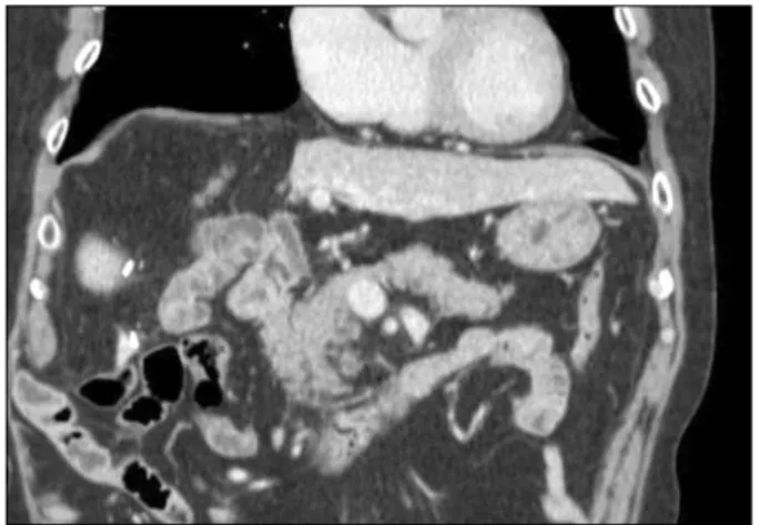
Fig. 1. Coronal view of the CT scan shows a well enhancing tumor in the common bile duct (arrow).
하여 본원 소화기내과에서 간세포암을 발견하고 S4, S8 계통 적 아구역 절제술을 시행받았다. 수술 후 병리결과는 크기 4.5×4.0×4.0 cm, Edmondson-Steiner 3등급, 담관 절제연 침범으로 보고되었다. 경과관찰 중 2년 전 간내 재발 소견으 로 경피적 간동맥 화학색전술(transarterial chemoemboili- zation, TACE)을 시행하였다. 과거력에서 간세포암 외 고혈 압이 있었으며 만성 B형간염, C형간염은 없었다. 음주, 흡연 은 부인하였고 간염과 간세포암의 가족력도 없었다. 혈압은 120/84 mmHg, 맥박은 분당 84회, 호흡은 분당 20회였으며 체온은 36.5oC였다. 복부는 편평하였고 장음은 정상적이었다.
복부 종괴는 촉지되지 않았으며 상복부 압통이나 반발통은 없 었다. 다른 신체검사와 계통적 문진에서 이상 소견은 없었다.
말초혈액검사에서 백혈구 수 8,500/μL, 혈색소 17.1 g/dL, 혈소판 수 100,000/μL, 총 빌리루빈 2.2 mg/dL (0.2-1.2 mg/dL), 직접 빌리루빈 0.7 mg/dL (0-0.5 mg/dL), AST/
ALT 50/60 IU/L, ALP 170 IU/L (30-115 IU/L), 알부민 4.1 g/dL (3.3-5.2 g/dL)였다. Prothrombin time 87% (80- 120%), PT-INR 1.09 (0.8-1.2)로 정상범위에 있었으며 혈청 검사상 AFP가 16.4 IU/mL (0.85-9.4 IU/mL)로 증가하였다.
Child-Pugh score는 A였다. 다른 혈액검사상 이상 소견은 없 었다.
복부 CT에서 근위 총담관에 1×2 cm 크기의 폴립 모양의 종괴 관찰되었으며 좌측 간내담관의 확장이 관찰되었다(Fig.
1). 종괴는 동맥기에서 조영증강되었으며 지연기에서 저음영 을 보이는 전형적인 간세포암 소견이었다. 폐쇄성 황달이 있 어 ERCP를 시행 후(Fig. 2), 담즙 배액을 위해 7 Fr catheter (nasal biliary drainage sets; Wilson Cook Medical Inc., Winston-Salem, NC, USA)로 내시경 경비 담관배액술(en- doscopic naso-biliary drainage, ENBD)을 시행하였다. ERCP 시행 시 근위 총담관의 원형 종괴에 대해 조직검사를 시행하였 고, 조직검사결과는 간세포암으로 보고되었다(Fig. 3). TACE 후 수술을 계획하고 선택적 간동맥 혈관조영술을 시행하였으 며 뚜렷하게 조영증강되는 병변이 없었으나 아드리아마이신 (adriamycin)과 리피오돌(lipiodol)을 혼합하여 간동맥내 화 학요법(transarterial chemoinfusion, TACI)을 시행하였으 며, gel-foam으로 색전술은 시행하지 않았다. 이후 ENBD를 제거하고 내시경역행담즙배액술(Endoscopic retrograde bi- liary drainage, ERBD)을 시행하여 10 Fr plastic stent 거치
Cho YY, et al. Disappearance of Intrahepatic Bile Duct HCC after ERCP and TACI
323
Vol. 63 No. 5, May 2014 Fig. 3. Histopathologic examination is compatible with hepato-
cellular carcinoma (H&E, ×100).
Fig. 4. Coronal view of the follow-up CT scan shows disappearance of common bile duct tumor after endoscopic retrograde biliary drainage removal.
후 퇴원하였다. 시술 1개월 후 혈청 검사에서 AFP는 5.7 IU/mL (0.85-9.4 IU/mL)로 감소하였다. 시술 6주 후 추적관 찰로 시행한 복부 CT에서 이전에 보이던 담관내 종괴가 사라 졌으며 간내 종괴는 관찰되지 않아 수술을 시행하지 않고 plastic stent를 제거하였다. 시술 5개월 후 시행한 복부 CT 에서 여전히 담관내 종괴는 관찰되지 않았다(Fig. 4). 현재 환 자는 23개월째 재발 없이 추적관찰 중이다.
고 찰
담관내 간세포암은 드물게 발생하며 연구자들은 담관내 간 세포암의 발생 비율을 전체 간세포암의 0.53%에서 9%까지 보고하고 있다.2,3 이번 증례에서처럼 간내 종괴가 없이 담관 내에만 발생하는 증례도 보고되고 있다.4-6 이번 증례는 수술 후 간내 재발 없이 발생한 담관내 간세포암이 조직검사, ENBD, TACI와 ERBD 이후 완전히 제거되었으며, 제거 후 환자가 장기 생존하고 있다는 점을 보고한 증례이다.
일반적인 담관내 간세포암 치료의 목표는 담관 내강을 폐 쇄하여 폐쇄성 황달 등의 문제를 일으키는 담관내 종양을 완 전히 제거하는 것이다. 과거에는 담관내 간세포암의 예후가 극히 불량할 것으로 생각하였으나 적극적인 수술적 치료를 통 하여 예후가 호전되고 있다. Satoh 등7은 671명의 간절제술 을 시행한 환자군을 분석하였다. 이 가운데 17명의 환자가 담 관내 간세포암이었으며 담관내 간세포암이 없는 군과 비교하 여 생존율의 차이가 없다고 하였다. Esaki 등5은 38명의 담관 내 간세포암 환자의 예후를 분석하였다. 평균 생존기간은 31 개월이었으며 간내 재발이 없는 경우 1년, 3년, 5년 생존율이 각각 89%, 65%, 59%이며 간내 재발이 있는 경우에는 각각 68%, 20%, 13%였다. 다변량 분석에서 간내 재발이 없는 경
우 생존율이 약 2.9배(95% CI 1.213-6.918) 증가하여, 간내 재발이 없는 경우 좋은 예후를 보였다.
Xiangji 등8은 담관내 간세포암이 있는 184명에 대한 후향 적 분석에서 간절제와 담관내 간세포암 제거를 시행한 군의 중앙 생존 기간이 다른 치료를 시행한 군에 비하여 우월하다 는 점을 들어 수술적 제거가 담관내 간세포암의 치료에서 가 장 중요하다고 하였다. 또한 수술이 불가능한 경우에 TACE 로 담관내 간세포암의 혈행 공급을 중단시키는 것이 예후를 향상시킨다고 하였다. Wang 등9의 국내 연구에서는 10예의 담관내 간세포암 환자의 예후를 분석하였는데, 4예에서는 간 엽절제술과 간외담도 절제술을, 2예에서는 간엽절제술만을 시 행하였으며 4예에서는 담관배액술을 시행하고 필요 시 TACE 를 시행하였다. 담관배액술을 시행한 군 중에 1예에서는 ERCP로 혈종과 괴사조직을 제거한 후 TACE를 시행하여 시 술 24개월이 지난 시점까지 생존하고 있었다. Ebara 등10은 TACE만으로 담관내 간세포암을 치료한 증례를 보고하였다.
저자들은 담관내 간세포암이 있는 환자에서 간엽절제술과 담 관배액술을 시행한 후 간내 재발이 없이 담관내 간세포암이 재발한 증례를 보고하였다. 이 환자에서 TACE를 4차례 시행 하여 완전관해를 얻었으며 환자는 5년간 재발 없이 장기 생존 하고 있다.
이번 증례에서 저자들은 ENBD를 통하여 담즙 정체를 해 소한 후 TACE를 시행하였으며 조영증강되는 병변이 없어서 TACI를 시행하고 색전술은 시행하지 않았다. 이후 ERBD로 전환하여 추적관찰하였고, 치료 반응이 불완전한 경우에 수술 을 고려하였다. 하지만 보고한 바와 같이 조직검사, ENBD, TACI, ERBD 이후 담관내 간세포암이 제거되어 수술을 시행 하지 않았고 현재 23개월째 간세포암이 재발하지 않고 환자 는 장기 생존하고 있다. 과거 증례보고에서 간내 재발이 없이
324
조영윤 등. 내시경역행담췌관조영술과 간동맥내 화학요법 후 소실된 담관내 간세포암The Korean Journal of Gastroenterology
담관내에만 간세포암이 재발한 경우는 보고되었으나 모두 수 술적 절제를 시행하거나 TACE를 시행하였으며 이번 증례에 서처럼 내시경 이후에 담관내 간세포암이 완전히 제거된 경우 는 보고되지 않았다.
담관 폐쇄의 기전은 아직까지는 정확하게 밝혀져 있지 않 지만4,11 현재 세 가지로 발생기전이 추정되고 있다. 첫째 담관 을 침범한 간세포암이 담관내로 직접 침윤하는 것, 둘째 담관 내강으로 성장한 종양이 떨어져 나가는 것, 마지막으로 원발 암에서의 출혈로 종양 혈전이 담관내에 생성되는 것이 세 가 지 가설이다. 이런 기전에 근거하여 많은 경우 담관내 간세포 암은 담관의 점막에 부착하며, 점막하층까지는 침범하지 않아 수술 시 쉽게 제거된다. 따라서 수술 시 간세포암이 존재하는 반대편 간엽까지 담관 침범이 있는 경우에도 담관내의 종양을 쉽게 제거할 수 있다.4,12 이번 증례에서는 이러한 종양의 특성 으로 조직검사, ENBD와 ERBD 과정에서 물리적인 자극으로 종양이 제거된 것으로 생각된다. 또한 간동맥내로 항암제가 투여되어 일부 종양이 괴사되었을 가능성이 있고, 이러한 이 유로 종양이 쉽게 제거되었을 것으로 생각된다.
담관내 간세포암의 표준 치료는 근치적 절제술을 시행하는 것이다. 하지만 담관내 간세포암은 점막하로 잘 침범하지 않 는 특성을 가지고 있으며 이번 증례의 경우 재발 없이 장기생 존하고 있다는 점에서, 간기능 저하 등으로 수술이 불가능한 담관내 간세포암 환자에서 TACE와 담관내시경이나 ERCP를 통한 적극적인 담관내 종양 제거 병용을 고려해야 할 것이다.
REFERENCES
1. Lin TY, Chen KM, Chen YR, Lin WS, Wang TH, Sung JL. Icteric type hepatoma. Med Chir Dig 1975;4:267-270.
2. Huang JF, Wang LY, Lin ZY, et al. Incidence and clinical outcome of icteric type hepatocellular carcinoma. J Gastroenterol Hepa-
tol 2002;17:190-195.
3. Qin LX, Ma ZC, Wu ZQ, et al. Diagnosis and surgical treatments of hepatocellular carcinoma with tumor thrombosis in bile duct:
experience of 34 patients. World J Gastroenterol 2004;10:
1397-1401.
4. Abe T, Kajiyama K, Harimoto N, Gion T, Shirabe K, Nagaie T.
Intrahepatic bile duct recurrence of hepatocellular carcinoma without a detectable liver tumor. Int J Surg Case Rep 2012;
3:275-278.
5. Esaki M, Shimada K, Sano T, Sakamoto Y, Kosuge T, Ojima H.
Surgical results for hepatocellular carcinoma with bile duct in- vasion: a clinicopathologic comparison between macroscopic and microscopic tumor thrombus. J Surg Oncol 2005;90:226- 232.
6. Makino T, Nakamori S, Kashiwazaki M, et al. An icteric type hep- atocellular carcinoma with no detectable tumor in the liver: re- port of a case. Surg Today 2006;36:633-637.
7. Satoh S, Ikai I, Honda G, et al. Clinicopathologic evaluation of hepatocellular carcinoma with bile duct thrombi. Surgery 2000;128:779-783.
8. Xiangji L, Weifeng T, Bin Y, et al. Surgery of hepatocellular carci- noma complicated with cancer thrombi in bile duct: efficacy for criteria for different therapy modalities. Langenbecks Arch Surg 2009;394:1033-1039.
9. Wang HJ, Kim JH, Kim JH, Kim WH, Kim MW. Hepatocellular car- cinoma with tumor thrombi in the bile duct. Hepatogastroenter- ology 1999;46:2495-2499.
10. Ebara C, Yamazaki S, Moriguchi M, et al. Complete remission by transarterial infusion with cisplatin for recurrent bile duct tumor thrombus of hepatocellular carcinoma: report of a case. World J Surg Oncol 2013;11:78.
11. Qin LX, Tang ZY. Hepatocellular carcinoma with obstructive jaun- dice: diagnosis, treatment and prognosis. World J Gastroenterol 2003;9:385-391.
12. Tada K, Kubota K, Sano K, et al. Surgery of icteric-type hepatoma after biliary drainage and transcatheter arterial embolization.
Hepatogastroenterology 1999;46:843-848.