서 론
피부의 가장 외층인 표피는 외부 환경으로부터 인체를 보호 할 뿐 아니라, 수분 증발 및 손 실을 억제하여 피부의 건조화를 방지한다. 표피의 인체보호기능은 지질 혼합체로 이루어져있는 표피장벽에 의해 수행되는데, ceramide (Cer, 50%), 콜레스테롤 (25%), 자유지방산 (10%)이 표피장벽의 주요 구성 지질이다.1)
Cer는 장벽 항상성 유지에 중요한 역할을 하는 대표적인 지질이 며, sphingolipids 대사과정에서 sphingosine (So), sphinganine (Sa)과 같은 sphingoid base와 긴 유리 지방산의 축합에 의해 서 생성된다.2) So와 Sa의 합성은 serine과 palmitoyl-coA의 축 합반응으로 시작되며, 이들은 여러 단계를 거쳐 Cer로 전환되 거나 또는 인산기가 결합됨으로써 Sphingosine-1-Phosphate (S1P), Sphinganine-1-Phosphate (Sa1P)로 전환된다. 최근 표피 의 sphingoid base나 인산화 대사체에 대한 연구에서 So의 항
실크 단백질의 식이 공급이 아토피 피부염 동물 모델 NC/Nga Mice 표피의 스핑고이드 베이스 및
인산화물 함량 변화에 미치는 영향*
김영애
1†· 송은화1†· 신경오2· 이용문2· 조윤희1§
· 이용문2· 조윤희1§
경희대학교 동서의학대학원 의학영양학과,1 충북대학교 약학대학2
Dietary effect of silk protein on epidermal levels of free sphingoid bases and phosphate metabolites in NC/Nga mice
*Kim, Youngae
1†ㆍ Song, Eun-hwa
1†ㆍ Shin, Kyoungoh
2ㆍ Lee, Yongmoon
2ㆍ Cho, Yunhi
1§1Department of Medical Nutrition, Graduate School of East-West Medical Science, Kyung Hee University, Yongin 449-701, Korea
2College of Pharmacy, Chungbuk National University, Cheongju 361-763, Korea
ABSTRACT
In our previous studies, dietary supplements of silk protein, sericin, and fibroin, were beneficial for improving epider- mal levels of ceramides, which are the major lipids for maintaining the epidermal barrier. In this study, we investigated the dietary effects of silk protein on epidermal levels of free sphingoid bases and their phosphates such as C18 sphingosine (So), C18 sphinganine (Sa), C18 sphingosine-1-phosphate (S1P), and C18 sphinganine-1-phosphate (Sa1P), which are either synthet- ic substrate or degradative metabolites of ceramides. Forty-five male NC/Nga mice, an animal model of atopic dermatitis (AD), were divided into three groups: group CA was an atopic control and fed a control diet, group S was fed a 1% sericin diet, and group F was fed a 1% fibroin diet. Fifteen male BALB/c mice served as group C (control group) and were fed the control diet. All mice were fed with diets and water ad libitum for 10 weeks. Sa in group CA was lower than that in group C, but So in group CA was similar to that in group C. So and Sa were higher in groups S and F than those in group CA; So level was even higher than that in group C, and Sa level was similar to that of group C. The So/Sa ratio in group CA, which is reported to increase in AD, was significantly higher than that of group C. The So/Sa ratio was lower in groups S and F than that in group CA, and decreased further in group F. However, S1P and Sa1P in groups S and F were similar to those in group CA. Taken together, we demonstrated that silk protein, sericin and fibroin dietary supplements, increased So and Sa levels, and decreased the So/Sa ratio. (Korean J Nutr 2012; 45(2): 113 ~ 120)
KEY WORDS: silk protein, sphingosine, sphinganine, NC/Nga mice, atopic dermatitis.
DOI 10.4163/kjn.2012.45.2.113
접수일:2011년 12월 14일 / 수정일:2012년 1월 3일 / 채택일:2012년 2월 9일
*This study was supported by a grant of the National Research Foundation (2010-0016554) in Republic of Korea.
§To whom correspondence should be addressed.
E-mail: [email protected]
†These authors contributed egually to this work.
© 2012 The Korean Nutrition Society
This is an Open Access article distributed under the terms of the Creative Commons Attribution Non-Commercial License (http://creative- commons.org/licenses/by-nc/3.0/) which permits unrestricted non-commercial use, distribution, and reproduction in any medium, provided the original work is properly cited.
균작용,3) S1P의 상처 치유 효능4) 등이 보고되고 있다.
건선염과 아토피 피부염은 표피 장벽 이상으로 나타나는 대 표적인 피부 질환으로 피부 건조화가 주요 증상이다.5) 현행 치 료법은 호르몬 제재6)나 자외선 요법,7) 의학적 치료8)가 시행되고 있으나 이들의 부작용에 대한 보고들과 더불어 피부 건강은 영 양과도 밀접한 관련이 있다는 인식 변화에 따라 특정 식이소재 의 피부에 대한 효능 연구가 국내외로 활발히 진행되고 있다.9) 특히 여러 식품 소재 중 실크 단백질은 세리신 (Sericin)과 피 브로인 (Fibroin)으로 구성되며, sericin이 fibroin의 표면을 감 싸고 있는 구조로 피부 건조화를 개선하는 기능성 식이소재로 각광받고 있다. 세리신은 그 자체로 자연보습인자 역할을 하는 serine을 전체 아미노산의 ∼30%수준으로 함유하고 있으며 그 외 다른 극성 아미노산으로 구성되어 있다. 반면 피브로인은 ser- ine 함유량이 12%로 세리신에 비해 낮지만, serine으로 상호전 환 될 수 있는 glycine을 44% 함유하고 있다.
이런 피부친화형 소재인 실크 단백질을 이용하여 기능성 화 장품, 미용 비누 등이 개발되어 있고,10) 실크단백질의 수분보유 능력,11) 주름방지,12) 피부암 억제 효과,13) 상처치유 효과14)가 보 고되었다. 그러나 이들 실크 단백질의 피부에 대한 효능은 대부 분 도포에 의한 것으로 실크 단백질의 섭취에 의한 피부 효능 보고는 본 연구자의 사전연구에서 제한적으로 이루어져 있다.
Ceramide의 표피장벽유지 기능 및 아토피 피부염, 건선염을 비 롯한 여러 피부 질환에서의 Cer 농도 감소에 대한 보고와15-20) 더불어 사전 연구에서는 실크단백질, 특히 세린신의 식이 공급 이 아토피 피부염 동물 모델인 NC/Nga mice 표피에서 Cers 함 량을 증가시킴을 보고하였다.28) 그러나 실크 단백질의 식이 공급 과 세라마이드 기본 구성 골격인 sphingoid base 및 인산화 대 사체와의 연관성에 대한 연구는 이루어지지 았다. 따라서 본 연 구에서는 실크단백질, 세린신 또는 피브로인의 식이공급이 아토 피 동물 모델 NC/Nga mice 표피에서 세라마이드의 기본 골격 인 So와 Sa의 함량 변화를 초래하는지 알아보고자하였으며, 또한 So와 Sa의 함량 변화가 이들 인산대사체인 S1P와 Sa1P 의 함량 변화와 병행되는지 또한 검증하고자 하였다.
재료 및 방법
재료 및 시약
D-erythro-So, D-erythro-Sa and C17 carbonD-erythro- S1P은 Avanti Polar Lipid (Alabaster, AL, USA)로부터 공급 받았다. Alkaline phosphatase (APase, Calf Intestinal)는 New England Biolabs (10,000 unit/ml, Ipswich, MA, USA) 에서 구입하였다. HPLC grade-methanol과 chloroform은 J.T Baker (Gross-Gerau, Germany)와 Merck (Darmstadt, Ger- many)에서 공급받았다. Triethylamine (TEA)과 O-Phtha- laldehyde (OPA)는 Sigma (St. Louis, MO, USA)에서 구입하
였다. BioRad protein assay kit와 bovine serum albumin (BSA) 은 BioRad Laboratories (Richmond, CA, USA)에서 구입하 였다. Diapase II와 HBSS (Hank’s Balanced Salt Solution)은 Roche (Mannheim, Germany)와 Gibco (Invitrogen Inc. Carls
-
bad, CA, USA)에서 구입하였다. 다른 시료들은 Sigma에서 구 입하였다.실크 단백질 시료 준비
실크단백질은 가잠 (Bombyx. mori)을 상엽으로 사육하여 얻은 누에고치를 정련하여 준비하였다. 실크단백질 sericin은 부분적으로 수정된 일반 정련 과정에 의해12) 추출되었는데, 먼저 누에고치 100 g 당 3리터의 물을 첨가하고 110℃에서 5 시간 가열한 후, 필터링 (0.45 μm, Sartorims, Germany) 및 동결 건조 과정을 거쳐 평균 분자량 (MW) 64,000내외의 실 크단백질 세리신을 회수하였다. 세리신 단백질이 제거된 생 사에 50 배량의 5%의 마르셀 비누 및 3%의 Na2CO3 혼합 용 액을 첨가하고 90℃에서 40분간 처리한 후, 증류수로 3번 정도 같은 작업을 반복하여 충분히 수세하였다. 이 과정 후 얻어진 silk 단백질 fibroin의 용해는 Madyarov 등21)의 방법에 따라 제조하 였다. 필터링 (0.45 μm, Sartorius, Germany)을 하여 잔유물을 제거한 후, 동결건조 과정을 거쳐 평균분자량 105,000 내외의 실크단백질 fibroin을 얻었다.21)
실험동물의 사육
5주령의 수컷 BALB/c mice 10마리와 5주령의 수컷 NC/
Nga mice 30마리를 SLC Japan (Shizuoka, Japan)으로부터 공급받았다. 사육실의 온도는 22 ± 2℃, 습도는 60 ± 5%를 유 지하였고, 매일 광주기와 암주기가 12시간이 되도록 조절하였 다. NC/Nga mice는 아토피 피부염 동물 모델로 공기 청정 시설 이 없는 일반 환경에서 자연적으로 아토피 피부염을 유발시켰 다.22) 실험동물은 모두 적응기 동안 AIN-93G의 정상식이 및 물을 ad libitum 방법으로 공급하였고, 그 후 NC/Nga mice는 난괴법으로 3군으로 나누었다. 즉 실크단백질을 첨가하지 않 은 정상식이를 10주간 공급받은 CA군 (음성대조군), sericin (S) 과 및 fibroin (F) 건조 분말을 각각 전체 식이무게 1% 수준으 로 식이에 혼합하여 10주간 공급하는 S군과 F군을 실험군으로 하였다. 또한 실크단백질을 첨가하지 않은 정상식이를 10주간 공급받은 BALB/c mice를 정상대조군으로 (control 군: C군) 하였다. 각 실험군의 식이조성은 Table 1과 같다. NC/Nga mice는 BALB/c mice에 유전자 조작을 통하여 공기청정 시설 이 없는 환경에서 아토피 피부염이 자연 유발되도록 한 동물 모델이기에22) 정상식이를 공급한 BALB/c mice를 정상대조군 으로 하였다.
체중, 식이 섭취량, FER 평가 측정
체중 및 식이 섭취량은 매주 월요일, 오전의 일정 시간에 측 정하였다. The food efficiency ratio은 1주간의 체충 증가량을
동일기간 섭취한 식이량으로 나누어 [{gain of body weight (g)/week}/{amount of food intake (g)/week}] 산출하였다.
표피 샘플 준비
10주간의 식이 공급 후, 동물을 희생하고 얻어진 피부조직은 Dispase II와 HBSS 혼합용액 (1 : 1 = v/v), 4℃에서 16시간 동안 처리하여 표피를 분리하였다.22)
단백질 정량
분리한 표피를 PBS와 함께 polytron으로 분쇄하고 얻어진 상층액은 bovine serum albumin (BSA)를 표준으로 한 Bio- Rad Protein Assay를 이용하여 595 nm에서 흡광도를 측정 하여 단백질을 정량하였다.23)
표피 Sphingoid base 추출
얻어진 표피조직에 25 mM HCl 및 1 M NaCl이 함유된 buf- fer를 가한 후 ultrasonic dismembrator (Fisher Scientific 550, USA)을 이용하여 분쇄하였다. 0.24%의 HCl이 함유된 메탄올 (1 mL), chloroform과 1 M NaCl 혼합용액 2 mL (1 : 1, v/v), 3 N NaOH (100 mL)을 연속적으로 첨가한 후, 3,000 × g, 5분 동안 원심분리하여 (GYROZEN 416G, Republic of Korea), sphingoid bases를 함유한 하층액을 추출하였다. 0.15 M KOH (1.6 mL)를 함유한 메탄올을 첨가하여 37℃에서 한 시간 동안 incubation한 후 동량의 chloroform, alkaline water (2 mL) 및
2 N NH4OH (400 μL)을 연속적으로 가하였다. 다시 3,000 × g에 서 5분 동안 원심분리 후 얻어진 하층액은 0.02N NH4OH (pH10)으로 3번 세척한 후 질소가스 아래서 건조시켰다.24)
표피 Sphingoid bases-1-phosphates 탈인산화 및 추출
sphingoid base-1-phosphates은 Alkaline Phosphatase (APase)에 의해서 탈인산화되어 sphingoid bases로 전환된다.25) 메탄올에 녹인 100 pmol C17S1P에 150 μL 1 M NaCl, 350 μL methanol, 300 μL chloroform, 35 μL 3 N NaOH을 첨가 하여 교반한 후 240 × g, 5분 동안 원심 분리 (MICRO 17R Model, Hanil Science Industrial, Inchun, Korea)하였다. 상 층액에 10 units APase와 99 μL of reaction buffer (200 mM Tris -HCl (pH 7.4), 75 Mm MgCl2 2 M glycin buffer, pH 9.0) 을 혼합하고, 37℃에서 1시간 동안 교반 (SH2000-DX Model, Thermo shaking incubator, FINEPCRⓇSeoul, Korea)하였 다. 600 μL chloroform을 첨가하여 30분간 교반하고, 240 × g 에서 3분간 원심분리하였다. 얻어진 하층액은 800 μL alkaline water로 3번 세척한 후 vacuum system에서 건조 시킨 후 보 관하였다.HPLC를 이용한 표피 sphingoid base 정량
125 μL methanol을 첨가하여 55℃에서 15분간 incubation 하고, 실온에서 용해된 lipid을 20 μL OPA reagent와 반응 한 후 실온에서 20분 동안 교반하였다. OPA 유도체샘플을
Table 1. Composition of experimental diets (g/kg dry diet)
Ingredient Group1)
C CA S F
Casein2) 230 230 220 220
Sericin - - 10 -
Fibroin - - - 10
L-cystine 3 3 3 3
Corn oil 100 100 100 100
Cellulose 50 50 50 50
Vitamin mix3) 10 10 10 10
Mineral mix4) 35 35 35 35
Sucrose 200 200 200 200
Corn starch 372 372 372 372
1) Group C: BALB/c mice were fed with a control diet as normal control; group CA, S and F: NC/Nga mice were fed with a control diet as atopic control (group CA) or diets containing 1% sericin extract (group S) or 1% fibroin extract (group F) 2) Casein (nitrogen
×6.25), 870 g/kg 3) mix #310025(Dytes Inc, Bethlehem, PA, USA): Niacin 3 g/kg, Calcium pantothenate 1.6 g/kg, Pyridoxine HCl 0.06 g/kg, Thiamine HCl 0.6 g/kg, Riboflavin 0.6 g/kg, Folic acid 0.2 g/kg, Biotin 0.2 g/kg, Vitamin E Acetate (500 IU/g) 15 g/kg, Vi- tamin B12 (0.1%) 2.5 g/kg, Vitamin A Palmitate (500,000 U/g) 0.8 g/kg, VitaminD3 (400,000 IU/g) 0.25 g/kg, Vitamin K1/DextroseMix (10 mg/g) 7.5 g/kg and Sucrose 967.23 g/kg 4) Salt mix composition: AIN-93 salt mix #210025 (Dytes Inc, Bethlehem, PA, USA): Calci- um Carbonate 357 g/kg, Potassium Phosphate (monobasic) 196 g/kg, Potassium Citrate H2O 70.78 g/kg, Sodium Chloride 74 g/
kg, Potassium Sulfate 46.6 g/kg, Magnesium Oxide 24 g/kg, Ferric Citrate U. S. P 6.06 g/kg, Zinc Carbonate 1.65 g/kg, Manganous Carbonate 0.63 g/kg, Cupric Carbonate 0.3 g/kg, Potassium Iodate 0.01 g/kg, Sodium Selenate 0.01025 g/kg, Ammonium Par- amolybdate 4H2O 0.00795 g/kg, Sodium Metasilicate 9H2O 1.45 g/kg, Chromium Potassium Sulfate12H2O 0.275 g/kg, Lithium Chlo- ride 0.0714 g/kg, Boricacid 0.0815 g/kg, Sodium Fluoride 0.0635 g/kg, Nickel Carbonate 0.0318 g/kg, Ammonium Vanadate 0.066 g/kg, and Finely powdered sucrose221.026 g/kg
HPLC을 이용하여 분석하였다. HPLC 분석은 Jasco (Tokyo, Japan) Model PU-980 pump, AS-1559 autosampler system, Waters SunfireTM C18 column (Reverse-phase, 5 μM; 4.6 mm i.d. × 150 mm)을 이용하였다. Mobile phase는 methanol: de
-
ionized distilled water (92 : 8, v/v) 혼합하여 사용하였고, flow rate은 1 mL/min로 조절하였다. OPA derivatives은 Jasco FP-920 spectrofluorometer을 이용하여 excitation wavelength of 340 nm과 emission wavelength of 455 nm에 서 측정하였다.25-27) sphingoid bases 농도는 다양한 농도의 external standards를 이용하여 calibration curves로 정량하 였다. 또한 sphingoid base-1-phosphates 농도는 internal standard인 C17 S1P peak area를 이용하여 측정하였다. 측정 된 sphingoid base 및 인산화 대사체의 농도는 nmol sphin- goid bases/μg protein로 나타내었다.통계분석
실험 결과의 자료는 SPSS statistical procedures for Win- dows (SPSS version 18.0, SPSS Inc. Chicago, IL, USA)을 이 용하여 통계 처리하였다. 결과는 각 군별 평균과 표준오차로 나타내었고 각 실험군간의 비교는 one way ANOVA로 분석한 후 Duncan’s multiple range tests로 p < 0.05 수준에서 검증 하였다.
결 과
체중, 식이 섭취량, FER 평가 측정
실험동물의 평균 체중 증가량 및 식이 섭취량은 Table 2에 제시하였다. 체중 증가량은 군간 유의적 차이가 없었으며 BALB/
c로 구성된 C군의 식이 섭취량은 NC/Nga mice로 구성된 CA, S, F군에 비해 유의적으로 낮았다. 그러나 C군을 포함한 모든 실험군의 식이 효율 [체중증가량 (g)/식이섭취량 (g)]은 유의 적 차이가 없었다.
의 식이 공급이 표피의 sphingoid base 농도에 미치는 효과
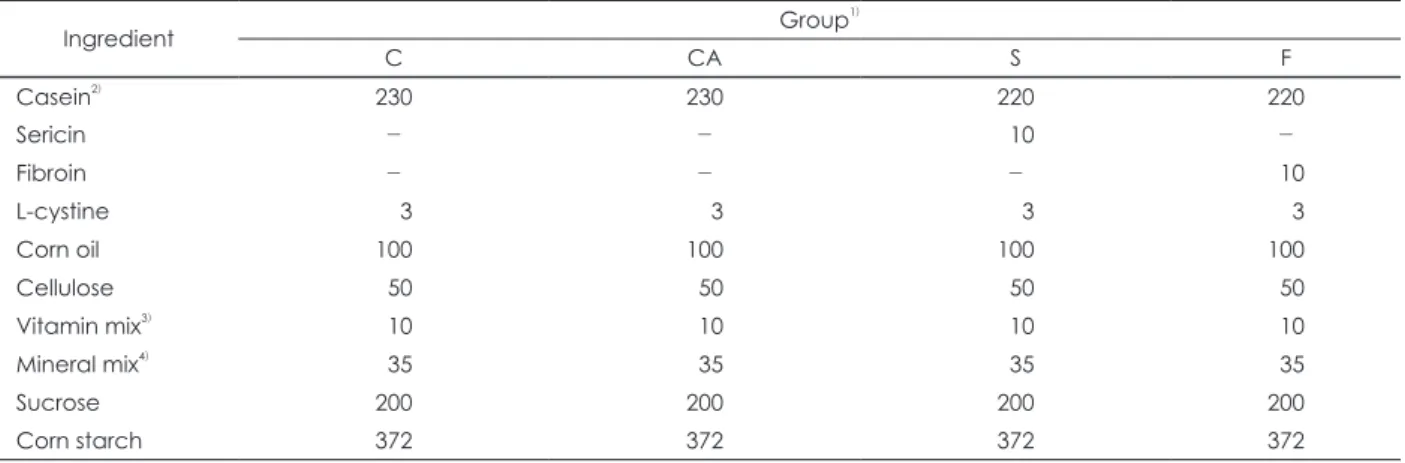
C, CA, S, F군 표피의 So과 Sa 농도는 Fig. 1에 제시하였다.
CA군의 So 농도는 C군에 못 미쳤으나 그 감소는 미비하였다.
S군과 F군의 So 농도는 CA군에 비해서 유의적으로 높게 나타 났으며 C군과는 유사하였다. S군과 F군간의 So 농도는 유사하 였다. CA군의 Sa 농도는 C군에 비해서 현저히 낮았으며, F군의 Sa 농도는 C군과 유사하였으나 S군의 Sa농도는 F군 수준에는 못 미쳤다.
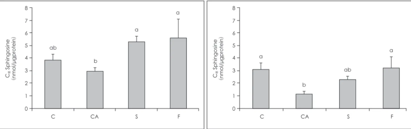
실크 단백질의 식이 공급이 표피의 So/Sa ratio에 미치는 효과
CA군의 So/Sa ratio은 C군에 비해서 현저히 높았으며, 이는 CA군의 현저한 Sa농도 감소에 기인한 것으로 파악된다. S군 및 F군의 So/Sa ratio는 모두 CA군 보다 낮았는데, S군은 C군보 다 높은 반면 F군은 So/Sa ratio이 C군과 유사하였다.실크 단백질의 식이 공급이 표피의 Sphingoid base-1-phosphates 함량에 미치는 효과
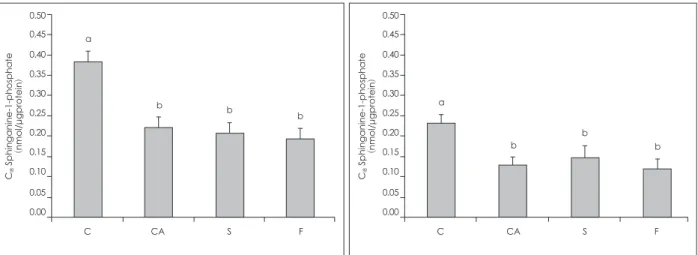
C군의 S1P 및 Sa1P의 농도는 다른 군에 비해서 현저히 높았 으나, CA군을 비롯한 S군 및 F군에서의 이들 농도는 유사하 였다.
고 찰
사전 연구에서 실크단백질의 식이 공급이 NC/Nga mice 표 피의 Cers를 증가시켰으며,28) 본 연구에서 실크단백질의 식이 공급이 Cers의 기본 골격인 free sphingoid bases와 인산화 대 사체들의 함량 변화에 미치는 영향을 살펴보았다. 1% 수준의 sericin 및 fibroin을 10주간 식이 공급한 결과 BALB/c mice (group C)는 NC/Nga mice (group CA, S and F)보다 식이 섭 취량은 적었으나, 체중 변화는 군간 차이가 없었으며 FER도 군 간 유의성이 없었다 (Table 2).
Sphingoid bases의 기능 및 표피 장벽 유지에 대한 관련성 은 최근 연구들에 의해 보고되고 있으나,3,4) 아토피 피부염과 관련하여 식이섭취에 대한 변화 보고는 전무한 실정이다. 본 실 험 결과는 표피의 sphingoid bases는 NC/Nga mice의 AD- like lesions의 진전과 연관성이 있는 것으로 나타났다. 정상대 조군과 비교하였을 때 아토피 대조군인 CA군의 Sa농도는 현저 히 감소하였다 (Fig. 1). 비록 Sa 농도와 AD의 직접적인 연관성
Table 2. Weight gain, food intake and FER of groups for 10 weeks
Group1)
C CA S F
Weight gain (g/week) 0.37 ± 0.052)a 0.47 ± 0.08a 0.51 ± 0.11a 0.51 ± 0.13a Food intake (g/week) 19.26 ± 0.41b 32.10 ± 1.13a 30.49 ± 0.30a 30.25 ± 0.59a FER3) 0.020 ± 0.003a 0.014 ± 0.002a 0.017 ± 0.003a 0.017 ± 0.004a 1) Group C: BALB/c mice were fed with a control diet; group CA, S and F: NC/Nga mice were fed with a control diet (group CA) or diets containing 1% sericin extract (group S) or 1% fibroin extract (group F) for 10 weeks 2) Values are expressed as means ± SEM (n = 10). Means with different alphabetical letters in the same row are significantly different according to duncan’s multiple range test at p < 0.05 3) FER; Food Efficiency Ratio = gain of body weight (g)/amount of food intake (g)
에 대한 연구는 보고되어 있지 않으나, 본 연구에서 Sa 농도의 감소는 AD에서의 de novo sphingolipid synthesis 억제로 인 한 Sa 합성 감소와 간접적으로 연관이 있을 것으로 여겨진다.29) 하지만 CA 군의 So농도는 C군과 유사하며, AD환자의 표피에 서 So농도의 감소가 나타난 사전 연구와 일치하지 않았다.30) 대조적으로 1% silk protein이 함유된 식이를 공급한 S군과 F 군의 So 및 Sa농도는 CA군에 비해서 높으며, C 군과 유사하였 다 (Fig. 1). 이러한 결과는 실크단백질의 식이 공급이 표피 장 벽과 보습에 중요한 지질인 Cer31)의 분해산물이자 합성 또는 재 합성 기질인 So와 Sa 함량을 증가시켜 AD-like lesions의 발 달을 방해하는 것으로 제안된다. 또한 다른 연구에 의해 So의 antimicrobial activity32)과 Sa의 microbial adherence33)가 알 려졌다. 이러한 선행 연구를 통해 본 실험의 결과 역시 So와 Sa 가 피부 표면의 미생물 감염에 대항하는 역할을 할 수 있는 것
으로 사료된다.
CA군에서 So/Sa ratio은 Sa 함량의 현저한 감소와 함께 C군 에 비해서 현저히 높았다 (Fig. 2). 이는 So/Sa ratio가 아토피 피 부염에서 증가된다고 보고된 선행 연구와 일치하며,35) CA군에 서 So/Sa ratio의 증가는 So 함량의 감소보다는 소수성 성질을 가진 Sa36)의 급격한 감소로 인하여 세포막의 투과성이 증가하 여 allergens, bacteria와 같은 외부 물질의 침입으로 인한 피부 막의 파괴로 인하여 유도 된 것으로 사료된다. CA군의 현저한 Sa 함량 감소와 대조적으로 S군과 F군의 Sa 함량은 C군과 유 사한 수준으로 증가되어 있었는데, 특히 F군의 Sa 함량 증가 더 욱 컸다. 이는 S군과 F군의 완만한 So 함량 증가와 더불어 S군 과 F군의 So/Sa ratio을 CA군에 비해서 유의적으로 감소시키 는 결과를 초래하였다 (Fig. 2). 아토피 피부염 유도 또는 실크단 백질의 식이 공급에 의한 So 및 Sa 함량의 절대적인 변화도 의 미있는 지표이나, 세포막 투과성을 감소시키는 Sa 기능을 감안 하면36) So/Sa ratio는 세포막이 투과성과 관련된 AD 발병 및 치 료 효과의 중요한 지표가 될 수 있을 것으로 사료된다. 아토피 피부염 유발은 표피의 So 및 Sa 함량, 특히 Sa 함량의 현저한 감소를 초래하였으며, 실크단백질, sericin, fibroin의 식이 공급 은 아토피 피부염 유발에 의해 현저히 야기된 Sa 감소를 다소 완화시켜 그 결과 So/Sa ratio 감소를 초래하였다.
Sphingoid base-1-phosphates은 sphingosine kinase (SK)36) 에 의해서 sphingoid base부터 생성되는데, AD와 관련된 대사 과정은 크게 밝혀지지 않았다. 본 실험 결과 CA군의 S1P, Sa1P 농도가 C군에 비해서 유의적으로 낮게 나타났다 (Fig. 3). SalP 의 기능은 아직 명확하지 않으나, 세포이동을 촉진하는 중요한 인자 및 상처회복의 조절 인자인 S1P의 감소로 CA군의 세포막 의 회복을 지연하는 것으로 사료된다. 또한 CA군과 S, F군의 S1P, Sa1P농도는 유사한 것으로 나타났다 (Fig. 3). 이 결과는 S, F
Fig. 1. Levels of C18 Sphingoid bases in the epidermis of mice. C: BALB/c mice were fed with a control diet, group CA, S and F: NC/
Nga mice were fed with a control diet (group CA) or diets containing 1% sericin extract (group S) or 1% fibroin extract (group F) for 10 weeks. Sphingoid bases were extracted from epidermal homogenates of each groups and analyzed using HPLC. Values are presented as Means ± SEM (n = 10). Means with different alphabetical letters are significantly different according to duncan’s mul- tiple comparison at p < 0.05.
8 7 6 5 4 3 2 1 0
8 7 6 5 4 3 2 1 0
CI8 Sphingosine
(nmol/μgprotein
) CI8 Sphingosine
(nmol/μgprotein
)
C CA S F C CA S F ab
b a
b a
ab a
a
Fig. 2. Ratio of C18So/Sa in the epidermis of mice. Group C: BALB/
c mice were fed with a control diet, group CA, S and F: NC/Nga mice were fed with a control diet (group CA) or diets containing 1% sericin extract (group S) or 1% fibroin extract (group F) for 10 weeks. Values are presented as Means ± SEM (n = 10). Means with different alphabetical letters are significantly different ac- cording to duncan’s multiple range test at p < 0.05.
4.5 4.0 3.5 3.0 2.5 2.0 1.5 1.0 0.5 0
C18 So/Sa ratio
C CA S F C
a
b
bc
군의 So와 Sa 농도가 변화한 경우 이들의 인산화대사체인 S1P 및 Sa1P 함량이 감소 또는 증가의 변화를 수반할 것이라고 예 상한 본 실험의 연구 목적에 부합하지 않는다. SK과 sphingo- sine phosphatase (SPPase)에 의해서 상호 전환되는 반응인 S1P과 So 또는 Sa1p과 Sa의 관계에서 Sphingoid-1-phosphates 의 농도는 아토피 피부염에서 크게 감소하나 세포와 조직의 항 상성을 유지하기 위하여 조절됨이 보고되었느나,37,38) Sphin- goid-1-phosphates의 농도는 실크 단백질인 sericin이나 fibroin 의 식이 공급에 의해서는 변화가 초래되지 않았다. Sphingoid base는 세라마이드의 분해 산물로 SK 효소에 의한 인산화 대 사체로 전환될 뿐 아니라 sphingosine phosphatase (SPPase) 에 의해서 다시 sphingoid base로 재전환되므로,36) 실크 단백질 의 식이 공급에 의해 변화가 초래되지 않은 Sphingoid-1-phos- phates의 농도에 대한 해석은 SK 및 Sppase mRNA과 단백 질발현 및 활성 변화 측정과 함께 추후 심도있게 이루어져야 한다.
실크 단백질은 수분보유능력,11) 주름방지,12) 피부암 억제 효 과,13) 상처치유효과14) 등과 관련하여 피부 건강에 관련된 bio- material과39) 기능성 화장품으로 개발되어 있다. 이러한 영향은 serine의 amino acid composition에 의해 나타난다. 세리신은 그 자체로 자연보습인자 역할을 하는 serine이 전체 아미노산 중에서 30%만큼 함유하고 있으며 피브로인은 serine 함유량이 12%로 세리신에 비해 낮지만, serine으로 상호전환 될 수 있는 glycine을 44%만큼 함유하고 있다. 따라서 이러한 고함량의 serine을 함유한 실크단백질을 피부 친화적인 소재로 강조할 수 있다.40) 실크단백질의 효능에 관련된 연구는 진행되고 있으나, 연구형태를 제외한41) 실크단백질의 식이 섭취에 의한 효능 연구
와 sphingoid base와 그 대사체의 피부와의 연관성에 대한 연구 는 아직 제한적이다. 그러므로 본 연구는 실크단백질의 식이공 급이 미치는 영향에 대한 것으로 의의를 갖는다. 1% silk pro- tein, sericin 및 fibroin의 식이 공급은 Cer의 분해산물이자 합 성 또는 재합성하는 기질인 So와 Sa 함량을 증가시키고, 세포 막 투과성의 중요한 지표인 So/Sa ratio 감소를 초래하는 긍정 적인 효능을 나타내었다.
요약 및 결론
최근 sphingolipid 대사과정에서 유도되는 sphingoid bases 와 그 대사체가 Cer와 마찬가지로 표피장벽기능 및 수분유지 에 중요한 역할을 나타내고 있다. 본 연구에서는 아토피 피부염 동물 모델인 NC/Nga mice에 실크 단백질 sericin과 fibroin 을 식이 공급 후 피부의 free sphingoid bases와 그 phosphates 조성 변화를 분석하였으며 결과 10주간의 실험 기간 동안 NC/
Nga mice에 실크 단백질 sericin과 fibroin을 식이 공급한 S군 및 F군의 식이효율은 아토피 피부염 대조군 (CA군)과 유의적 차이가 없었다. CA군에서는 So와 Sa농도가 C군에 비해 감소 하였는데 특히 Sa 감소가 더욱 현저하였다. 실크 단백질 seri- cin과 fibroin을 식이 공급한 S군 및 F군에서는 So, Sa 농도가 증가하였으며, 특히 fibroin을 식이 공급한 군에서 현저히 증가 하였다. 그 결과 So/Sa ratio은 CA군에서 크게 증가하였으며, S군 및 F군에서는 CA 군에 비해 낮았는데, 특히. fibroin을 식 이 공급한 군에서 더욱 낮았다. 실크 단백질 sericin과 fibroin 을 식이 공급이 S1P과 Sa1P에는 영향을 주지 않았다.
Fig. 3. Levels of C18 Sphingoidbase-1-phosphates in the epidermis of mice. Group C: BALB/c mice were fed with a control diet, group CA. S and F: NC/Nga mice were fed with a control diet (group CA) or diets containing 1% sericin extract (group S) or 1% fi- broin extract (group F) for 10 weeks. Sphingoid base-1-phosphates of epidermal homogenates were first dephosphorylated by alka- line phosphatase at 37ºc then separated sphingoid bases were extracted and analyzed using HPLC. Values are presented as means ± SEM (n = 10). Means with different alphabetical letters are significantly different according to duncan’s multiple range test at p < 0.05.
0.50 0.45 0.40 0.35 0.30 0.25 0.20 0.15 0.10 0.05 0.00 CI8 Sphinganine-1-phosphate
(nmol/μgprotein
)
C CA S F a
b b
b
0.50 0.45 0.40 0.35 0.30 0.25 0.20 0.15 0.10 0.05 0.00
CI8 Sphinganine-1-phosphate
(nmol/μgprotein
)
C CA S F a
b
b
b
이상의 결과를 종합해볼 때 NC/Nga mice에서 실크단백질 의 식이 공급은 표피 장벽과 보습에 중요한 지질인 Cer의 분해 산물이자 합성 또는 재합성하는 기질인 So, Sa 함량을 증가와 병행하여 아토피 피부염에서 증가된다고 보고된 So/Sa ratio를 감소시켰다. 궁극적으로 실크단백질은 표피장벽 기능에 긍정적 인 영향을 보임으로써 향후 아토피 피부염 개선을 위한 기능성 소재로써 활용 가능성이 높다고 사료된다.
Literature cited
1) Coderch L, López O, de la Maza A, Parra JL. Ceramides and skin function. Am J Clin Dermatol 2003; 4(2): 107-129
2) Bartke N, Hannun YA. Bioactive sphingolipids: metabolism and function. J Lipid Res 2009; 50 Suppl: S91-S96
3) Bibel DJ, Aly R, Shinefield HR. Antimicrobial activity of sphin- gosines. J Invest Dermatol 1992; 98(3): 269-273
4) Vogler R, Sauer B, Kim DS, Schäfer-Korting M, Kleuser B.
Sphingosine-1-phosphate and its potentially paradoxical effects on critical parameters of cutaneous wound healing. J Invest Der- matol 2003; 120(4): 693-700
5) Elias PM, Wood LC, Feingold KR. Epidermal pathogenesis of inflammatory dermatoses. Am J Contact Dermat 1999; 10(3):
119-126
6) Horn EJ, Domm S, Katz HI, Lebwohl M, Mrowietz U, Kragbal- le K; International Psoriasis Council. Topical corticosteroids in psoriasis: strategies for improving safety. J Eur Acad Dermatol Venereol 2010; 24(2): 119-124
7) Majoie IM, Oldhoff JM, van Weelden H, Laaper-Ertmann M, Bousema MT, Sigurdsson V, Knol EF, Bruijnzeel-Koomen CA, de Bruin-Weller MS. Narrowband ultraviolet B and medium- dose ultraviolet A1 are equally effective in the treatment of mod- erate to severe atopic dermatitis. J Am Acad Dermatol 2009;
60(1): 77-84
8) Warren RB, Griffiths CE. Systemic therapies for psoriasis: meth- otrexate, retinoids, and cyclosporine. Clin Dermatol 2008; 26(5):
438-447
9) Kim J, Kim H, Jeong do H, Kim SH, Park SK, Cho Y. Compara- tive effect of gromwell (Lithospermum erythrorhizon) extract and borage oil on reversing epidermal hyperproliferation in guinea pigs. Biosci Biotechnol Biochem 2006; 70(9): 2086-2095 10) Lee KG, Yeo JH, Lee YW, Kweon HY, Kim JH. Bioactive and
skin-compatible properties of silk sericin. Korean J Sericult Sci 2001; 43(2): 109-115
11) Padamwar MN, Pawar AP, Daithankar AV, Mahadik KR. Silk sericin as a moisturizer: an in vivo study. J Cosmet Dermatol 2005;
4(4): 250-257
12) Kato N, Sato S, Yamanaka A, Yamada H, Fuwa N, Nomura M.
Silk protein, sericin, inhibits lipid peroxidation and tyrosinase ac- tivity. Biosci Biotechnol Biochem 1998; 62(1): 145-147 13) Zhaorigetu S, Yanaka N, Sasaki M, Watanabe H, Kato N. Silk
protein, sericin, suppresses DMBA-TPA-induced mouse skin tu- morigenesis by reducing oxidative stress, inflammatory responses and endogenous tumor promoter TNF-alpha. Oncol Rep 2003;
10(3): 537-543
14) Nagai N, Murao T, Ito Y, Okamoto N, Sasaki M. Enhancing effects of sericin on corneal wound healing in rat debrided corneal epithe- lium. Biol Pharm Bull 2009; 32(5): 933-936
15) Choi MJ, Maibach HI. Role of ceramides in barrier function of healthy and diseased skin. Am J Clin Dermatol 2005; 6(4): 215-223
16) Farwanah H, Raith K, Neubert RH, Wohlrab J. Ceramide profiles of the uninvolved skin in atopic dermatitis and psoriasis are com- parable to those of healthy skin. Arch Dermatol Res 2005; 296(11):
514-521
17) Imokawa G, Abe A, Jin K, Higaki Y, Kawashima M, Hidano A.
Decreased level of ceramides in stratum corneum of atopic derma- titis: an etiologic factor in atopic dry skin? J Invest Dermatol 1991; 96(4): 523-526
18) Meguro S, Arai Y, Masukawa Y, Uie K, Tokimitsu I. Relationship between covalently bound ceramides and transepidermal water loss (TEWL). Arch Dermatol Res 2000; 292(9): 463-468 19) Motta S, Monti M, Sesana S, Caputo R, Carelli S, Ghidoni R. Ce-
ramide composition of the psoriatic scale. Biochim Biophys Acta 1993; 1182(2): 147-151
20) Reiter LV, Torres SM, Wertz PW. Characterization and quanti- fication of ceramides in the nonlesional skin of canine patients with atopic dermatitis compared with controls. Vet Dermatol 2009; 20(4): 260-266
21) Madyarov S, Lee KG, Yeo JH, Nam J, Lee YW. Improved meth- od for the preparation of silk fibroin hydrolysates. Korean J Seri- cult Sci 1999; 41(2): 108-115
22) Suto H, Matsuda H, Mitsuishi K, Hira K, Uchida T, Unno T, Oga- wa H, Ra C. NC/Nga mice: a mouse model for atopic dermatitis.
Int Arch Allergy Immunol 1999; 120 Suppl 1: 70-75
23) Takagi Y, Nakagawa H, Yaginuma T, Takema Y, Imokawa G. An accumulation of glucosylceramide in the stratum corneum due to attenuated activity of beta-glucocerebrosidase is associated with the early phase of UVB-induced alteration in cutaneous barrier function. Arch Dermatol Res 2005; 297(1): 18-25
24) Doering T, Holleran WM, Potratz A, Vielhaber G, Elias PM, Su- zuki K, Sandhoff K. Sphingolipid activator proteins are required for epidermal permeability barrier formation. J Biol Chem 1999;
274(16): 11038-11045
25) Min JK, Yoo HS, Lee EY, Lee WJ, Lee YM. Simultaneous quan- titative analysis of sphingoid base 1-phosphates in biological sam- ples by o-phthalaldehyde precolumn derivatization after dephos- phorylation with alkaline phosphatase. Anal Biochem 2002; 303 (2): 167-175
26) Flamand N, Justine P, Bernaud F, Rougier A, Gaetani Q. In vivo distribution of free long-chain sphingoid bases in the human stra- tum corneum by high-performance liquid chromatographic anal- ysis of strippings. J Chromatogr B Biomed Appl 1994; 656(1):
65-71
27) Caligan TB, Peters K, Ou J, Wang E, Saba J, Merrill AH Jr. A high-performance liquid chromatographic method to measure sphingosine 1-phosphate and related compounds from sphingo- sine kinase assays and other biological samples. Anal Biochem 2000; 281(1): 36-44
28) Park KH, Choi YS, Kim HA, Lee KG, Yeo JH, Jung DH, Kim SH, Cho YH. Dietary effect of silk protein on ceramide synthesis and the expression of ceramide metabolic enzymes in the epider- mis of NC/Nga mice. J Korean Soc Food Sci Nutr 2007; 36(5):
554-562
29) Macheleidt O, Kaiser HW, Sandhoff K. Deficiency of epidermal protein-bound omega-hydroxyceramides in atopic dermatitis. J Invest Dermatol 2002; 119(1): 166-173
30) Arikawa J, Ishibashi M, Kawashima M, Takagi Y, Ichikawa Y, Imokawa G. Decreased levels of sphingosine, a natural antimi- crobial agent, may be associated with vulnerability of the stratum corneum from patients with atopic dermatitis to colonization by Staphylococcus aureus. J Invest Dermatol 2002; 119(2): 433-439 31) Elias PM, Menon GK. Structural and lipid biochemical corre- lates of the epidermal permeability barrier. Adv Lipid Res 1991;
24: 1- 26
32) Melnik B. Disturbances of antimicrobial lipids in atopic derma- titis. J Dtsch Dermatol Ges 2006; 4(2): 114-123
33) Bibel DJ, Aly R, Shinefield HR. Inhibition of microbial adher- ence by sphinganine. Can J Microbiol 1992; 38(9): 983-985 34) Loiseau N, Moradian S, Elias PM, Holleran WM, Uchida Y. Ce-
ramide metabolites in epidermal permeability barrier function and atopic dermatitis [abstract]. J Invest Dermatol 2009; 129 Suppl 1: S68. Abstract no. 405
35) Stewart ME, Downing DT. Free sphingosines of human skin in- clude 6-hydroxysphingosine and unusually long-chain dihydro- sphingosines. J Invest Dermatol 1995; 105(4): 613-618 36) Hait NC, Oskeritzian CA, Paugh SW, Milstien S, Spiegel S. Sphin-
gosine kinases, sphingosine 1-phosphate, apoptosis and diseases.
Biochim Biophys Acta 2006; 1758(12): 2016-2026
37) Gómez-Muñoz A. Ceramide 1-phosphate/ceramide, a switch be-
tween life and death. Biochim Biophys Acta 2006; 1758(12): 2049- 38) Prieschl EE, Csonga R, Novotny V, Kikuchi GE, Baumruker 2056
T. The balance between sphingosine and sphingosine-1-phos- phate is decisive for mast cell activation after Fc epsilon receptor I triggering. J Exp Med 1999; 190(1): 1-8
39) Mondal M, Trivedy K, Nirmal Kumar S. The silk proteins, seri- cin and fibroin in silkworm, Bombyx mori Linn., -a review. Casp J Environ Sci 2007; 5(2): 63-76
40) Voegeli R, Meier J, Blust R. Sericin silk protein: unique structure and properties. Cosmet Toilet 1993; 108: 101-108
41) Aramwit P, Sangcakul A. The effects of sericin cream on wound healing in rats. Biosci Biotechnol Biochem 2007; 71(10): 2473- 2477