Biomedical Science Letters 2018, 24(3): 253~262 https://doi.org/10.15616/BSL.2018.24.3.253 eISSN : 2288-7415
Inhibitory Effects of PD98059, SB203580, and SP600125 on α- and δ -granule Release and Intracellular Ca
2+Levels in Human Platelets
Hyuk-Woo Kwon
†,*Department of Biomedical Laboratory Science, Far East University, Eumseong 27601, Korea
Platelets are activated at sites of vascular injury via several molecules, such as adenosine diphosphate, collagen and thrombin. Full platelet aggregation is absolutely essential for normal hemostasis. Moreover, this physiological event can trigger circulatory disorders, such as thrombosis, atherosclerosis, and cardiovascular disease. Therefore, platelet function inhibition is a promising approach in preventing platelet-mediated circulatory disease. Many studies reported the involvement of mitogen-activated protein kinases (MAPKs) signaling pathways in platelet functions. However, these studies were limited. Thus, we examined MAPK signaling pathways in human platelets using specific MAPK inhibitors, such as PD98059, SB203580, and SP600125. We observed that these inhibitors were involved in calcium mobilization and influx in human platelets. They also suppressed thrombin-induced α- and δ-granule release. These results suggest that PD98059, SB203580, and SP600125 exhibit Ca2+ antagonistic effects.
Key Words: MAPK signaling pathways, Calcium mobilization, Calcium influx, Granule release
INTRODUCTION
Mitogen-activated protein kinases (MAPKs) are a family of serine/threonine protein kinases that are divided into three subgroups: extracellular signal-regulated kinases (ERKs), c-Jun N-terminal kinases (JNKs), and p38. Studies have detected MAPKs in human platelets and have shown them to be phosphorylated by thrombin for full activation (Kramer et al., 1995; Bugaud et al., 1999; Nadal-Wollbold et al., 2002).
Furthermore, the roles of MAPKs have been studied in hemostasis and thrombosis (Adam et al., 2008). In platelets, ERKs have been extensively investigated among MAPKs.
ERKs may be activated via growth factors, receptor tyrosine
kinases and G protein-coupled receptors in various cell types (van Blesen et al., 1995). Its signaling process is initiated by the small G protein Ras, which activates the Raf-1/MAPK/
ERK (MEK)/ERK phosphorylation pathway (Kyriakis et al., 1992). ERK2 is reportedly activated by the dual-specificity kinase, MEK1/2, and protein kinase C (PKC)-dependent pathway in platelets (Börsch-Haubold et al., 1995; Börsch- Haubold et al., 1996). Regarding the ERK1/2 upstream mole- cule specific inhibitors of conventional PKCs (Ro-318425 and Ro-318220) reportedly inhibit MEK1/2 and ERK1/2 (Börsch-Haubold et al., 1995; Nadal-Wollbold et al., 2002).
Furthermore, H-Ras and Raf-1 in the platelets do not in- fluence ERK1/2 activation (Nadal-Wollbold et al., 2002). A specific PKCδ inhibitor (Rottlerin) also reportedly suppresses
Original Article
Received: May 28, 2018 / Revised: August 29, 2018 / Accepted: August 29, 2018
*Professor.
†Corresponding author: Hyuk-Woo Kwon. Department of Biomedical Laboratory Science, Far East University, 76-32, Daehak-gil, Gamgok-myeon, Eumseong-gun, Chungcheongbuk-do 27601, Korea.
Tel: +82-43-880-3801, Fax: +82-43-880-3876, e-mail: [email protected]
○CThe Korean Society for Biomedical Laboratory Sciences. All rights reserved.
○CCThis is an Open Access article distributed under the terms of the Creative Commons Attribution Non-Commercial License (http://creativecommons.org/licenses/by-nc/3.0/) which permits unrestricted non-commercial use, distribution, and reproduction in any medium, provided the original work is properly cited.
the thrombin-induced phosphorylation of MEK1/2, ERK1/2, and p38 (Yacoub et al., 2006). Src kinase inhibitor PP2 and phospholipase C inhibitor U73122, have also been shown to inhibit the von Willebrand factor induced ERK2 phos- phorylation (Ramakrishnan et al., 2001). Considering these previous reports, it appears that ERK2 may be regulated by PKCα and PKCδ. Furthermore, the study of JNK1 in platelets has identified, a key role for it in platelet function and thrombosis (Adam et al., 2010). JNK1-deficient mice were shown to reduce thromboxane A2 (TXA2) production despite normal platelet counts, ultrastructures, and expression of adhesive receptors such as GPIbα, GPVI, and glycopro- tein IIb/IIIa (αIIb/β3). Moreover, JNK1-/- showed diminished ERK2 levels after thrombin stimulation, which suggested that JNK1 may be an upstream molecule of ERK2 or at least associated with each other (Adam et al., 2010). Thrombin- activated ERK2 and JNK1 were also reportedly down- regulated by αIIb/β3 inhibition, which suggested that the activation probably involved a common pathway (Bugaud et al., 1999).
p38 is activated by thrombin (Kramer et al., 1995) and the phospholipases Cβ, and PKCδ, are known as upstream mediators (Yacoub et al., 2006). Activated p38 increases the phosphorylation of cytosolic phospholipase A2, resulting in arachidonic acid release and TXA2 production; however, SB203580 inhibits TXA2 production (Kramer et al., 1996).
Furthermore, PD98059 and SB203580, specific inhibitors of ERK1/2 and p38, block COX-1 directly and SB203580 further inhibits the conversion of PGH2 to TXA2 (Börsch- Haubold et al., 1998), which means that MAPKs may in- fluence endogenous enzyme activity leading to TXA2 pro- duction. Various studies have revealed the MAPK functions in platelets; however, their complete regulatory functions have been elusive. Therefore, we investigated MAPK sig- naling pathways in human platelet using specific inhibitors.
MATERIALS AND METHODS
MaterialsThrombin was obtained from Chrono-Log Corporation (Havertown, PA, USA). Fura 2-acetoxymethyl (Fura 2-AM) and BAPTA-AM were obtained from Invitrogen Molecular
Probes (Eugene, OR, USA). Anti-phosphor-IP3-receptor type I (Ser1756), anti-phosphor-p38, anti-phosphor-ERK (1/2), anti- phosphor-JNK (1/2), anti-p38, anti-ERK (1/2), anti-JNK (1/2), anti-rabbit IgG-horseradish peroxidase conjugate, and lysis buffer were obtained from Cell Signaling (Beverly, MA, USA). PD98059 (ERK inhibitor), SB203580 (p38 inhibitor), SP6000125 (JNK inhibitor) and anti-β-actin were purchased from Santa Cruz Biotechnology (Santa Cruz, CA, USA).
Thapsigargin was purchased from Cayman Chemical. Poly- vinylidene difluoride (PVDF) membrane and enhanced che- miluminesence solution (ECL) were purchased from General Electric Healthcare (Buckinghamshire, UK).
Preparation of washed human platelets
Human platelet-rich plasma (PRP) was obtained from the Korean Red Cross Blood Center (Changwon, Korea), and centrifuged for 10 min at 1,300 g. The platelets were then washed twice with washing buffer (138 mM NaCl, 2.7 mM KCl, 12 mM NaHCO3, 0.36 mM NaH2PO4, 5.5 mM glucose, and 1 mM Na2EDTA, pH 6.5), and resuspended in suspen- sion buffer (138 mM NaCl, 2.7 mM KCl, 12 mM NaHCO3, 0.36 mM NaH2PO4, 0.49 mM MgCl2, 5.5 mM glucose, 0.25% gelatin, pH 6.9). The platelets concentrations were adjusted to a final concentration of 5 × 10⁸/mL. All afore- mentioned procedures for platelet activity were performed at 25℃. Experimental approval (PIRB12-072) was obtained from the Public Institutional Review Board at the National Institute for Bioethics Policy (Seoul, Korea).
Determination of platelet aggregation
Platelets (108/mL) were preincubated with or without substances in the 2 mM CaCl2 for 3 min at 37℃ followed by thrombin stimulation (0.05 U/mL). The aggregation was performed for 5 min using an aggregometer (Chrono-Log Corporation). Platelet aggregation rate was determined as an increase in light transmission. MAPK inhibitors were dis- solved in dimethyl sulfoxide (DMSO) to a final concentration of 0.1%.
Western blots for analyzing of ERK1/2-, p38-, and JNK1/2-phosphorylation
Platelet aggregation was stopped by adding 1× lysis
buffer. The platelet lysates were then measured using a BCA protein assay kit (Pierce Biotechnology, IL, USA). Proteins (15 μg) were analyzed via SDS-PAGE (6%, 1.5 mm), and PVDF membranes were used for protein transfer. The dilu- tions for the primary and secondary antibodies were 1:1,000 and 1:10,000, respectively. The membranes were visualized using the ECL solution.
Determination of Ca2+ mobilization and influx
PRP was incubated with 5 μM of Fura 2-AM at 37℃ for 60 min. The washed platelets (108/mL), loaded with Fura 2-AM, were prepared using the procedure described above preincubated with MAPK inhibitors for 3 min at 37℃ in the presence of 100 μM of EGTA, and stimulated with thrombin (0.05 U/mL) for Ca2+ mobilization and thapsigargin (1 μM) for Ca2+ influx. After thapsigargin stimulation, 2 mM of calcium was added at 3 min. Fura 2-AM fluorescence was measured using a spectrofluorometer (SFM-25; BioTeek Instruments, Italy) at an excitation wavelength that changed every 0.5 s from 340 to 380 nm; and an emission wave- length set at 510 nm. The [Ca2+]i values were calculated using the Grynkiewicz method (Grynkiewicz et al., 1985).
Determination of p-selectin release
Washed human platelets (108/mL) were preincubated with or without substances in the presence of 2 mM CaCl2 for 3 min at 37℃ followed by thrombin stimulation (0.05 U /mL). The platelets were reconstituted in 250 μL of ice-cold phosphate-buffered saline (PBS; pH 7.4) and incubated with 10 μL of Alexa Fluor 488 anti-human CD62P in PBS (pH 7.4) containing 0.09% sodium azide and 0.2% bovine serum albumin (BSA) for 60 min at 4℃ in a dark room.
The platelets were then washed thrice with ice-cold PBS and resuspended in 0.5% paraformaldehyde in PBS. The platelet-bound Alexa Fluor 488 anti-human CD62P was determined using flow cytometry (BD Biosciences, San Diego, CA, USA) and the data was analyzed using the BD cellQuestTM software.
Determination of ATP and serotonin release
Washed platelets (108/mL) were stimulated with thrombin (0.05 U/mL) with or without MAPK inhibitors in the pre-
sence of 2 mM CaCl2. The reaction was terminated and cen- trifuged at 200 ×g for 10 min at 4℃, and the supernatant was used for ATP and serotonin release assay. ATP and serotonin release were measured using a Synergy HT Multi- Mode Microplate Reader (BioTek Instruments, Winooski, VT, USA).
Statistical analyses
The experimental results are indicated as mean ± standard deviation accompanied by the number of observations. The data was determined via analysis of variance (ANOVA).
Significant differences among the group means were com- pared using the Newman-Keuls method. Statistical analysis was performed using SPSS 21.0.0.0 (SPSS, Chicago, IL, USA). P < 0.05 was considered to be statistically significant.
RESULTS
Effects of MAPK inhibitors on thrombin-induced human platelet aggregation
Based on a previous report, 0.05 U/mL of thrombin was used as it maximally aggregated human platelets (Shin et al., 2015). The thrombin-induced aggregation rate was 92.5
± 1.2%, however, PD98059, SB203580 and SP600125 at 20 μM could not inhibit thrombin-induced platelet aggre- gation (Fig. 1A). Thus, we used 40 μM of the MAPK in- hibitors on thrombin-induced platelet aggregation. As shown in Fig. 1A, PD98059, SB203580 and SP600125 at 40 μM suppressed human platelet aggregation. Therefore, we used these concentrations to evaluate anti-platelet effects.
Effects of MAPK inhibitors on ERK1/2-, p38, and JNK1/2-phosphorylation
MAPK phosphorylation is known to be positively regu- lated in human platelet aggregation. Thus, we examined if PD98059, SB203580, and SP600125 inhibited the MAPK phosphorylation. Compared to unstimulated platelets throm- bin (0.05 U/mL) elevated ERK2-, p38-, and JNK1-phos- phorylation (Fig. 1B, 1C, and 1D). However, 40 μM of PD98059, SB203580, and SP600125 reduced ERK2-, p38-, and JNK1-phosphorylation. Furthermore, 0.1% DMSO did not influence phosphorylation (Fig. 1B, 1C, and 1D).
Effects of MAPK inhibitors on [Ca2+]i mobilization
We investigated the effect of PD98059, SB203580 and SP600125 on Ca2+ antagonistic activity. As shown in Fig.
2A, thrombin-increased Ca2+ mobilization from 100.5 ± 1.3 nM to 835.9 ± 39.7 nM in the presence of 2 mM CaCl2. However, we eliminated the indirect effects of Ca2+ influx to accurately measure [Ca2+]i mobilization by using EGTA (100 μM), which was recorded as 370.5 ± 1.1 nM (Fig.
2B). Thrombin-induced [Ca2+]i mobilization suppressed PD- 98059, SB203580, and SP600125 (Fig. 2C, 2D, and 2E).
The percentage inhibition by 40 μM of MAPK inhibitors were 33.6%, 36.2%, and 57.4%, respectively (Table 1).
Effects of MAPK inhibitors on IP3RI-phosphorylation
IP3 receptor type I (IP3RI) phosphorylation leads to the inhibition of [Ca2+]i mobilization (Quinton and Dean, 1992).
Therefore, phosphorylation of IP3RI is used to evaluate Ca2+-antagonistic effects. As shown in Fig. 3, all MAPK inhibitors strongly increased IP3RI phosphorylation, which proved the involvement of MAPK signaling pathways in [Ca2+]i mobilization.
Effects of MAPK inhibitors on [Ca2+]i influx
Thapsigargin is an inhibitor of the sarco/endoplasmic reticulum Ca2+ ATPase, which is used to trigger Ca2+ influx Fig. 1. Effects of MAPK inhibitors on thrombin-induced human platelet aggregation and MAPK phosphorylation. (A) Effects of MAPK inhibitors on thrombin-induced human platelet aggregation. (B) Effects of PD98059 on ERK-phosphorylation. (C) Effects of SB- 203580 on p38-phosphorylation. (D) Effects of SP600125 on JNK-phosphorylation. Platelet aggregation and Western blots were carried out as described in the "Materials and Methods" section. The data is expressed as mean ± standard deviation (n = 4). *P < 0.05 versus thrombin- stimulated human platelets.
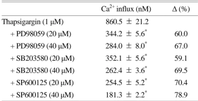
from extracellular spaces. after adding 2 mM CaCl2, 1 μM of thapsigargin triggers Ca2+ influx from 101.5 ± 1.2 nM (the basal level) to 860.5 ± 21.2 nM (Fig. 4A). However, 40 μM of PD98059, SB203580, and SP600125 inhibits Ca2+
influx (Fig. 4B, 4C, and 4D). The percentage inhibition by 40 μM of MAPK inhibitors were observed to be 67.0%,
69.5%, and 78.9%, respectively (Table 2).
Effects of MAPK inhibitors on p-selectin expression
In order to verify the inhibitory mechanism of PD98059, SB203580 and SP600125 on α-granule release, we investi- gated p-selectin expression. PD98059, SB203580, and SP- Fig. 2. Effects of MAPK inhibitors on [Ca2+]i mobilization. (A) Thrombin-induced [Ca2+]i mobilization. (B) Thrombin-induced [Ca2+]i mobilization in the presence of EGTA (100 μM). (C) Effects of PD98059 on [Ca2+]i mobilization in the presence of EGTA (100 μM). (D) Effects of SB203580 on [Ca2+]i mobilization in the presence of EGTA (100 μM). (E) Effects of SP600125 on [Ca2+]i
mobilization in the presence of EGTA (100 μM). [Ca2+]i mobili- zation was determined as described in the "Materials and Methods"
section. The data is expressed as mean ± standard deviation (n = 3).
*P < 0.05 versus thrombin-stimulated human platelets.
600125 (40 μM each) strongly inhibited p-selectin expression (Fig. 5A and 5B), which reflected their inhibitory effect on
[Ca2+]i levels. The intracellular Ca2+ chelator, BAPTA-AM also strongly suppressed thrombin-induced p-selectin ex- pression (Fig. 5A and 5B).
Effects of MAPK inhibitors on ATP and serotonin release
Since the MAPK inhibitors decreased the thrombin- Table 1. Effects of MAPK inhibitors on thrombin-induced [Ca2+]i
mobilization
[Ca2+]i mobilization
(nM) Δ (%)
Thrombin (0.05 U/mL) 370.5 ± 1.1
+ PD98059 (20 μM) 259.1 ± 2.3* 30.1 + PD98059 (40 μM) 246.1 ± 1.1* 33.6 + SB203580 (20 μM) 274.5 ± 2.4* 25.9 + SB203580 (40 μM) 236.5 ± 4.3* 36.2 + SP600125 (20 μM) 228.5 ± 3.3* 38.3 + SP600125 (40 μM) 157.8 ± 0.7* 57.4 Data from Fig. 2. Δ (%) = [(thrombin + substances) - thrombin] / thrombin × 100.
Data is expressed as mean ± standard deviation (n = 4).
*P < 0.05 versus thrombin-stimulated human platelets.
Fig. 4. Effects of MAPK inhibitors on Ca2+ influx. (A) Thapsigargin-induced Ca2+ influx in the presence of EGTA (100 μM). (B) Effects of PD98059 on Ca2+ influx in the presence of EGTA (100 μM). (C) Effects of SB203580 on Ca2+ influx in the presence of EGTA (100 μM).
(D) Effects of SP600125 on Ca2+ influx in the presence of EGTA (100 μM). Ca2+ influx was determined as described in the "Materials and Methods" section.
Fig. 3 Effects of MAPK inhibitors on IP3RI-phosphorylation.
Western blots were analyzed as described in the "Materials and Methods" section. The data is expressed as mean ± standard devia- tion (n = 3).
elevated [Ca2+]i levels, we investigated the involvement of the MAPK signaling pathways in the inhibition of ATP and serotonin release from δ-granules. Thrombin (0.05 U/mL)
strongly increased ATP (Fig. 6A) and serotonin release (Fig.
6B); however, all the MAPK inhibitors suppressed ATP and serotonin release. Moreover, BAPTA-AM decreased ATP and serotonin release, indicating the involvement of intra- cellular Ca2+ levels in granule release (Fig. 6A and 6B).
DISCUSSION
Complete platelet aggregation is essential for hemostatic plug formation. This physiological event is achieved by free cytosolic Ca2+. Thus, inhibiting intracellular Ca2+ is useful in preventing circulatory disorders. Elevation of [Ca2+]i by ago- nists is dependent on its mobilization from the endoplasmic reticulum and influx from extracellular spaces. Thrombin, a platelet agonist, is known to stimulate platelet aggregation by binding to the Gq-coupled proteinase-activated receptor, which activates phospholipase Cβ (PLCβ). The activated PLCβ hydrolyzes phosphatidylinositol 4, 5-bisphosphate Table 2. Effects of MAPK inhibitors on thapsigargin-induced
Ca2+ influx
Ca2+ influx (nM) Δ (%)
Thapsigargin (1 μM) 860.5 ± 21.2
+ PD98059 (20 μM) 344.2 ± 5.6* 60.0 + PD98059 (40 μM) 284.0 ± 8.0* 67.0 + SB203580 (20 μM) 352.1 ± 5.6* 59.1 + SB203580 (40 μM) 262.4 ± 3.6* 69.5 + SP600125 (20 μM) 254.5 ± 5.2* 70.4 + SP600125 (40 μM) 181.3 ± 2.2* 78.9 Data from Fig. 4. Δ (%) = [(thapsigargin + substances) - thapsigargin]
/ thrombin × 100.
Data is expressed as mean ± standard deviation (n = 4).
*P < 0.05 versus thapsigargin-stimulated human platelets.
Fig. 5. Effects of MAPK inhibitors on p-selectin expression.
(A) Flow cytometry histograms representing p-selectin expression.
a, Intact platelets (base); b, Thrombin (0.05 U/mL); c, Thrombin (0.05 U/mL) + DMSO (0.1%); d, Thrombin (0.05 U/mL) + PD- 98059 (40 μM); e, Thrombin (0.05 U/mL) + SB203580 (40 μM);
f, Thrombin (0.05 U/mL) + SP600125 (40 μM); g, Thrombin (0.05 U/mL) + BAPTA-AM (10 μM). (B) Effects of MAPK inhi- bitors on thrombin-induced p-selectin expression (%). Determi- nation of p-selectin expression was carried out as described in the
"Materials and Methods" section. The data is expressed as mean ± standard deviation (n = 3). *P < 0.05 versus thrombin-stimulated human platelets.
(PIP2) to inositol 1, 4, 5-trisphosphate (IP3) and diacylgly- cerol (Berridge and Irvine, 1984; Cattaneo, 2005; Guidetti et al., 2008; Jennings, 2009). Moreover, IP3 mobilizes free cytosolic Ca2+ from dense tubular systems by binding to IP3 receptor type I (IP3RI). The increased Ca2+ activates Ca2+/ calmodulin-dependent kinase and the DG-dependent kinase.
Myosin light chain kinase (MLCK) is the most representa- tive Ca2+/calmodulin-dependent enzyme and phosphorylates myosin light chain (20 kDa) in platelets. The phosphory- lation of myosin light chain is associated with the activation of platelets and subsequent release of platelet granules.
Therefore, the increase of calcium plays an important role in inducing the granule secretion (i.e., dense body and α- granule) and platelet aggregation (Nishikawa et al., 1980;
Kaibuchi et al., 1982).
IP3RI function is inhibited by its phosphorylation and IP3RI phosphorylation involves in the inhibition of [Ca2+]i mobilization (Quinton and Dean, 1992). Therefore, phos- phorylating IP3RI is very useful for evaluating the Ca2+- antagonistic effect of substances or compounds. As shown in Fig. 2, all MAPK inhibitors inhibited the thrombin- induced [Ca2+]i mobilization and also showed strong IP3RI
(Ser1756) phosphorylation (Fig. 3).
Stromal interaction molecule 1 (STIM1) has been identi- fied as a key element of the store-operated Ca2+ entry mech- anism. STIM1 is a transmembrane protein with an N- terminal EF hand domain (Varga-Szabo et al., 2009). Upon Ca2+ release in the endoplasmic reticulum, STIM1 recognizes the depletion of Ca2+ and facilitates Ca2+ influx. Recently, STIM1 was reported to have phosphorylation sites on the serine/threonine and tyrosine residues (Lopez et al., 2012;
Lang et al., 2013), and thapsigargin was reported to trigger STIM1 tyrosine phosphorylation in human platelets (Lopez et al., 2012). Moreover, both ERK1/2 and the Src family of tyrosine kinases were reported to be involved in STIM1 phosphorylation (Elvers et al., 2012; Lopez et al., 2012). In HEK293 cells, ERK1/2 mediates STIM1 phosphorylation at Ser575, Ser608 and Ser621 during Ca2+ store depletion, and that Ca2+ entry and store refilling restore phosphory- lation to basal levels (Pozo-Guisado et al., 2013). As shown in Fig. 4B, PD98059 (an ERK inhibitor) suppressed Ca2+
influx, thereby associating the ERK signaling pathway with Ca2+ influx. SB203580 and SP600125 also suppressed the thapsigargin-induced [Ca2+]i influx (Fig. 4C and 4D). These Fig. 6. Effects of MAPK inhibitors on serotonin-, and ATP-release. (A) Effects of MAPK inhibitors on serotonin release in thrombin- activated platelets. (B) Effects of MAPK inhibitors on ATP release in thrombin-activated platelets. Determination of ATP and serotonin release was carried out as described in the "Materials and Methods" section. The data is expressed as mean ± standard deviation (n = 3). * P
< 0.05 versus thrombin-stimulated human platelets.
results suggested that the JNK and p38 signaling pathways were also involved in Ca2+ influx. To date, the relationship between stim1 and MAPKs has been reported in various cells and ERK is most closely related. However, in our experiments, it was suggested that p38 and JNK also down- regulated thapsigargin-induced calcium influx in human platelets, which means that p38 and JNK may interact with STIM1. Thus, the specific interaction between JNK, p38, and STIM1 in human platelets will be tested in the future.
MAPK inhibitors showed a potent calcium inhibitory effect (20, 40 μM) but did not suppress at low concentrations in human platelet aggregation reactions (Fig. 1A). Because the platelet aggregation reaction is the final reaction, a var- iety of signal transduction processes trigger the final platelet aggregation reaction. MAPK inhibitors could affect the de- tailed signaling of platelets, but the inhibitory effects may be limited. Therefore, MAPK inhibitors (20 μM) could not have a strong effect on the final platelet aggregation inhibition.
The markers of α- and δ-granule release used in this study are p-selectin, ATP, and serotonin. p-selectin is located in the inner membrane of α-granule, and ATP, serotonin are located in the δ-granule. Therefore, we examined the MAPK signaling pathways that were associated with α- and δ- granule release. Thrombin-elevated p-selectin expression (Fig.
5A, 5B) and ATP and serotonin release (Fig. 6A, 6B). How- ever, these reactions were inhibited by MAPK inhibitors.
Because [Ca2+]i is the most important regulator of α- and δ-granule release, p-selectin expression, ATP and serotonin release are also dependent on [Ca2+]i levels. This result re- flects that BAPTA-AM (10 μM) treated platelets significantly downregulated the p-selectin expression (Fig. 5A-g, 5B), ATP, and serotonin release (Fig. 6A, 6B). Therefore, these inhibitory effects on α- and δ-granule release by MAPK inhibitors are mediated by downregulation of [Ca2+]i mobili- zation and [Ca2+]i influx.
In conclusion, the precise mechanism of the MAPK sig- naling pathway is still not fully understood. However, our results suggest that the MAPK signaling pathway plays a role in α- and δ-granule release by down-regulating Ca2+
mobilization and influx. Our results also suggest that the MAPK signaling pathway is involved in the regulation of calcium and granule release.
ACKNOWLEDGEMENT
This study was supported by a grant (NRF-2011-0012143 to Hwa-Jin Park) from the Basic Science Research Program via the National Research Foundation of Korea (NRF) funded by the Ministry of Education, Science and Techno- logy, Korea.
CONFLICT OF INTEREST
The authors declare no conflicts of interest.
REFERENCES
Adam F, Kauskot A, Nurden P, Sulpice E, Hoylaerts MF, Davis RJ, Bryckaert M. Platelet JNK1 is involved in secretion and thrombus formation. Blood. 2010. 115: 4083-4092.
Adam F, Kauskot A, Rosa JP, Bryckaert M. Mitogen-activated protein kinases in hemostasis and thrombosis. Journal of Thrombosis and Haemostasis. 2008. 6: 2007-2016.
Berridge MJ, Irvine RF. Inositol trisphosphate, a novel second mes- senger in cellular signal transduction. Nature. 1984. 312: 315 -321.
Börsch-Haubold AG, Kramer RM, Watson SP. Cytosolic phos- pholipase A2 is phosphorylated in collagen-and thrombin- stimulated human platelets independent of protein kinase C and mitogen-activated protein kinase. Journal of Biological Chemistry. 1995. 270: 25885-25892.
Börsch-Haubold AG, Kramer RM, Watson SP. Inhibition of mitogen- activated protein kinase kinase does not impair primary acti- vation of human platelets. Biochemical Journal. 1996. 318:
207-212.
Börsch-Haubold AG, Pasquet S, Watson SP. Direct inhibition of cyclooxygenase-1 and-2 by the kinase inhibitors SB 203580 and PD 98059 SB 203580 also inhibits thromboxane synthase.
Journal of Biological Chemistry. 1998. 273: 28766-28772.
Bugaud F, Nadal-Wollbold F, Lévy-Toledano S, Rosa JP, Bryckaert M. Regulation of c-jun-NH2 terminal kinase and extracellular- signal regulated kinase in human platelets. Blood. 1999. 94:
3800-3805.
Cattaneo M. The P2 receptors and congenital platelet function defects. In Seminars in thrombosis and hemostasis. 2005. 31:
168-173.
Elvers M, Herrmann A, Seizer P, Münzer P, Beck S, Schönberger T, Gawaz M. Intracellular cyclophilin A is an important Ca2+
regulator in platelets and critically involved in arterial thrombus
formation. Blood. 2012. 120: 1317-1326.
Grynkiewicz G, Poenie M, Tsien RY. A new generation of Ca2+ in- dicators with greatly improved fluorescence properties. Journal of Biological Chemistry. 1985. 260: 3440-3450.
Guidetti GF, Lova P, Bernardi B, Campus F, Baldanzi G, Graziani A, Balduini C, Torti M. The Gi-coupled P2Y12 receptor regulates diacylglycerol-mediated signaling in human platelets. Journal of Biological Chemistry. 2008. 283: 28795-28805.
Jennings LK. Role of platelets in atherothrombosis. The American Journal of Cardiology. 2009. 103: 4A-10A.
Kaibuchi K, Sano K, Hoshijima M, Takai Y, Nishizuka Y. Phos- phatidy linositol turnover in platelet activation; calcium mobili- zation and protein phosphorylation. Cell Calcium. 1982. 3:
323-335.
Kramer RM, Roberts EF, Strifler BA, Johnstone EM. Thrombin induces activation of p38 MAP kinase in human platelets.
Journal of Biological Chemistry. 1995. 270: 27395-27398.
Kramer RM, Roberts EF, Um SL, Börsch-Haubold AG, Watson SP, Fisher MJ, Jakubowski JA. p38 mitogen-activated protein kinase phosphorylates cytosolic phospholipase A2 (cPLA2) in thrombin-stimulated platelets Evidence that proline-directed phosphorylation is not required for mobilization of arachidonic acid by cPLA2. Journal of Biological Chemistry. 1996. 271:
27723-27729.
Kyriakis JM, App H, Zhang XF, Banerjee P, Brautigan DL, Rapp UR, Avruch J. Raf-1 activates MAP kinase-kinase. Nature.
1992. 358: 417-421.
Lang F, Münzer P, Gawaz M, Borst O. Regulation of STIM1/
Orai1-dependent Ca2+ signalling in platelets. Thrombosis and Hhaemostasis. 2013. 110: 925-930.
Lopez E, Jardin I, Berna-Erro A, Bermejo N, Salido GM, Sage SO, Redondo PC. STIM1 tyrosine-phosphorylation is required for STIM1-Orai1 association in human platelets. Cellular Sig- nalling. 2012. 24: 1315-1322.
Nadal-Wollbold F, Pawlowski M, Lévy-Toledano S, Berrou E, Rosa JP, Bryckaert M. Platelet ERK2 activation by thrombin is dependent on calcium and conventional protein kinases C but not Raf-1 or B-Raf. FEBS Letters. 2002. 531: 475-482.
Nishikawa M, Tanaka T, Hidaka H. Ca2+-calmodulin dependent phosphorylation and platelet secretion. Nature. 1980. 287: 863
-865.
Pozo-Guisado E, Casas-Rua V, Tomas-Martin P, Lopez-Guerrero AM, Alvarez-Barrientos A, Martin-Romero FJ. Phosphory- lation of STIM1 at ERK1/2 Target sites regulates the inter- action with the microtubule plus-end binding protein EB1. J Cell Sci. 2013. 126: 3170-3180.
Quinton TM, Dean WL. Cyclic AMP-dependent phosphorylation of the inositol-1,4,5-trisphosphate receptor inhibits Ca2+ re- lease from platelet membranes. Biochemical and Biophysical Research Communications. 1992. 184: 893-899.
Ramakrishnan V, DeGuzman F, Bao M, Hall SW, Leung LL, Phillips DR. A thrombin receptor function for platelet glycoprotein Ib-IX unmasked by cleavage of glycoprotein V. Proceedings of the National Academy of Sciences of the United States of America. 2001. 98: 1823-1828.
Shin JH, Kwon HW, Cho HJ, Rhee MH, Park HJ. Inhibitory effects of total saponin from Korean Red Ginseng on [Ca2+]i mobilization through phosphorylation of cyclic adenosine monophosphate-dependent protein kinase catalytic subunit and inositol 1, 4, 5-trisphosphate receptor type I in human platelets.
Journal of Ginseng Research. 2015. 39: 354-364.
van Blesen T, Hawes BE, Luttrell DK, Krueger KM, Touhara K, Porfflri E, Lefkowitz RJ. Receptor-tyrosine-kinase-and Gβγ- mediated MAP kinase activation by a common signalling pathway. Nature. 1995. 376: 781-784.
Varga-Szabo D, Braun A, Nieswandt B. Calcium signaling in platelets. Journal of Thrombosis and Haemostasis. 2009. 7:
1057-1066.
Yacoub D, Théorêt JF, Villeneuve L, Abou-Saleh H, Mourad W, Allen BG, Merhi Y. Essential role of protein kinase Cδ in platelet signaling, αIIbβ3 activation, and thromboxane A2 re- lease. Journal of Biological Chemistry. 2006. 281: 30024 -30035.
https://doi.org/10.15616/BSL.2018.24.3.253
Cite this article as: Kwon HW. Inhibitory Effects of PD98059, SB203580, and SP600125 on α- and δ-granule Release and Intracellular Ca2+ Levels in Human Platelets.
Biomedical Science Letters. 2018. 24: 253-262.