Odontogenic keratocyst is a distinctive form of develop- mental odontogenic cyst because of its specific histopatho- logic features, an aggressive biologic behavior and a high recurrence rate.1It has been recently proposed to be named
“keratocystic odontogenic tumor” (KCOT) rather than a cyst by the World Health Organization and defined as a benign uni-or multicystic intraosseous neoplasm of odonto- genic origin with a characteristic lining of parakeratinized stratified squamous epithelium.1-3The most common loca- tion of KCOT is posterior body of mandible and ramus.4-6 KCOT demonstrates a well-defined radiolucent area with smooth and often corticated margins while large lesion may appear multilocular.1Most of KCOTs (83.5%) have been found to be unilocular, and only 16.5% multilocular.1 KCOT can occur alone or as part of the nevoid basal cell
carcinoma syndrome (Gorlin syndrome). Gorlin syndrome is an autosomal-dominant disorder that has various clini- cal manifestations such as multiple basal cell carcinomas of the skin, multiple KCOTs of the jaws, skeletal abnor- malities, eye abnormalities, and central nervous system abnormalities.4The KCOTs associated with nevoid basal cell carcinoma syndrome displayed different characteristics from non-syndromic cases.3
Treatment of KCOTs remains controversial. The chal- lenges lie in minimizing both the risk of recurrence and the surgical morbidity.5,7Simple enucleation with or with- out curettage and marsupilization are conservative treat- ments of KCOT. Aggressive treatment includes peripheral ostectomy, chemical curettage using Carnay’s solution and resection.6Long-term follow up is mandatory for success- ful treatment.5
KCOT has a propensity to grow in an antero-posterior direction within medullary cavity of bone, therefore it may be very large before detection since swelling may not be dominant. This feature may be useful in differential clini- cal and radiographic diagnosis because dentigerous and radicular cysts of comparable size are usually associated
─ 61 ─
Keratocystic odontogenic tumor: case report with CT and ultrasonography findings
A. Pinar Sumer, Mahmut Sumer*, Peruze Celenk, Murat Danaci**, Ömer Gunhan***
Department of Dentomaxillofacial Radiology, Faculty of Dentistry, University of Ondokuz Mayis, Samsun, Turkey
*Department of Oral and Maxillofacial Surgery, Faculty of Dentistry, University of Ondokuz Mayis, Samsun, Turkey
**Department of Radiology, Faculty of Medicine, University of Ondokuz Mayis, Samsun, Turkey
***Department of Pathology, Gulhane Military Medicine Academy, Ankara, Turkey ABSTRACT
Keratocystic odontogenic tumor (KCOT) is a benign odontogenic tumor with a potentially aggressive and infiltrative behavior. KCOT is most commonly occurred in mandible and demonstrate a unilocular, round, oval, scalloped radio- lucent area, while large lesions may appear multilocular. An important characteristic of KCOT is its propensity to grow in an antero-posterior direction within medullary cavity of bone causing minimal expansion. Definitive diagno- sis relies on histological examination. In this report, a KCOT that had an expansion both buccal and lingual cortical bone is described including its features in computed tomography and ultrasonographic exams. The lesion was removed surgically via an intraoral approach under local anesthesia and histologically reported as a KCOT. (Imaging Sci Dent 2012; 42 : 61-4)
KEY WORDS : Odontogenic Cysts; Computed Tomography, X-Ray; Ultrasonography
*The study was presented as a poster presentation in the 5thACBID International Congress of Oral and Maxillofacial Surgery Society, Turkey at May 25-29, 2011 held in Antalya, Turkey
Received November 30, 2011; Revised December 28, 2011; Accepted January 21, 2012 Correspondence to : Prof. A. Pinar Sumer
Department of Dentomaxillofacial Radiology, Faculty of Dentistry, University of Ondokuz Mayis, 55139 Kurupelit, Samsun, Turkey
Tel) 90-362-312-1919, Fax) 90-362-457-6032, E-mail) [email protected]
Imaging Science in Dentistry 2012; 42 : 61-4 http://dx.doi.org/10.5624/isd.2012.42.1.61
Copyright ⓒ 2012 by Korean Academy of Oral and Maxillofacial Radiology
This is an Open Access article distributed under the terms of the Creative Commons Attribution Non-Commercial License (http://creativecommons.org/licenses/by-nc/3.0) which permits unrestricted non-commercial use, distribution, and reproduction in any medium, provided the original work is properly cited.
Imaging Science in Dentistry∙pISSN 2233-7822 eISSN 2233-7830
with bony expansion.1,4Definitive diagnosis relies on his- tological examination. In this report, a KCOT that had caused an expansion of both buccal and lingual cortical bone is described with its features in computed tomography and ultrasonographic exams.
Case Report
A 30-year old woman presented with a painless swelling in the left mandibular posterior area. The patient had noticed this swelling for approximately 8 months. The patient’s medical and familial history was noncontributory. Clinical intraoral examination showed the buccal and lingual bony
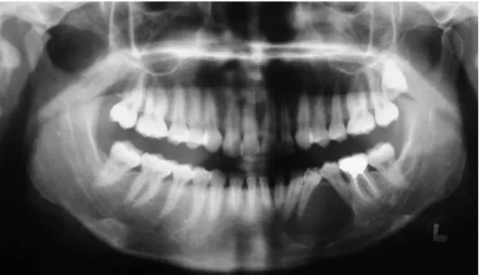
expansion around the left mandibular canine and first and second premolars (Fig. 1). The teeth involved in the lesion were vital with electrical vitality test except the first pre- molar. Panoramic radiograph revealed a well-defined uni- locular radiolucency extending from the distal aspect of the mandibular left lateral incisor to the distal area of the left second premolar (Fig. 2). The lesion displaced the roots of the mandibular left canine and the first and second pre- molars.
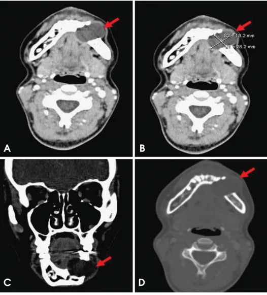
Axial computed tomographic (CT) images showed an ovoid well-defined scalloped cystic lesion 28×18 mm in diameter without bone septa and calcifications. The lesion caused expansion and destruction buccally and lingually.
There was a soft tissue extension without invasion through the cortical perforation. There was no contrast enhancement in the center of the lesion but marked in periphery. Coronal CT images showed an expansive process with scalloped border in mandibular corpus (Fig. 3).
Ultrasonography (US) examination showed a 26.5×20 mm sized, oval, ill-defined dens cystic content with no evidence of internal vascularization. It had a posterior enhancement (Fig. 4).
The lesion was surgically removed via an intraoral app- roach under local anesthesia and histologically reported as a KCOT (Fig. 5). The patient was currently at the fifth month of follow-up without a recurrence.
Discussion
An important characteristic of KCOT is its propensity to grow along the internal aspect of the jaws, causing minimal expansion.1,4Because a considerable number of KCOTs are
─ 62 ─ Keratocystic odontogenic tumor: case report with CT and ultrasonography findings
Fig. 1.Intraoral photograph shows buccal and lingual expansion.
Fig. 2.Panoramic radiograph shows a well-defined unilocular radioluc- ency in the left mandibular body region.
asymptomatic and their clinical signs often fail to appear, these lesions are mostly detected in the late stage, which occasionally allow them to reach a large size.4,7,8Some- times they are detected only by incidental radiographic findings.7,9It was reported that swelling and pain were more frequent in East Asians, whereas KCOT discovered as an incidental finding was more frequent in the Latin American reports.3 Boffano et al5analyzed 261 KCOTs and reported that 37.2% of patients presented with symp- toms and 62.8% were asymptomatic with their lesions found incidentally. In the present case, the KCOT caused an unusual clinical sign of buccal and lingual cortical expansion despite its small size.
The radiographic appearance of KCOT may resemble that of many other odontogenic lesions. If it is associated with a crown of an unerupted tooth, it may be indistinguish- able radiographically from dentigerous cyst.1,8The scalloped margin and multilocular appearance of KCOT may resem- ble an ameloblastoma but the greater propensity to expand
of ameloblastoma may help for differentiation of these lesions. KCOT may show similarity to an odontogenic myxoma because of the mild expansion and multilocular appearance.4
Complete radiographic examination is essential in suspi- cion of KCOT because of its aggressive behavior and high recurrence rate. CT can be used to determine the extent and location of any cortical perforations with soft tissue extension.4In the present case, axial CT images demon- strated a significant expansion and perforation of the buccal and lingual cortical plates of the mandible. Generally a significant expansion may occur in the upper ramus and coronoid process, however the buccal and lingual cortical plates of the mandible revealed slight expansion only.
US is noninvasive and cost-effective, and recommended as a complementary imaging method for intraosseous lesions of the jaw.10,11However, it is not useful in the pre- sence of intact and thick cortical bone and usually the growth of KCOT is larger in mesial-distal direction than in
─ 63 ─
A. Pinar Sumer et al
Fig. 3. Computerize tomography (CT) images. A. The axial CT scan (soft tissue window with contrast) shows a low density lesion that caused expansion and destruction buccally and lingually. B. The axial CT scan shows an ovoid well-defin- ed scalloped border 28×18 mm in diameter cystic lesion. C. The coro- nal CT scan shows a hypodens lesion located in left mandibular corpus. D.
The axial CT scan (bone window) shows the absence of buccal and lingual cortical outlines. There was no contrast enhancement in the lesion.
A B
C D
the buccal-lingual, maintaining the vestibular and lingual/
palatinal bones intact.10 Therefore, US examination could be inconclusive in some KCOT lesions.10,11In the present case, US examination was effectively used in the assess-
ment of the lesion due to the significant buccal and lingual cortical expansion. US examination showed dense cystic content with no evidence of internal vascularization. This finding was consistent with findings by Lauria et al10and Sumer et al.11
In conclusion, a keratocystic odontogenic tumor that revealed an unusual expansion of buccal and lingual cor- tical plates of the mandible was described with its features on CT and US exams.
References
1. Neville BW, Damm DD, Allen CM, Bouquot JE. Oral and maxillofacial pathology. 3rd ed. St. Louis: Saunders; 2009. p.
683-7.
2. Phillpsen HP. Keratocystic odontogenic tumour. In: Barnes L, Eveson JW, Reichart P, Sidransky D. WHO classification of tumors: pathology and genetics of head and neck tumors. 3rd ed. Lyon: IARC Press; 2005. p. 306-7.
3. MacDonald-Jankowski DS. Keratocystic odontogenic tumour:
systematic review. Dentomaxillofac Radiol 2011; 40 : 1-23.
4. White SC, Pharoah MJ. Oral radiology: principles and inter- pretation. 6th ed. St. Louis: Mosby; 2009. p. 351-5.
5. Boffano P, Ruga E, Gallesio C. Keratocystic odontogenic tumor (odontogenic keratocyst): preliminary retrospective review of epidemiologic, clinical, and radiologic features of 261 lesions from University of Turin. J Oral Maxillofac Surg 2010; 68 : 2994-9.
6. Morgan TA, Burton CC, Qian F. A retrospective review of treatment of the odontogenic keratocyst. J Oral Maxillofac Surg 2005; 63 : 635-9.
7. Li TJ. The odontogenic keratocyst: a cyst, or a cystic neoplasm?
J Dent Res 2011 ; 90: 133-42.
8. Cawson RA, Odell EW. Cawson’s essentials of oral pathology and oral medicine. 7th ed. London: Churchill Livingstone;
2002.
9. Grasmuck EA, Nelson BL. Keratocystic odontogenic tumor.
Head Neck Pathol 2010; 4 : 94-6.
10. Lauria L, Curi MM, Chammas MC, Pinto DS, Torloni H. Ultra- sonography evaluation of bone lesions of the jaw. Oral Surg Oral Med Oral Pathol Oral Radiol Endod 1996; 82 : 351-7.
11. Sumer AP, Danaci M, Ozen Sandikci E, Sumer M, Celenk P.
Ultrasonography and Doppler ultrasonography in the evaluation of intraosseous lesions of the jaws. Dentomaxillofac Radiol 2009; 38 : 23-7.
─ 64 ─ Keratocystic odontogenic tumor: case report with CT and ultrasonography findings
Fig. 5.Histopathologic examination shows a parakeratinized epithe- lium with a clear basal layer and a wavy appeared luminal surface.
The cystic lumen contains keratin and the rete was flat (H&E stain,
×200).
Fig. 4.Ultrasound image shows complex cyst with dens internal echoes with a posterior enhancement.