Effect of Masticatory Muscle Pain Control by Morphine
Sang-Hoon Yoo, D.M.D.,M.S.D.1, Min-Jae Kim, D.D.S.1, Joo-Yeon Chang, D.M.D.1, Soo-Kyung Kang, D.M.D.,M.S.D.,Ph.D.1, Q-Schick Auh, D.M.D.,M.S.D.,Ph.D.1, Jung-Pyo Hong, D.M.D.,M.S.D.,Ph.D.1,2, Yang-Hyun Chun, D.M.D.,M.S.D.,Ph.D.1,2
Department of Orofacial Pain and Oral Medicine, School of Dentistry, Kyung Hee University1 Institute of Oral biology, School of Dentistry, Kyung Hee University2
This study was designed to evaluate the pain control effect by morphine injection to masticatory muscle pain patients.
Patients with masticatory muscle pain visited the Department of Oral Medicine, Kyung Hee University Dental Hospital were recruited to this study and diagnosed by RDC/TMD. Experimental group were divided into three group; saline injection group(n=10), lidocaine injection group(n=10) and morphine injection group(n=10). Evaluation list was the subjective pain evaluation(visual analogue scale, Mc Gill pain questionnaire, pain drawing) and the objective pain evaluation(pressure pain threshold, pressure pain tolerance) and evaluation time was injection before, after 10min, 30min, 60min and then it was analyzed statistically.
The results were as follows :
1. The subjective pain evaluation and the objective pain evaluation were significantly different statistically in within subject effects(p<0.001).
2. The subjective pain drawing evaluation(p<0.001) were significantly different statistically in between subject effects.
3. The objective pressure pain threshold evaluation(p=0.025) were significantly different statistically in between subject effects.
4. The morphine injection group(p=0.001) were more significantly different than the saline injection group statistically in the subject pain drawing evaluation.
Therefore, it was considered that the morphine injection was effective to pain control for masticatory muscle pain patients within 60 minute.
Key words: Masticatory Muscle Pain, Morphine sulfate, Temporomandibular Disorders
Corresponding author : Yang-Hyun Chun
Department of Orofacial Pain and Oral Medicine Kyung Hee University, School of Dentistry
Hoegi-dong 1, Dongdaemun-ku, Seoul 130-701, Korea Tel: 82-2-958-9359
Fax: 82-2-968-2043 E-mail: [email protected] Received: 2012-06-05 Accepted: 2012-06-30
Ⅰ. INTRODUCTION
Pains are perceived, after activation of first afferent nerves by peripheral stimulations generate electrical impulses that transmitted to central nervous system through serial neuro-pathway. Pain developing stimulations, such as tissue damages or inflammation, release pain neurotransmitters. These neurotransmitters activate receptors located on cell membrane, which cause excitatory action potential.
Eventually, because activation of peripheral pain
receptors initiate pain pathway, peripheral pain receptors play an important role. Thus, wide and varied studies on peripheral tissue are underway so as to regulate pain by peripheral pain receptors.
A typical drug of pain control and analgesic effect is opioid. Opioids are still the most powerful drugs for severe pain but their use is hampered by side effects such as respiratory depression, nausea, constipation, addiction and tolerance.1) But after discovering peripherally acting opioid agonist, we expect peripheral analgesic effect of opioid without central side effects.1-3) Opioid receptor synthesized at dorsal root ganglion and migrated along the neuronal axon to peripheral and central nerve terminal.4-6) It has an analgesic effect on post-operation pain or chronic pain.7)
Especially peripheral analgesic effect is very effective under inflammatory state.8) There are studies of applying opioid when oral surgery so it can reduce post operation pain,9) and 5 mg, 10 mg morphine sulfate were applied in TMJ due to reduce pain.10) As a result, this study was designed to evaluate the pain control effect by morphine sulfate injection to masticatory muscle pain patients.
Ⅱ. SUBJECTS AND METHODS 1. Subjects
The subjects participated in this study were volunteers among outpatients of Department of Oral medicine, Kyung Hee University Dental Hospital during the period from August to November 2010.
This study was conducted in Department of Oral medicine, Kyung Hee University Dental Hospital after receiving approval from Institutional review board of Kyung Hee University Dental Hospital.
The participants were informed about the details of this experiment and signed the consent paper after reading it. And all the participants are limited that Visual Analog Scale(VAS) is over 50 and age between 20 to 55 who are diagnosed to Axis I:Group Ia Myofascial Pain by Research Diagnostic
Criteria for Temporomandibular Disorders(RDC/
TMD). The subjects were excluded who had systemic musculoskeletal pain, systemic arthritis, malignant tumor, hypertension, diabetes mellitus, cardiovascular disease and pregnant women or chronic analgesic or psychiatric drug user.
2. Methods
One researcher divided randomly the subjects into three groups 10 persons each who had diagnosed according to RDC/TMD.
Another researcher injected 0.2 ml drug with 27G subcutaneous needle and 1.0 ml disposable syringe(SOFJECⓇ ; HWAJINMEDICAL, KOREA) into each subjects in 10 seconds, and injection point was the most painful area on unilateral masticatory muscle in palpation.
The other researcher ordered randomly the sequence of injected drugs , as a result, double blind procedure about the injected drug was done to both researcher and subjects.
Initially classified subject groups were morphine sulfate(15mg/1ml; BCworld, KOREA) 3.0 mg injection group, lidocaine HCl(2%/20 ml; Huons, KOREA) injection group and saline(NaCl 9 g/1000 ml; JWPharmaceutical, KOREA) injection group.
The subjective pain and objective pain to each drugs were evaluated at just before the injection, 10 minutes after the injection, 30 minutes after injection and 60 minutes after the injection.
3. Pain Evaluation
1) Subjective Pain Evaluation
The methods used to evaluate subjective pain in this study were visual analogue scale(VAS test), McGill Pain Questionnaire(MGQ test) and Pain drawing(PD test).
In the VAS test, subjects were asked to mark their pain with marking pen(namepenF®, Monami, KOREA) on 100 mm straight line according to pain extent; start point of 100 mm straight line with no pain, end point with the strongest pain that could
imagine. The result was converted into numbers according to the percentage.
In MGQ method, generally used McGill Pain Questionnaire in Korean version was applied to research the patients pain according to the questionnaire. The patient's subjective pain was digitalized and the data of the questionnaire was calculated by average of total score.
Lastly, the PD, subjective pain evaluation method, was a method that let the patients mark their pain area themselves with marking pen. The marking area was squared, then marking squares were counted, scored and converted to percentage.
2) Objective Pain Evaluation
The methods used to evaluate objective pain were pressure pain threshold(PPT) test and pressure pain tolerance(PPTol) test.
The PPT test used in this study was to estimate the pressure pain threshold around the most painful masticatory area before and after the injection with pressure pain measuring instrument(Wagner Instruments, Greenwich CT, USA), then converted into numbers.
Also PPTol was applied same area and used same pressure pain measuring instrument to calculate the pain limit of same pressure and converted into numbers.
We keep the patient's masticatory system as relaxed position as possible without tooth contact to use the pressure pain measuring instrument. The pressure was applied to muscle vertically with 11mm diameter probe by 30kPa/s velocity, the measured kgf value was divided by area of the probe and converted to kPa value. The subjects were asked to rise their left hand at the moment they felt the first pain and intolerable pain, then the values were recorded and calculated for the PPT and PPTol.
4. Statistical Analysis
The results from three subjective pain evaluation tests and two objective pain evaluation tests were
done the repeated measures ANOVA test using descriptive statistics and Greenhouse-Geisser method. At first, we verified the effects through within subject effects, between subject effects and interaction effects. When there were effects, we tried Post Hoc through multiple comparisons using Tukey HSD method. Every statistics significance level was p<0.05 and study scores were analyzed with PASW Statistics(SPSS version 18.0).
Ⅲ. RESULTS 1. Descriptive statistics result
The subjects consisted of 16 males and 14 females, total 30 people and age range was 20 to 52years, average age was 29.4±4.5years. Before the injection group average was 62.3±4.34, 10min after the injection group average was 57.9±10.91, 30 min after the injection group average was 54±14.17, 60min after the injection group was 47.0±17.37. The average and the standard deviation of time line group was in Table 1.
2. Subjective Pain Evaluation Result
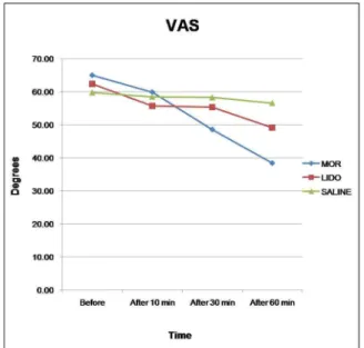
The results of VAS test as a subjective pain evaluation method show that morphine injection group decreased the most with time, followed by lidocaine injection group, but the saline injection group had little changed. Especially, these tendencies was prominent from 10 minutes after the injection to 30 minutes, and in 60 minutes after the injection, the differences between groups were lasted.(Fig. 1)
The results of MGQ test which is questionnaire -styled subjective pain evaluation method show that morphine injection group decreased steeply the most with time, lidocaine injection group decreased obviously after 10 minutes, but saline injection group had little changed.(Fig. 2)
The results of PD test that let the patients mark their pain area themselves as a subjective pain evaluation method show similar pattern in morphine
Type Mean SD N
Before
LIDO 62.3000 4.32178 10
MOR 65.0000 .00000 10
SALINE 59.7000 5.14350 10
Sum 62.3333 4.34172 30
After10min
LIDO 55.6000 5.05964 10
MOR 59.8000 18.18302 10
SALINE 58.4000 4.19524 10
Sum 57.9333 10.91640 30
After30min
LIDO 55.3000 7.48406 10
MOR 48.5000 22.88255 10
SALINE 58.2000 3.48967 10
Sum 54.0000 14.16893 30
After60min
LIDO 49.1000 5.87745 10
MOR 38.3000 26.98580 10
SALINE 56.5000 4.85913 10
Sum 47.9667 17.37315 30
SD; standard deviation, LIDO; lidocaine HCl 2%/20 ml, MOR; morphine sulfate 15 mg/1 ml, SALINE; NaCl 9 g/1000 ml
Table 1. The results of descriptive statistics data by PASW Statistics
subject effects F df1 df2 P-value
VAS within 16.132 1.8 48.3 0.000 *
between 0.718 2 27 0.497
MGQ within 14.286 1.8 48.8 0.000 *
between 1.466 2 27 0.249
PD within 10.939 1.9 51.8 0.000 *
between 10.45 2 27 0.000 *
* p < 0.05, VAS; visual analogue scale, MGQ; McGill pain questionnaire, PD; pain drawing
Table 2. The results of Repeated Measures ANOVA test with Greenhouse-Geisser application by the subjective pain evaluation
Fig. 1. The mean of VAS in every groups. MOR;
morphine sulfate 15mg/1ml, LIDO; lidocaine HCl 2%/20ml, SALINE; NaCl 9g/1000ml
injection group and lidocaine injection group with time, but a quite different pattern in saline injection group. Especially, difference between saline injection group and morphine injection group, and difference between saline injection group and lidocaine injection group were remarkable.(Fig. 3)
The results of VAS test, MGQ test and PD test which are subjective pain evaluation methods were analyzed statistically, and were summarized in Table 2. The VAS(p<0.001) and MGQ test(p<
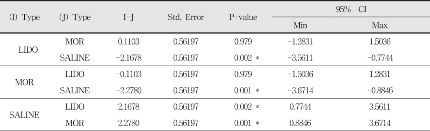
0.001) were significantly different statistically in within the groups. PD test was significantly different statistically in within the group(p<0.001) and between the groups(p<0.001). As a result, VAS, MGQ and PD test, all were significantly different statistically in within subject effects, but only PD test was significantly different statistically in between subject effects.
Thus we tried Post Hoc to the PD test which was significantly different statistically in within and between subject effects, and summarized in Table 3. Inspecting for the lidocaine injection group in priority, morphine injection group was not significantly different statistically but saline
Fig. 2. The mean of total average of McGill Pain Questionnaire in every groups. MOR;
morphine sulfate 15mg/1ml, LIDO; lidocaine HCl 2%/20ml, SALINE; NaCl 9g/1000ml
Fig. 3. The mean of percentage of Pain Drawing in every groups. MOR; morphine sulfate 15mg /1ml, LIDO; lidocaine HCl 2%/20ml, SALINE;
NaCl 9g/1000ml
injection group was significantly different(p=0.002).
Inspecting for morphine injection group in priority,
(I) Type (J) Type I-J Std. Error P-value 95% CI
Min Max
LIDO MOR 0.1103 0.56197 0.979 -1.2831 1.5036
SALINE -2.1678 0.56197 0.002 * -3.5611 -0.7744
MOR LIDO -0.1103 0.56197 0.979 -1.5036 1.2831
SALINE -2.2780 0.56197 0.001 * -3.6714 -0.8846
SALINE LIDO 2.1678 0.56197 0.002 * 0.7744 3.5611
MOR 2.2780 0.56197 0.001 * 0.8846 3.6714
*p < 0.05, CI; confidence interval, LIDO; lidocaine HCl 2%/20ml, MOR; morphine sulfate 15mg/1ml, SALINE; NaCl 9g/1000ml
Table 3. The results of Post Hoc Multiple Comparisons by Tukey HSD for pain drawing
both lidocaine injection group and saline injection group were not significantly different statistically.
Inspecting for the saline injection group in priority, both lidocaine injection group(p=0.002) and mor- phine injection group(p=0.001) were significantly different statistically.
3. Objective Pain Evaluation Result
The results of PPT test as a objective pain evaluation method show that morphine injection group, lidocaine injection group and saline injection group had similarly increasing tendencies with time by 30 minutes. But the saline injection group showed low threshold range compared to morphine injection group and lidocaine injection group. In lidocaine injection group and morphine injection group, partially coinciding tendencies are showed with time from 30 minutes after the injection to 60 minutes.(Fig. 4)
The PPTol test which is evaluating objective endurance to pain induced by pressure shows relatively variable tendencies with time. Slightly and consistently increased change ranges are showed in lidocaine injection group and saline injection group, but in morphine injection group, the change range is a little large with time.(Fig. 5)
The results of PPT test and PPTol test which
Fig. 4. The mean of pressure pain threshold, PPT in every groups. MOR; morphine sulfate 15mg /1ml, LIDO; lidocaine HCl 2%/20ml, SALINE;
NaCl 9g/1000ml
are objective pain evaluation methods were analyzed statistically, and were summarized in Table 4. The PPT(p<0.001) and PPTol test(p<
0.001) were significantly different statistically in within the groups. However, the PPT test was significantly different statistically in between the groups(p=0.025), too. As a result, both PPT and
Fig. 5. The mean of pressure pain tolerance, PPTol in every groups. MOR; morphine sulfate 15mg/1ml, LIDO; lidocaine HCl 2%/20ml, SALINE; NaCl 9g/1000ml
subject effects F df1 df2 P-value
PPT within 30.662 2.5 66.8 0.000 *
between 4.239 2 27 0.025 *
PPTol within 9.024 2.4 65.9 0.000 *
between 0.953 2 27 0.398
* p < 0.05, PPT; pressure pain threshold, PPTol; pressure pain tolerance
Table 4. The results of Repeated Measures ANOVA test with Greenhouse-Geisser application by the objective pain evaluation
(I) Type (J) Type I-J Std. Error P-value 95% CI
Min Max
LIDO MOR 8.5000 12.45254 0.776 -22.3750 39.3750
SALINE 34.7750 12.45254 0.025 * 3.9000 65.6500
MOR LIDO -8.5000 12.45254 0.776 -39.3750 22.3750
SALINE 26.2750 12.45254 0.107 -4.6000 57.1500
SALINE LIDO -34.7750 12.45254 0.025 * -65.6500 -3.9000
MOR -26.2750 12.45254 0.107 -57.1500 4.6000
*p < 0.05, CI; confidence interval, LIDO; lidocaine HCl 2%/20ml, MOR; morphine sulfate 15mg/1ml, SALINE; NaCl 9g/1000ml
Table 5. The results of Post Hoc Multiple Comparisons by Tukey HSD for pressure pain threshold
PPTol test were significantly different statistically in within subject effects, but only PPT test was significantly different statistically in between subject effects.
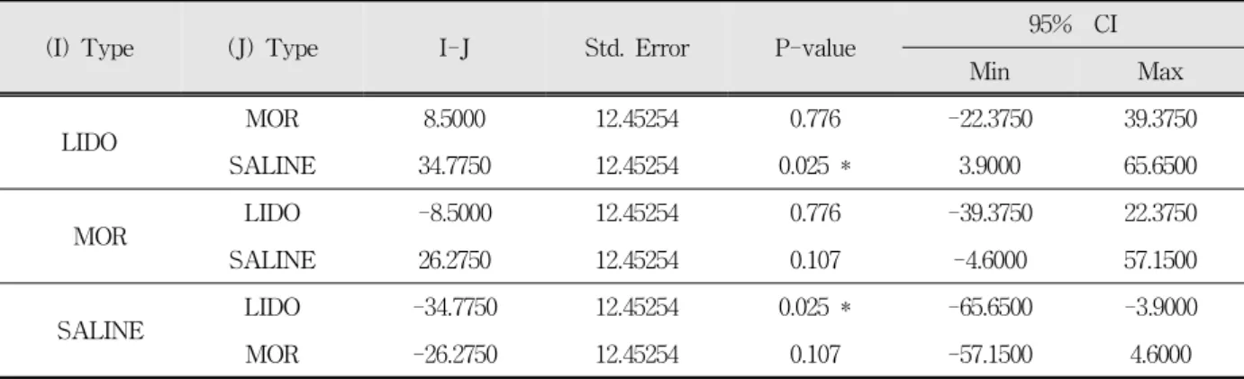
Thus we tried Post Hoc to the PPT test which was significantly different statistically in within and between subject effects, and summarized in Table 5. Inspecting for the lidocaine injection group in priority, morphine injection group was not significantly different statistically but saline injection group was significantly different(p=0.025).
Inspecting for morphine injection group in priority, both lidocaine injection group and saline injection group were not significantly different statistically.
Inspecting for the saline injection group in priority, lidocaine injection group was significantly different statistically(p=0.025), but morphine injection group was not significantly different statistically.
Ⅳ. DISCUSSION
The glutamate which is representative pain inducing material secretes at the first afferent nerve fiber terminal, and the receptor which is located at the nociceptor terminal activates when there was a glutamate secreted by nerve cell, Schwan cell, mast cell due to tissue damage or inflammation.11-14) There are glutamate receptors which is ionic NMDAR(N-methyl D-aspartate receptor), AMPAR (α-amino-3-hydroxy-5-methyl-4-isoxazolepropioni c acid receptor) and metabotropic glutamate receptors(mGluR). When the first afferent nerve fiber terminal secretes the glutamate, post synaptic nerve cell receptor senses and activated. The study of Chun et al,15)which was the influence of terminal AMPA receptor to the muscle nociceptive effect and c-fos activation proposed that GluR1 and GluR2, AMPA subunits, develops in the trigeminal ganglion neurons and masseter afferent nerve cell bodies. As a result, acute muscle pain partially mediated by AMPA receptor which is located in the terminal and when several terminal glutamate receptor subunit is blocked it might be reduce the muscle pain and central nerve activation more effectively.15) In the study of Ro et al,16) the animal model of hypertonic saline(HS) infusion protocol causes peripheral release of glutamate, and that blockade of peripheral NMDA receptors signifi- cantly reduces HS-induced nocifensive behavior and central neuronal activation.
In the study of investigating the effects of ketamine, which is antagonist of glutamate, on chronic myofascial pain and mandibular function in temporomandibular disorder patients, ketamine did not play a major role in the pathophysiology of chronic myofascial temporomandibular disorder pain. Although there was a minor effect of ketamine on maximum voluntary jaw opening, local administrating may not be promising treatment for these patients.17) If there was an increase of glutamate in intermedia, it could cause chronic pain and we can find a higher glutamate if there was a chronic non-inflammatory pain on human tendon
and muscle.18,19)
Not only glutamate but also capsaicin is known as a major pain inducing material and a powerful stimulant to capsaicin transient receptor potential cation channel, subfamilly V, member 1(TRPV1). If capsaicin was injected in human masseter muscle, it induced powerful local hyperalgesia, referred pain and mechanical hypersensitivity.20-22) In animal experiment with rat masseter muscle, capsaicin caused powerful pain reaction and long lasting mechanical hyperalgesia.23-25)
Chun and Ro suggested that intramuscular capsaicin in rat masseter muscle induced significant increase of trigeminal caudalis(Vc) neuron response and that the blockade of peripherally localized mGluR5 can effectively attenuate muscular hypersensitivity.26)In the study of investigating the interaction between glutamate and capsaicin in inducing muscle pain and sensitization in humans, pain reduced more effectively when glutamate injection after capsaicin injection than glutamate injection after saline injection. These findings indicate that intramuscular administrations of glutamate and capsaicin interact and influence pain and sensitization of muscle nociceptors.27)
Opioids are still the most powerful drugs for severe pain but their use is hampered by side effects such as respiratory depression, nausea, constipation, addiction and tolerance.1) But after discovering peripheral opioid receptors, we expect peripheral analgesic effect without central side effects.1-3) Opioid peptide-containing circulating leukocytes extravasate upon activation of adhesion molecules, and corticotropin-releasing factor (CRF), chemokines or noradrenaline can elicit opioid release by activating their respective receptors on leukocytes. Exogenous opioids or endogenous opioid peptides bind to opioid receptors that are synthesized in dorsal root ganglia and transported along intra-axonal microtubules to peripheral and central terminals of sensory neurons results in antinociceptive effects.1)
Three kinds of opioid receptor such as μ, δ, κ - opioid receptor synthesized at dorsal root
ganglion28-34) and they are distributed to not only central but also peripheral through first afferent neuron.35-36) Opioid effect at peripheral is not easly detected in normal tissue. But when inflammatory reaction occurs, opioid receptor already appears at peripheral nerve in minutes to hours.5,37,38) Functional roll of peripheral pain blocking μ-opioid receptor is well known by pain regulating model with peripheral activating drug or low dose opioid agonists.39)
Jessell and Iversen40) discovered that opioid analgesics inhibit substance P release from primary afferent nerve fiber at trigeminal nerve, so they found that opioid receptor present at trigeminal nucleus. Tegeder et al.41) discovered that low dose of morphine-6-b-glucuronide local injection in human muscle had analgesic effect caused by concentric or eccentric muscle contraction. Bakke et al.42) found that local morphine injection to temporomandibular joint could inhibit jaw muscle activation by inflammatory stimulation and this fact explains the regulating function of opioid receptor in temporomandibular joint. Ro et al.8) investigated whether inflammation in the orofacial muscle alters μ-opioid receptor mRNA and protein expressions in trigeminal ganglia, and assessed the contribution of peripheral μ-opioid receptors under acute and inflammatory muscle pain conditions with rats.
They concluded that activated peripheral μ-opioid receptor in inflammatory muscle blocked pain more effectively and this amplified μ-opioid receptor are contributed by μ-opioid receptor synthesizing rate at trigeminal ganglia. Eisenberg et al.43)showed the peripheral and systemic antinociceptive effect of morphine local injection on formalin-induced facial pain behavior. They concluded that activation of peripheral opioid receptor contributed to reduce nociceptive stimulation and hyperalgesia. Local administration of μ-opioid receptor accelerator in animal research, Houghton et al.44)discovered the blockade of bony pain, Catheline et al.45) and Truong et al.46) discovered the blockade of neuropathic pain, and Ko et al.47)said the diminution of hyperalgesia and allodynia related to
inflammatory pain in monkeys.
Same as the animal research, peripheral administration of opioid has a good analgesic effect in human study. Activated peripheral opioid receptor has a powerful analgesic effect to chronic rheumatoid osteoarthritis, inflammatory toothache and post-operative visceral pain.48-53) Modi et al9) discovered buprenorphine with bupivacaine for intraoral nerve blocks to provide postoperative analgesia in patients after minor oral surgery could not prolong the anesthetic time but the time of postoperative analgesic duration could prolonged three times. Analgesic effect of peripheral opioid receptor is known more effective when inflammatory state but, in none inflammatory knee joint surgery morphine administration has an analgesic effect.54-56) In the study of Ziegler et al.10) analgesic effect of intra-articular morphine in patients with temporomandibular joint disorders, morphine 10mg had the longest and prominent analgesic effect. From this study they had an analgesic effect on non inflammatory patient so they concluded sufficient dose of morphine could more effective on inflammatory patients.
Analgesic effect of peripheral opioid receptor has been studied steadily, but clinical study on human masticatory muscle has not been performed actively. So in this study we injected low dose of morphine to masseter muscle of myofascial pain patients who were diagnosed from RDC/TMD and we confirmed the analgesic effects in 60 minutes, compared the results with lidocaine and saline injection.
The results of VAS test show that morphine injection group decreased the most, followed by lidocaine injection group with time. But the saline injection group had little changed.(Fig. 1) The results of MGQ test show that morphine injection group decreased steeply the most with time, lidocaine injection group decreased obviously after 10 minutes, but saline injection group had little changed.(Fig. 2) The results of PD test show similar pattern in morphine injection group and lidocaine injection group with time, but a quite
different pattern in saline injection group.
Especially, difference between saline injection group and morphine injection group, and difference between saline injection group and lidocaine injection group were remarkable.(Fig. 3)
The VAS(p<0.001) and MGQ test(p<0.001) were significantly different statistically in within the groups. PD test was significantly different statisti- cally in within the group(p<0.001) and between the groups(p<0.001). As a result, VAS, MGQ and PD test, all were significantly different statistically in within subject effects, but only PD test was significantly different statistically in between subject effects.(Table 2) Thus we tried Post Hoc to the PD test which was significantly different statistically in within and between subject effects.
Inspecting for the lidocaine injection group in priority, morphine injection group was not significantly different statistically but saline injection group was significantly different(p=0.002).
Inspecting for morphine injection group in priority, both lidocaine injection group and saline injection group were not significantly different statistically.
Inspecting for the saline injection group in priority, both lidocaine injection group(p=0.002) and morphine injection group(p=0.001) were significantly different statistically.(Table 3)
The results of PPT test show that morphine injection group, lidocaine injection group and saline injection group had similarly increasing tendencies with time by 30 minutes.(Fig. 4) But the saline injection group showed low threshold range compared to morphine injection group and lidocaine injection group. The PPTol test shows relatively variable tendencies with time.(Fig. 5) The PPT(p<0.001) and PPTol test(p<0.001) were significantly different statistically in within the groups. However, the PPT test was significantly different statistically in between the groups(p= 0.025), too. As a result, both PPT and PPTol test were significantly different statistically in within subject effects, but only PPT test was significantly different statistically in between subject effects.
(Table 4) Thus we tried Post Hoc to the PPT test
which was significantly different statistically in within and between subject effects. Inspecting for the lidocaine injection group in priority, morphine injection group was not significantly different statistically but saline injection group was significantly different(p=0.025). Inspecting for morphine injection group in priority, both lidocaine injection group and saline injection group were not significantly different statistically. Inspecting for the saline injection group in priority, lidocaine injection group was significantly different statistically (p=0.025), but morphine injection group was not significantly different statistically.(Table 5)
Therefore, it was considered that the morphine injection was effective to pain control for masticatory muscle pain patients within 60 minutes.
It could be further investigation on time extension.
Ⅴ. CONCLUSIONS
The present study was designed to evaluate the pain control effect by morphine injection to masticatory muscle pain patients who were recruited to this study and diagnosed by Research Diagnostic Criteria for Temporomandibular Disorders(RDC/TMD). Experimental group were divided into three groups; saline injection group(n=10), lidocaine injection group(n=10) and morphine injection group(n=10).
Evaluation list was the subjective pain evaluation(visual analogue scale, Mc Gill pain questionnaire, pain drawing) and the objective pain evaluation(pressure pain threshold, pressure pain tolerance) and evaluation time was injection before, after 10min, 30min, 60min and then it was analyzed statistically.
The results were as follows :
1. The subjective pain evaluation and the objective pain evaluation were significantly different statistically in within subject effects(p<0.001).
2. The subjective pain drawing evaluation(p<0.001) were significantly different statistically in between subject effects.
3. The objective pressure pain threshold evaluation
(p=0.025) were significantly different statistically in between subject effects.
4. The morphine injection group(p=0.001) were more significantly different than the saline injection group statistically in the subject pain drawing evaluation.
Therefore, it was considered that the morphine injection was effective to pain control for masticatory muscle pain patients within 60 minutes.
It could be further investigation on time extension.
REFERENCES
1. Stein C, Lang LJ. Peripheral mechanisms of opioid analgesia. Current Opinion in Pharmacology 2009;9:3- 8.
2. Stein C, Hassan AHS, Przewlocki R, Gramsch C, Peter K, Herz A. Opioids from immunocytes interact with receptors on sensory nerves to inhibit nociception in inflammation. Proc Natl Acad Sci USA 1990;87:5935-9.
3. Przewlocki R, Hassan AHS, Lason W, Epplen C, Herz A, Stein C. Gene expression and localization of opioid peptides in immune cells of inflamed tissue:
functional role in antinociception. Neuroscience 1992;
48:491-500.
4. Fields HL, Emson PC, Leigh BK, Gilbert RF, Iversen LL. Multiple opiate receptor sites on primary afferent fi bres. Nature. 1980;284:351-3.
5. Stein C. Peripheral mechanisms of opioid analgesia.
Anesth Analg. 1993;76:182-91.
6. Machelka H. Targeting of opioid-producing leuko- cytes for pain control. Neuropeptides. 2007;41:355-63.
7. Boogaerts J, Lafont N. Mechanism of action and clinical use of opioids administered by the peripheral perineural route. Cah Anesthesiol. 1991;39:91-5.
8. Nũnéz S, Lee JS, Zhang Y, Bai G, Ro JY. Role of peripheral mu-opioid receptors in inflammatory orofacial muscle pain. Neuroscience 2007;146: 1346- 54.
9. Modi M, Rastogi S, Kumar A, Buprenorphine with bupivacaine for intraoral nerve blocks to provide postoperative analgesia in outpatients after minor oral surgery. J Oral Maxillofac Surg. 2009 Dec;67(12):
2571-6.
10. Ziegler CM, Wiechnik J, Mühling J. Analgesic effects of intra-articular morphine in patients with
temporomandibular joint disorders: a prospective, double-blind, placebo-controlled clinical trial. J Oral Maxillofac Surg. 2010 Mar;68(3):622-7.
11. Piani D, Frei K, Do KQ, Cuenod M, Fontana A, Murine brainmacrophages induce NMDA receptor mediated neurotoxicity in vitro by secreting glutamate. Neurosci. Lett. 1991;133:159-162.
12. Parpura V, Liu F, Jeftinija KV, Haydon PG, Jeftinija SD. Neuroligand-evoked calcium-dependent release of excitatory amino acids from Schwann cells. J.
Neurosci. 1995;15: 5831-5839.
13. DeGroot J, Zhou S, Carlton SM, Peripheral glutamate release in the hindpaw following low and high intensity sciatic stimulation. NeuroReport. 2000;11:4 97-502.
14. Lawand NB, McNearney T, Westlund KN, Amino acid release into the knee joint: key role in nociception and inflammation. Pain. 2000;86:69-74.
15. Chun YH, Frank D, Lee JS, Zhang Y, Auh QS, Ro JY.
Peripheral AMPA receptors contribute to muscle nociception and c-fos activation. Neuroscience Research. 2008;62:97-104.
16. Ro JY, Capra NF, Lee JS, Masri R, Chun YH.
Hypertonic saline-induced muscle nociception and c-fos activation are partially mediated by peripheral NMDA receptors. European Journal of Pain.
2007;11:398-405.
17. Castrillon EE, Cairns BE, Ernberg M, Wang K, Sessle BJ, Arendt-Nielsen L, Svensson P. Effect of peripheral NMDA receptor blockade with ketamine on chronic myofascial pain in temporomandibular disorder patients: a randomized, double-blinded, placebo-controlled trial. J Orofac Pain. 2008 Spring;22 (2):122-30.
18. Alfredson H, Forsgren S, Thorsen K, Lorentzon RJ. In vivo microdialysis and immunohistochemical analyses of tendon tissue demonstrated high amounts of free glutamate and glutamate NMDAR1 receptors, but no signs of inflammation, in Jumper's knee. Orthop Res.
2001;19:881-886.
19. Rosendal L, Larsson B, Kristiansen J, Peolsson M, Sogaard K, Kjaer M, Sorensen J, Gerdle B. Increase in muscle nociceptive substances and anaerobic metabolism in patients with trapezius myalgia:
microdialysis in rest and during exercise. Pain. 2004;
112:324-334.
20. Arima T, Svensson P, Arendt-Nielsen L. Capsaicin- induced muscle hyperalgesia in the exercised and non-exercised human masseter muscle. J. Orofac.
Pain. 2000;14:213–223.
21. Arima T, Arendt-Nielsen L, Minagi S, Svensson P.
Effect of capsaicin-evoked jaw-muscle pain on intramuscular blood-flow. Arch. Oral Biol. 2009;54:
241-249.
22. Wang K, Arendt-Nielsen L, Svensson P. Capsaicin- induced muscle pain alters the excitability of thehumanjaw-stretch reflex. J. Dent. Res. 2002;81:
650-654.
23. Lee JS, Zhang Y, Ro JY. Involvement of neuronal, inducible, and endothelia nitric oxide synthases in capsaicin-induced muscle hypersensitivity. Eur. J.
Pain. 2009;13(9):924-928.
24. Ro JY, Lee JS, Capra NF, Zhang Y. Role of soluble guanylatecyclase in the trigeminal subnucleus caudalis incapsaicin-induced muscle hypersensitivity.
Brain Res. 2007;1184:141-148.
25. Ro JY, Lee JS, Zhang Y. Activation of TRPV1 and TRPA1 leads to muscle nociception and mechanical hyperalgesia. Pain. 2009;144:270-277.
26. Chun YH, Ro JY. Electrophysiological characterization of the rat trigeminal caudalis (Vc) neurons following intramuscular injection of capsaicin. Neuroscience Letters. 2010;469:289-293.
27. Arendt-Nielsen L, Svensson P, Sessle BJ, Cairns BE, Wanga K. Interactions between Glutamate and Capsaicin in Inducing Muscle Pain and Sensitization in Humans. Eur J Pain. 2008 July;12(5):661-670.
28. Mansour A, Fox CA, Burke S, Meng F, Thompson RC, Akil H, Watson SJ. Mu, delta, and kappa opioid receptor mRNA expression in the rat CNS: an in situ hybridization study. J Comp Neurol. 1994;350:412-438.
29. Minami M, Maekawa K, Yabuuchi K, Satoh M.
Double in situ hybridization study on coexistence of mu, delta and kappa OR mRNAs with preprotachykinin A mRNA in the rat dorsal root ganglia. Brain Res Mol Brain Res. 1995;30:203–-210.
30. Mennicken F, Zhang J, Hoffert C, Ahmad S, Beaudet A, O’'Donnell D. Phylogenetic changes in the expression of delta opioid receptors in spinal cord and dorsal root ganglia. J Comp Neurol. 2003;465:349-360.
31. Dado RJ, Law PY, Loh HH, Elde R. Immunofluo- rescent identification of a delta (delta)-opioid receptor on primary afferent nerve terminals. Neuroreport.
1993;5:341-344.
32. Arvidsson U, Riedl M, Chakrabarti S, Lee JH, Nakano AH, Dado RJ, Loh HH, Law PY, Wessendorf MW, Elde R. Distribution and targeting of a mu-opioid receptor (MOR1) in brain and spinal cord. J Neurosci.
1995;15:3328-3341.
33. Ji RR, Zhang Q, Law PY, Low HH, Elde R, Hokfelt T. Expression of mu-, delta-, and kappa-opioid receptor-like immunoreactivities in rat dorsal root ganglia after carrageenan-induced inflammation. J Neurosci 19951;5:8156-8166.
34. Coggeshall RE, Zhou S, Carlton SM. Opioid receptors on peripheral sensory axons. Brain Res. 1997;764:126- 132.
35. Hassan AH, Ableitner A, Stein C, Herz A. Infla- mmation of the rat paw enhances axonal transport of opioid receptors in the sciatic nerve and increases their density in the inflamed tissue. Neuroscience.
1993;55:185-195.
36. Mousa SA, Zhang Q, Sitte N, Ji R, Stein C.
beta-Endorphincontaining memory-cells and mu- opioid receptors undergo transport to peripheral inflamed tissue. J Neuroimmunol. 2001;115:71–78.
37. Schäfer M, Imai Y, Uhl GR, Stein C. Inflammation enhances peripheral mu-opioid analgesia, but not mu-opioid receptor transcription in dorsal root ganglia. Eur J Pharmacol. 1995;279:165-9.
38. Antonijevic I, Mousa SA, Schäfer M, Stein C.
Perineurial defect and peripheral opioid analgesia in inflammation. J Neurosci. 1995;15:165-72.
39. Stein C, Schafer M, Machelska H. Attacking pain at its source: new perspectives on opioids. Nat Med.
2003;9:1003-1008.
40. Jessell TM, Iversen LL. Opiate analgesics inhibit substance Prelease from rat trigeminal nucleus.
Nature. 1977;268:549–551.
41. Tegeder I, Meier S, Burian M, Schmidt H, Geisslinger G, Lotsch J. Peripheral opioid analgesia in experi- mental human pain models. Brain. 2003;126:1092- 1102.
42. Bakke M, Hu JW, Sessle BJ. Morphine application to peripheral tissues modulates nociceptive jaw reflex.
Neuroreport. 1998;9:3315–3319.
43. Eisenberg E, Vos BP, Strassman AM. The peripheral antinociceptive effect of morphine in a rat model of facial pain. Neuroscience. 1996;72:519–525.
44. Houghton AK, Valdez JG, Westlund KN. Peripheral morphine administration blocks the development of hyperalgesia and allodynia after bone damage in the rat. Anesthesiology. 1998;89:190–201.
45. Catheline G, Guilbaud G, Kayser V. Peripheral component in the enhanced antinociceptive effect of systemic U-69,593, a kappa opioid receptor agonist in mononeuropathic rats. Eur J Pharmacol. 1998;357:171
-178.
46. Truong W, Cheng C, Xu QG, Li XQ, Zochodne DW.
Mu opioid receptors and analgesia at the site of a peripheral nerve injury. Ann Neurol. 2003;53:366-375.
47. Ko MC, Butelman ER, Woods JH. The role of peripheral mu opioid receptors in the modulation of capsaicin-induced thermal nociception in rhesus monkeys. J Pharmacol Exp Ther. 1998;286:150-156.
48. Duckett JW, Cangiano T, Cubina M, Howe C, Cohen D. Intravesical morphine analgesia after bladder surgery. J Urol. 1997;157:1407-1409.
49. Likar R, Schafer M, Paulak F, Sittl R, Pipam W, Schalk H, Geissler D, Bernatzky G. Intraarticular morphine analgesia in chronic pain patients with osteoarthritis. Anesth Analg. 1997;84:1313-1317.
50. Rorarius M, Suominen P, Baer G, Pajunen P, Tuimala R, Laippala P. Peripherally administered sufentanil inhibits pain perception after postpartum tubal ligation. Pain. 1999;79:83-88.
51. Likar R, Sittl R, Gragger K, Pipam W, Blatnig H, Breschan C, Schalk HV, Stein C, Schafer M.
Peripheral morphine analgesia in dental surgery. Pain.
1998;76:145-150.
52. Likar R, Koppert W, Blatnig H, Chiari F, Sittl R, Stein C, Schafer M. Efficacy of peripheral morphine analgesia in inflamed, noninflamed and perineural tissue of dental surgery patients. J Pain Symptom Manage. 2001;21:330-337.
53. Eisenach JC, Carpenter R, Curry R. Analgesia from a peripherally active kappa-opioid receptor agonist in patients with chronic pancreatitis. Pain. 2003;101:89- 95.
54. Lombardi AV Jr, Berend KR, Mallory TH, Dodds KL, Adams JB. Soft tissue and intra-articular injection of bupivacaine, epinephrine and morphine has a beneficial effect after total knee arthroplasty. Clin Orthop Relat Res. 2004;428:125-130.
55. Grabowska-Gawel A, Gawel K, Hagner W, Biliński PJ. Morphine or bupivacaine in controlling posto- perative pain in patients subjected to knee joint arthroscopy. Ortop Traumatol Rehabil. 2003;5(6):
758-762.
56. Rosseland LA, Stubhaug A, Grevbo F, Reikerås O, Breivik H. Effective pain relief from intra-articular saline with or without morphine 2 mg in patients with moderate-to-severe pain after knee arthroscopy:
A randomized, double-blind controlled clinical study.
Acta Anaesthesiol Scand. 2003;47:732-738.
국문초록
Morphine에 의한 저작근 통증의 조절 효과
경희대학교 치의학전문대학원 안면통증구강내과학교실1, 경희대학교 구강생물학연구소2
유상훈1・김민재1・장주연1・강수경1・어규식1・홍정표1,2・전양현1,2
이 연구는 Morphine을 이용한 저작근통증의 조절 효과를 확인하기 위해서 시행되었으며, 경희대학교 치과대학병원 구강내 과에 내원한 환자 중 RDC/TMD로 진단된 지원자를 saline 주사군, lidocaine 주사군, morphine 주사군 각각 10명씩 배정하였 다. 통증부위에 주사 전, 주사 후 10분, 30분, 60분에 각각 주관적인 통증 평가인 시각유추척도검사, 맥길통증설문지검사 그리 고 통증부위표시검사와 객관적인 통증 평가인 압력통증역치검사와 압력통증한계검사를 실시하였다.
검사 후 평가된 자료를 통계 처리하여 다음과 같은 결과를 얻었다.
1. 주관적인 통증 평가와 객관적인 통증 평가 모두 집단 내 효과가 있었다. (p<0.001) 2. 주관적인 통증 평가인 통증부위표시검사(p<0.001)에서 집단 간 효과가 있었다.
3. 객관적인 통증 평가인 압력통증역치검사(p=0.025)에서 집단 간 효과가 있었다.
4. 주관적인 통증 평가인 통증부위표시검사는 morphine 주사군(p=0.001)이 saline 주사군에 비해서 효과가 있었다.
이상의 연구결과로 저작근에 통증이 있는 환자에게 morphine 주사 시 60분 이내에는 주관적인 평가에서 통증 조절 효과가 있었으며, 향후 시간 연장에 따른 지속적인 추가 연구가 필요 할 것으로 생각된다.
주제어: 저작근 통증, Morphine sulfate, 측두하악장애,