ISSN: 2233-601X (Print) ISSN: 2093-6516 (Online)
− 287 −
Received: November 10, 2016, Revised: February 22, 2017, Accepted: March 29, 2017, Published online: August 5, 2017
Corresponding author: Giacomo Sica, Division of Radiology, Department of Diagnostic Imaging, Monaldi Hospital, Via L. Bianchi, Naples, Italy
(Tel) 39-08119812582 (Fax) 39-08119812582 (E-mail) [email protected]
© The Korean Society for Thoracic and Cardiovascular Surgery. 2017. All right reserved.
This is an open access article distributed under the terms of the Creative Commons Attribution Non-Commercial License (http://creativecommons.org/
licenses/by-nc/4.0) which permits unrestricted non-commercial use, distribution, and reproduction in any medium, provided the original work is properly cited.
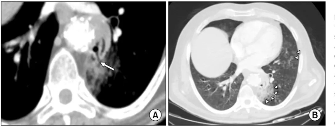
Aortopulmonary Fistula Presenting without an Endoleak after Thoracic Endovascular Aortic Repair
Giacomo Sica, M.D., Ph.D. 1 , Gaetano Rea, M.D. 1 , Giorgio Bocchini, M.D. 1 , Romilda Lombardi, M.D. 2 , Massimo Muto, M.D. 1 , Tullio Valente, M.D. 1
1
Division of Radiology, Department of Diagnostic Imaging, Monaldi Hospital,
2