Stroke Caused by Cerebral Air Embolism after Central Venous Catheter Removal:
A Case Report
중심정맥관 제거 후 대뇌공기색전증에 의해 발생한 뇌졸중: 증례 보고
Ki Eon Kwon, MD , Noh Hyuck Park, MD* , Seon-Jeong Kim, MD, Ji Yeon Park, MD
Department of Radiology, Myongji Hospital, Hanyang University College of Medicine, Goyang, Korea
Cerebral air embolism is a rare, potentially catastrophic iatrogenic complication of central ve- nous catheter removal. Cerebral air embolism can lead to serious neurological sequelae, result- ing from cerebral infarction. Early radiological diagnosis of cerebral air embolism is critical for emergent hyperbaric oxygen treatment. In this study, we report the case of a 68-year-old man who developed cerebral air embolism after the removal of a central venous catheter that was im- mediately diagnosed using brain CT and brain diffusion-weighted imaging.
Index terms Air Embolism; Central Venous Catheter; Cerebral Infarction;
Multidetector Computed Tomography; Magnetic Resonance Imaging
INTRODUCTION
A central venous catheter (CVC) is a useful and frequently used device for critically ill patients. CVCs are inserted for hemodynamic monitoring, hemodialysis, administra- tion of medication, parenteral nutrition, and poor peripheral venous access (1). Various complications associated with the use of a CVC can occur during its insertion, reten- tion, or removal (1). One of the most lethal complications is cerebral air embolism. We report a case of cerebral air embolism that occurred soon after CVC removal.
CASE REPORT
A 68-year-old man presented with sudden abdominal distension and pain. The pa-
Received May 29, 2018 Revised August 24, 2018 Accepted November 25, 2018
*Corresponding author Noh Hyuck Park, MD Department of Radiology, Myongji Hospital, Hanyang University College of Medicine, 55 Hwasu-ro 14beon-gil, Deogyang-gu, Goyang 10475, Korea.
Tel 82-31-810-7262 Fax 82-31-810-6537 E-mail [email protected] This is an Open Access article distributed under the terms of the Creative Commons Attribu- tion Non-Commercial License (https://creativecommons.org/
licenses/by-nc/4.0) which permits unrestricted non-commercial use, distribution, and reproduc- tion in any medium, provided the original work is properly cited.
ORCID iDs Noh Hyuck Park https://
orcid.org/0000-0003-4716-3491 Ki Eon Kwon
https://
orcid.org/0000-0001-5679-5381
tient was initially evaluated at another hospital with abdominal and pelvic CT. He was trans- ferred to our hospital through the emergency department. After reexamination of the previ- ous CT, a diagnosis of sigmoid colon cancer with mechanical colonic obstruction was made.
Emergent endoscopic colonic stent insertion was performed by the gastroenterologist. After colonic stent insertion, he was transferred to the intensive care unit (ICU) for close monitor- ing before elective surgical treatment. A CVC was inserted in the right internal jugular vein prior to the endoscopic colonic stent insertion. A week later, removal of the right internal jug- ular venous catheter was planned. His head was placed flat on the bed in the supine position.
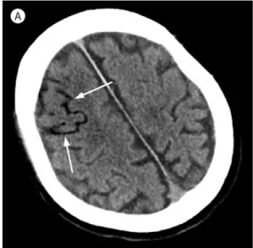
He was instructed to hold his breath when the internal jugular vein catheter was pulled out.
The exit site was compressed with gauze long enough for the bleeding to stop. An occlusive dressing was applied afterward. Soon after CVC removal, he had sudden onset of shortness of breath, loss of consciousness, and seizure-like movements. His blood pressure was 157/91 mm Hg and pulse rate was 100 beats per minute, with normal sinus rhythm. After oxygen administration at 10 L/min via mask, saturation was maintained at 98%. Emergent non-con- trast-enhanced brain CT (64-slice multidetector CT; LightSpeed VCT, GE Healthcare, Milwau- kee, WI, USA) revealed multifocal air densities (about -120 Hounsfield unit) along the right frontal sulcus (Fig. 1A). Subsequent diffusion-weighted image (DWI) of the brain (Signa HDxt 3T, GE Healthcare) revealed acute cerebral infarction in the right frontal lobe (Fig. 1B, C). He was diagnosed as having a cerebral air embolism and transferred to the ICU. Hyperbaric therapy was needed but was not available in our hospital. Thus, high-flow oxygen therapy was continued with closed monitoring. After a day, he remained drowsy and in stupor. He had intermittent myoclonic seizure and weakness of the left extremities. The day after the initial study, follow-up brain DWI was performed to evaluate the progression of infarction caused by cerebral air embolism and revealed increased extent of acute cerebral infarction in the right frontal lobe. In addition, newly developed acute cerebral infarction was observed in the left postcentral gyrus (Fig. 1D, E). Brain susceptibility-weighted imaging (SWI), found no intracerebral focal lesion that correlated with the cerebral air embolism. Transthoracic echocardiography revealed no visible intracardiac shunt. About 4 weeks later, he gradually regained consciousness and no longer had seizure episodes. He underwent low anterior re-
Fig. 1. A 68-year-old man who developed cerebral air embolism after the removal of a central venous catheter.
A. Initial non-contrast-enhanced brain CT shows mul- tifocal curvilinear areas of very low attenuation (ar- rows; about -120 HU) along the right frontal sulcus, suggesting air emboli.
A
section of the sigmoid colon cancer and showed much improvement in his general condition.
After 4 weeks of rehabilitation, his mental status was improved except for a mild confusion.
Moreover, the weakness of his left extremities was much improved after rehabilitation therapy.
DISCUSSION
Complications related to the use of CVCs include hemorrhage, pneumothorax, embolism, and infection (2). Among these complications, cerebral air embolism is uncommon but the Fig. 1. A 68-year-old man who developed cerebral air embolism after the removal of a central venous catheter.
B, C. Initial brain DWI with a B value of 1000 (B) and an ADC map (C) depicting an acute cerebral infarction in the right frontal lobe.
D, E. Short-term follow-up (a day after initial DWI) brain DWI with a B value of 1000 (D) and an ADC map (E) depicting increased extent of acute cerebral infarction in the right frontal lobe. In addition, newly developed acute cerebral infarction can be observed in the left postcentral gyrus.
ADC = apparent diffusion coefficient, DWI = diffusion-weighted image B
D
C
E
most lethal. Cerebral air embolism occurs as a consequence of air emboli obstructing the small cerebral arteries (3). Cerebral air embolism occur during CVC removal whenair emboli enter the arterial circulation. Two mechanisms lead to cerebral air embolism after CVC re- moval. One is the result of air emboli passing through an intracardiac right-to-left shunt, which is often called paradoxical cerebral air embolism (3). This intracardiac right-to-left shunt includes a patent foramen ovale (PFO) and congenital atrial septal defect. The inci- dence rate of PFO is reported to be approximately 35% (2). In our case, an intracardiac shunt was immediately suspected. However, the transthoracic echocardiogram was normal in our case. Another is a retrograde mechanism in which air emboli reflux through the internal jug- ular vein, leadings to venous cerebral air embolism. This can be induced when the patient is in a sitting position (2). Unlike the retrograde mechanism, paradoxical cerebral air embolism is an arterial embolism in which venous air crosses into the arterial system through cardiac or pulmonary shunts. Once the venous air has crossed into the arterial system, brain damage occurs when the air bubble move to a cerebral artery and causes acute cerebral infarction (4).
Except in intracardiac right-to-left shunts, transpulmonary passage has been suggested as the mechanism underlying paradoxical cerebral air embolism. Transpulmonary air passage occurs through pulmonary arteriovenous malformations and inducible intrapulmonary ar- teriovenous anastomoses (3). A case of cerebral air embolism caused by a change in air pres- sure from air travel, which is a particularly rare cause of stroke, has been reported (5). Stroke caused by cerebral air embolism can have severe neurological sequelae. Thus, early diagno- sis with immediate treatment is essential for better outcome. Brain CT is the most valuable diagnostic modality to show the air emboli in the brain. However, brain CT may not reveal air emboli in the cerebral cortex. Therefore, brain DWI should be conducted to evaluate the cerebral infarction. Unlike acute infarction caused by cardiac thrombi, cerebral air embo- lism usually involves the frontal cortical area with sparing of the deep subcortical area. This characteristic location of air emboli in the frontal cortical area is associated with the diame- ter and surface tension of air bubbles. The surface tension of air bubbles is inversely corre- lated with their diameter. Therefore, small air bubbles are more likely than larger air bubbles to obstruct the small-sized cerebral arteries in the cortical areas without rupture (6). In our case, acute cerebral infarction was confined to the right frontal cortical area on initial brain DWI and was correlated with air density noted in the brain CT. Air density in the brain CT is the most important clue to diagnosis of cerebral air embolism. However, these air bubbles rapidly dissolve in the blood. Therefore, air density in the brain can only be seen in early stages of cerebral air embolism. Gradient echo (GRE) MRI and SWI are sensitive for the de- tection of air bubbles in the brain which have dark signal intensity. In comparison with brain CT, dark signal intensity lesions on GRE or SWI may be diagnosed as air bubbles rather than hemorrhage (5). However, the SWI in our case was taken the day after the initial event and did not reveal the dark signal intensity lesion. Air bubbles may have already dissolved in the blood when the SWI was taken. During the insertion, use, or removal of a CVC, any neuro- logical symptoms should be investigated to detect a possible cerebral air embolism. In addi- tion, immediate imaging studies, including brain CT and DWI, are essential, as delayed im- aging may not show the presence of air in the cerebral vasculature. If cerebral air embolism is suspected initially, then the patient should be placed head down and lying on their left
side. This is called Durant’s maneuver (1). This maneuver keeps the air trapped within the heart away from the outflow of the right ventricle. In addition, hyperbaric oxygen therapy (HBOT) or 100% oxygen administration should be initiated as early as possible. HBOT initia- tion prior to 6 hours after an event shows better prognosis than those that treatment initiated later than 6 hours after an event. Early oxygen therapy reduces the size of air bubbles and in- creases the arterial oxygen tension; thus, air density in the cerebral cortex will disappear on brain CT (7). Therefore, residual air density in the cerebral cortex on follow up brain CT indi- cates a poor prognosis. However, the prevention of cerebral air embolism is most important to reduce the risks of morbidity and mortality. During CVC removal, a patient should be in supine position with the head down. Then, the patient is instructed to hold a breath at the end of inspiration. After CVC removal, gauze compression must be immediately performed to prevent outside air from entering the intravenous tract. Moreover, the patient should be in the supine position for at least half an hour after CVC removal. Behaviors that change the pressure in the intrathoracic cavity, such as coughing and shifting to an upright position must be avoided (2).
Among the many complications associated with CVC removal, stroke caused by cerebral air embolism is the most lethal and thus requires immediate treatment. Therefore, preven- tion of cerebral air embolism during CVC removal is most important. In addition, early diag- nosis may decrease the risk of death, and the radiologist has a critical role in diagnosis.
Conflicts of Interest
The authors have no potential conflicts of interest to disclose.
REFERENCES
1. Brockmeyer J, Simon T, Seery J, Johnson E, Armstrong P. Cerebral air embolism following removal of cen- tral venous catheter. Mil Med 2009;174:878-881
2. Zong Y. Cerebral air embolism after central venous catheter removal: a case report and literature review. J Anesth Crit Care Open Access 2014;1:00006
3. Eum DH, Lee SH, Kim HW, Jung MJ, Lee JG. Cerebral air embolism following the removal of a central venous catheter in the absence of intracardiac right-to-left shunting: a case report. Medicine (Baltimore) 2015;
94:e630
4. Malhotra K, Rayi A. Gyriform infarction in cerebral air embolism: imaging mimicker of status epilepticus. Ann Indian Acad Neurol 2017;20:313-315
5. Jung HS, Jeong HW, In HS. Cerebral air embolism in a patient with a tuberculous-destroyed lung during commercial air travel: a case report. J Korean Soc Radiol 2011;65:109-112
6. Jeon SB, Kim JS, Lee DK, Kang DW, Kwon SU. Clinicoradiological characteristics of cerebral air embolism.
Cerebrovasc Dis 2007;23:459-462
7. Blanc P, Boussuges A, Henriette K, Sainty JM, Deleflie M. Iatrogenic cerebral air embolism: importance of an early hyperbaric oxygenation. Intensive Care Med 2002;28:559-563
중심정맥관 제거 후 대뇌공기색전증에 의해 발생한 뇌졸중: 증례 보고
권기언 · 박노혁* · 김선정 · 박지연
대뇌공기색전증은 중심정맥관 제거 후 발생할 수 있는 의인성 합병증으로 드물게 발생하지 만 심각한 결과를 초래할 수 있다. 대뇌공기색전증은 대뇌경색으로 진행되어 심각한 신경학 적 후유증을 남길 수 있다. 영상의학적으로 대뇌공기색전증을 조기에 진단하는 것이 즉각적 인 고압산소치료를 위해 매우 중요하다. 본 증례 보고에서는 68세 남자 환자의 중심정맥관 제거 후 발생한 대뇌공기색전증의 뇌 전산화단층촬영과 뇌 확산강조영상을 통한 즉각적인 진단에 대해 보고하고자 한다.
한양대학교 의과대학 명지병원 영상의학과