거머리, Limnotrachelobdella sinensis의 기생으로 인한 붕어, Carassius auratus 아가미의 조직병리학적 관찰
박명애ㆍ김석렬ㆍ김명석ㆍ김정호*ㆍ박정준†1) 국립수산과학원 병리연구과, *강릉원주대학교 해양자원육성전공
Histopathological observation of the gill of the crucian carp, Carassius auratus by the leech, Limnotrachelobdella sinensis
Myoung Ae Park, Seok-Ryel Kim, Myoung-Sug Kim, Jeong-Ho Kim* and Jung Jun Park† Pathology Division, National Fisheries Research and Development Institute, Busan 619-902, Korea
*Faculty of Marine Bioscience and Technology Gangneung-Wonju Nat. Univ. Gangneung, Gangwon-do 210-702, Korea
On the inner side of each operculum of the crucian carp, Carassius auratus (n=10), the leech, Limnotrachelobdella sinensis of 1-4 individuals were parasitic. The leeches had approximately 41.0 mm in total length and 11 mm in width. These body was composed with anterior sucker, neck, trunk and posterior sucker and average length was 2.3 mm, 7.2 mm, 23.3 mm and 8.7 mm respectively. To both sides of the trunk lateral vesicle of 11 pair existed. When observed by SEM, anterior sucker was hemisphere shape and the mouth where proboscis comes out existed with the its center. Proboscis was connected the esophagus directly. Under light microscopy, bloodsucking gill of C. auratus showed lamella fusion, hypertrophy the epithelial cell of the filament and lamella, increased mucocytes and congested capillaries. On the other hand, necrotic and hydropic degeneration epithelial cell of the lamella, and infiltration of the macrophages from some individuals were suggested the secondary infection with the bacteria or virus after bloodsucking activity of the leech.
Key words :Limnotrachelobdella sinensis, Carassius auratus, Gill
2010년 3-4월 충청북도 청주시와 청원군 소재의 미호천과 병천천에 서식하는 자연산 붕어, Carassius auratus가 대량 폐사하였고, 살아있는 개체들도 상당 수가 유영력이 현저하게 떨어져 있었다. 대부분의 붕어들은 체표 및 지느러미에는 외견상 병변이 보이 지 않았으나, 모든 개체가 아가미뚜껑이 벌어져 있었 으며, 아가미뚜껑 내부에 길이 3-5 cm 가량의 거머리
†Corresponding Author : Jung Jun Park, Tel: 051-720-2483, Fax: 051-720-2498, E-mail. [email protected]
들이 기생하고 있는 상태로 발견되었다.
거머리는 환형동물문 (Phylum Annelida), 거머리 아강 (Subclass Hirudinae)에 속하며, 전세계적으로 약 230여 종이 보고되었다. 이들은 자웅동체로서 담수 또는 해수에 존재하면서 대부분 척추동물에 기생하 여 흡혈을 하며 살아간다 (Burreson and Allen 1978;
Möller and Anders, 1986; Janssen 1993).
어류 기생성 거머리는 담수어 및 해수어에서 여러 종이 보고되어 있고, 국내에서는 떡붕어 (Carassius
cuvieri)의 아가미에 기생하는 거머리를 Trachelobdella sp.로 분류하여 보고한 사례가 있다 (Park and Kim, 2002;
Rhee, 1989). 하지만, Nagasawa 등 (2009)은 국내 및 일본 에서 보고된 Trachelobdella sp.를 Limnotrachelobdella sinensis로 재명명하여 보고하였다. 따라서 본 연구의 거머리는 Nagasawa 등 (2009)의 보고에서 설명된 거 머리와 형태 및 크기 뿐 만아니라 숙주까지 동일하였 기 때문에 Limnotrachelobdella sinensis로 분류하였다.
기존 거머리의 분류 및 형태에 관한 연구들은 국내ㆍ외 에서 다수 보고되었지만 (Möller and Anders, 1986;
Fredric et al., 1998; Govedich and Bain, 2005), 거머리 에 의한 숙주의 조직병리학적인 연구는 미흡한 실정 이다 (Park and Kim, 2002; Nehili et al., 1994; Rhee, 1989).
이에 본 연구에서는 자연산 붕어, Carassius auratus의 아가미에 기생한 Limnotrachelobdella sinensis를 조직학적으로 분석하여, 거머리의 흡혈활 동에 의한 숙주의 조직병리학적인 증상들을 보고하 고자 한다.
재료 및 방법
시료 채집
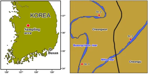
2010년 4월 충청북도 청원군 병천천의 지역 두 곳 (St. 1; 36°38′27″N, 127°20′52″, St. 2; 36°40′16″N, 127°21′22″E) 그리고 청주시 미호천 (St. 3; 36°39′31″
N, 127°23′02″E)에서 채집하였다 (Fig. 1).
Fig. 1. Sampling area.
각각의 지역에서 채집된 붕어, Carassius auratus는 평균 전장 36.8 cm이고, 투망을 이용하여 10개체를 채집하 였다. 붕어에 기생하고 있는 거머리, Limnotrachelobdella sinensis는 총 41개체를 채집하였다. 채집되어진 붕어 의 아가미와 아가미뚜껑 그리고 거머리는 70% 에탄 올, 10% 중성포르말린 그리고 1.0% glutaraldehyde로
현장에서 즉시 고정하였다.
조직학적 분석
실험실로 옮겨온 시료들은 모두 24시간 이내에 고정액을 교환하였다. 광학현미경으로 관찰하기 위 한 시료들은 관찰 부위를 1 cm2의 크기로 다듬고,
동일한 고정액으로 8시간 동안 재고정 하였다. 이후 시료들은 24시간 동안 흐르는 물에 수세한 후 일반적 인 파라핀 포매법을 이용하여 포매하였다. 포매된 시료들은 3 ㎛의 두께의 절편을 제작하여 Harris’s hematoxylin-0.5% eosin (H-E)와 Masson’s trichrome 염색을 시행하였다. 주사전자현미경 (SEM)으로 관 찰하기 위한 시료들은 0.5 cm의 크기로 다듬고, 1.0%
glutaraldehyde로 재고정하였다. 이후 0.1M phosphate buffer로 수세한 후 1% osmium tetroxide (OsO4)로 후 고정 하였다. 이후 에탄올을 이용하여 단계적으로 탈수 시키고, amyl acetate로 치환하였다. 이들 시료들 은 CO2 가스를 이용하여 건조 및 금이온 증착을 통하 여 SEM (JSM-7500F, Hitachi, Japan)으로 관찰하였다.
Masson’s trichrome 염색으로 나타나는 세포 및 구조 의 색을 정확히 표기하기 위해 Pantone® solid coated 의 고유번호를 ( )안에 표시하였다.
결과
거머리, Limnotrachelobdella sinensis에 감염된 붕 어, Carassius auratus는 유영이 느리고 체색의 변화 및 체표손상을 보이지 않았지만 아가미뚜껑이 양쪽 모두 벌어져 있었으며 (Fig. 2A), 아가미뚜껑의 표면 에는 은색의 구형 반점이 뚜렷하게 관찰되었다 (Fig.
2B). 이들 구형의 반점들은 거머리가 아가미뚜껑의 안쪽에 붙어있는 부분으로서 반점들이 보이는 병어 의 아가미뚜껑을 열어보면 안쪽에 반점의 수와 동일 한 개체의 거머리가 존재하고 있었다. 아가미뚜껑 한쪽에 3개체 이상의 거머리가 기생하고 있는 붕어 의 경우 아가미가 흰색으로 변하여 빈혈 증세를 보이 고 있었으며, 새엽의 말단부위는 괴사로 인하여 검은 색을 띠고 있었다 (Fig. 2C).
거머리들의 외형은 넓은 타원형이었으며, 전제적
인 색은 옅은 적갈색이었으나, 몸체의 측면에 존재하 는 측면 돌기 (lateral vesicle)들은 몸체보다 더 진한 적갈색을 띠고 있었다. 이들의 평균 길이는 41 mm (27-49 mm)였고, 폭은 11 mm (5.4-13.4 mm)로서 (Fig.
2D), 몸체는 전흡반 (anterio sucker), 목 (neck), 몸통 (trunk), 후흡반 (posterior sucker)으로 나뉘어졌다. 각 부분의 평균 길이는 각각 2.3 mm, 7.2 mm, 23.3 mm, 8.7 mm로 측정되었다. 몸통의 양옆에는 11쌍의 측면 돌기가 존재하였는데, 측면 돌기의 수는 거머리의 크기와 상관없이 모든 개체들이 동일하였다 (Fig.
3A). 전흡반과 후흡반 모두 배쪽을 향하고 있는 반구 모양이었다. 하지만 후흡반은 비교적 단단하였고, 전 흡반의 등쪽에는 2쌍의 안점 (eye)이 존재하고 있었 다 (Fig. 3B).
전흡반을 주사전자현미경 (SEM)으로 관찰한 결 과 전흡반의 표면은 다수의 주름으로 덮여 있으며, 배쪽으로 파여있는 반구모양을 하고 있었다 (Fig.
4A). 배쪽 전흡반의 중앙에는 지름 60 ㎛ 정도의 입이 관찰되었다. 이들 흡반의 표면에는 다수의 분비공이 존재하고 있었는데 분비공의 지름은 0.5-3.3 ㎛으로 다양하였다 (Fig. 4B).
광학현미경으로 관찰하기 위해 거머리를 longitudinal section 하였을 때 전흡반의 두께는 약 350 ㎛ 정도였 으며, 몸통의 앞부분에는 식도가 존재하고 있었다.
식도의 주변에는 Masson’s 삼중염색을 시행하였을 때 파란색 (2728c)과 보라색 (2735c, 271c)으로 염색되 는 타액선 (salivary gland)이 다수 존재하고 있었다 (Fig. 5A). 전흡반의 입에서 나오는 proboscis는 기저 부에 투명하게 보이는 턱으로 둘러싸여 있으며, 이들 의 지름은 약 250 ㎛으로써 중앙에는 숙주의 혈액을 흡입하는 구강 (buccsal cavity)이 존재하였다. Proboscis 는 지름 320 ㎛ 정도의 인두와 이어져 있었다(Fig. 5B).
거머리가 부착되어 있는 아가미뚜껑의 안쪽 부분
에는 후흡반 주위로 출혈이 나타나고, 거머리는 아가 미뚜껑의 내부에 단단히 고착되어 있었다 (Fig. 6A).
거머리가 부착되어있는 아가미뚜껑을 조직학적으 로 관찰하면 후흡반은 아가미뚜껑 내부의 상피층을 밀어내고 연골층에 부착되어 있으며, 후흡반에 의해 서 밀려난 아가미뚜껑의 내부상피층은 상피세포의 수종변성 (hydrophic degeneration)과 함께 울혈이 관 찰되었다 (Fig. 6B). 거머리가 1-2개체 정도 기생하고 있는 붕어 아가미는 새판의 상피세포들이 수종변성 및 비대되었다. 또한 점액세포 및 염세포의 감소가 동반되었다 (Fig. 6C). 일부 새판에서는 모세혈관의 팽창과 함께 새판 상피세포가 괴사되어 소실되는 조
직상이 관찰되었다. 또한 모세혈관 사이에 존재하는 pillar cell의 핵과 세포질이 응축되는 증상이 관찰되 었다 (Fig. 6D). 유영력이 현저하게 저하되고 수면위 에 떠있는 폐사 직전의 개체들은 새판 융합이 관찰되 었으며, 새엽의 중앙에 존재하는 연골조직이 파괴되 었다 (Fig. 7A). 모든 새판이 융합된 아가미 조직에서 는 새판상피세포의 융합과 함께 괴사된 상피세포도 관찰되었으며 (Fig. 7B), 파괴된 연골조직 내에는 다 량의 혈구가 관찰되었다 (Fig. 7C). 새판과 새엽의 융합이 심각하게 나타나는 아가미들은 각새골과 인 접한 부분에 괴사된 조직이 관찰되었다 (Fig. 7D).
Fig. 2. The Crucian carp, Carassius auratus infested with leech, Limnotrachelobdella sinensis. A: Dorsal view of the crucian carp, note the lifting operculum. B: Operculum, showing the round spot on the operculum. C: The infested gill, note the marked pallor of the gills. D: Leeches, showing the 3 leeches on the inner operculum.
Fig. 3. The morphology of the leech, Limnotrachelobdella sinensis. A: Dorsal and ventral view, note that divided into the anterior sucker (As), neck (N), trunk (T) and posterior sucker (Ps), and showing the 11 pair of lateral vesicles (Lv). B: Dorsal view of the anterior sucker, showing the 2 pair of eyes (Ey) of rectangle in A.
Fig. 4. Scanning electron micrograpy of the leech, Limnotrachelobdella sinensis. A: Anterior sucker (As).
B: Mouth (Mo).
Fig. 5. Light microscopy of the leech, Limnotrachelobdella sinensis. Masson’s trichrome stain. A: Anterior sucker (As) and trunk, showing the esophagus (Ep) and salivary gland (Sg). B: Proboscis (Pb), note the connected with the pharynx (Pr). Bc, buccal cavity; J, jaw.
Fig. 6. Pathological feature of the crucian carp, Carassius auratus. H-E stain. A:
The Crucian carp, infested with leech. B: Operculum, lifting of the epithelial layer (El) of the operculum by the posterior sucker (Ps) of rectangle in A-a. C and D: Lamella of rectagle in A-b. C: Hydrogenic degeneration (black arrowhead) and hypertrophic (white arrowhead) epithelial cell. D:
Hypertrophic epithelial cell of the lamella (black arrowhead) and dilated capillary (asterisk).
Fig. 7. Histopathological feature of the crucian carp, Carassius auratus. H-E stain.
A: Lamella (Gl), showing the partially lamella fusion. B: Lamella, showing the necrotic epithelial cell. C: Cartilage matrix, showing the congestion.
D: Lamella fusion. Gf, filament.
고 찰
Limnotrachelobdella sinensis는 2010년 4월에 채집 되었고 3월부터 하천의 붕어가 폐사하기 시작하였지 만, 중국과 일본에서는 주로 겨울과 봄에 L. sinensis의 피해사례가 보고된바 있다 (Yang, 1987; Ogawa et al., 2007). L. sinensis의 생활사는 아직 정확하게 알려 진바 없지만, Ogawa 등 (2007)은 이들이 1년생으로서 12월부터 이듬해 4월까지 어류에 기생하고, 그 이후 의 기간에는 감염성이 없는 상태로 존재한다고 설명 하였다.
일반적으로 거머리는 3급수의 지표생물로 지정되 어 있고, 몸은 32개의 체절로 이루어졌으며, 공통적으 로는 넓이에 비해 길이가 긴 모양이지만 몸체의 형태 에 따라서 5가지 (tapered, elongate, regionated, with
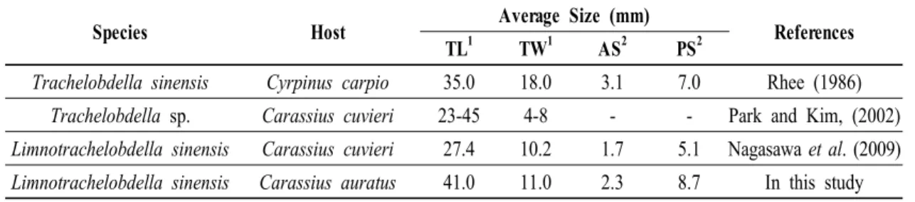
fin, vase-shaped)로 나눌 수 있다. Tapered와 elongate의 형태는 비슷하지만 tapered의 몸통부분이 좀더 넓고, 횡단면으로 관찰하면 tapered 형태는 반달형이지만 elongate 형태는 원형이다. 또한 거머리 몸통의 모양 및 넓이의 크기는 숙주를 통한 흡혈 양에 따라 변할 수 있다 (Stachowitsch, 1992). 따라서 본 연구에서 사용 된 L. sinensis는 tapered 형태에 속하였다. L. sinensis의 체색은 옅은 적갈색이었는데 이는 흡혈활동으로 인 하여 내장에 다량의 혈액이 존재하기 때문이다 (Nagasawa et al., 2009). 이들의 몸체는 전흡반, 목, 몸통, 후흡반으로 구분되었지만 몇몇 종은 전흡반이 존재하지 않고 입이 외부로 노출되며, proboscis 또한 모든 종이 가지고 있는 것은 아니다 (Stachowitsch, 1992). 표 1은 기존의 연구들에서 보고된 L. sinensis와 유사한 종의 각 부위별 크기를 나타냈다.
Species Host Average Size (mm)
References TL1 TW1 AS2 PS2
Trachelobdella sinensis Cyrpinus carpio 35.0 18.0 3.1 7.0 Rhee (1986) Trachelobdella sp. Carassius cuvieri 23-45 4-8 - - Park and Kim, (2002) Limnotrachelobdella sinensis Carassius cuvieri 27.4 10.2 1.7 5.1 Nagasawa et al. (2009) Limnotrachelobdella sinensis Carassius auratus 41.0 11.0 2.3 8.7 In this study
*AS, anterior sucker; PS, posterior sucker; TL, total length; TW, total width
*1, straight length; 2, diameter
Table 1. Average size of the leeches from the previously studies.
본 실험의 붕어 아가미를 광학현미경으로 관찰하 였을 때 유영력이 현저하게 떨어진 개체들은 새판 상피세포의 수종변성과 비대 그리고 모세혈관의 확 장이 관찰되었고, 수면위에 떠 있는 폐사 직전의 개체 들은 새판의 융합 및 상피세포의 괴사가 관찰되었다. 어류의 아가미는 수질 환경 뿐만 아니라 다양한 병원 체에 의해서 아주 민감하게 반응하는 중요한 기관으
로서 (Bhagwant and Elahee, 2002), 아가미 새판 상피 세포의 비대와 세포 표면의 작은 돌기형성 (microprojection)은 물리ㆍ화학적인 자극에 의해서 일차적으로 나타나는 증상이다. 특히 상피세포의 비 대는 생리학적인 기능의 증가, 즉 과기능 (hyperfunction) 의 결과로 인하여 세포의 크기 및 기능이 증대되는 현상이다 (Meyers and Hendricks, 1985; Takashima and
Hihiya, 1995). 이러한 세포의 비대는 새판 사이의 공간이 좁아짐에 따라 아가미로 들어온 물의 흐름을 방해할 수 있다 (Bhagwant and Elahee, 2002). 아가미 새판 상피세포의 증식과 융합은 화학적 요인 및 세균 과 기생충의 만성적인 감염에 의해서 일반적으로 발 생하는 병리학적 증상이며, 새엽 상피세포의 증가로 인한 새엽의 융합은 자유로운 가스교환의 장애를 유 발시키는 원인이 된다 (Gardner and Yevich 1970;
Skidmore and Tovell 1972). 또한 아가미 새판 상피세 포의 괴사 및 상피층의 붕괴는 물리ㆍ화학적인 자극 이 동시에 숙주 조직에 가해질 때 쉽게 나타난다 (Takashima and Hihiya, 1995).
L. sinensis가 숙주 아가미뚜껑 내부에 기생하면서 흡혈활동을 하게 되면 거머리의 두께 만큼 아가미뚜 껑이 열려지게 되며, 이로 인하여 아가미는 수중에 계속 노출되게 된다. 아가미 상피세포의 손상으로 인하여 부족한 산소를 보충하기 위해 붕어들은 수면 에 머무르게 되는데 이때 열려 있는 아가미뚜껑 때문 에 아가미와 공기가 직접적으로 맞닿으면 아가미 조 직의 손상은 더욱 심화되는 것으로 추정된다. 하지만 붕어 아가미 손상의 1차적인 원인은 L. sinensis의 흡 혈활동에 의한 물리적인 자극이며, 손상된 아가미 조직에 세균 또는 바이러스 같은 병원체가 2차적으 로 감염되어 가스교환 장애가 나타나고, 그로 인하여 산소공급이 수행되지 않아 빈혈에 의해서 폐사에 이 르는 것으로 생각되어진다.
요 약
10 개체의 붕어, Carassius auratus 아가미뚜껑의 한쪽면에 1-4개체의 거머리, Limnotrachelobdella sinensis들이 기생하고 있었다. 거머리들의 평균 길이 는 약 41.0 mm 정도였고, 폭은 11 mm였다. 이들의
몸체는 anterior sucker, neck, trunk, posterior sucker로 구성되어 있었고 각각의 평균 길이는 2.3 mm, 7.2 mm, 23.3 mm, 8.7 mm였다. 몸통의 양 옆에는 11쌍의 lateral vesicle이 존재하였다. Anterior sucker를 SEM으 로 관찰한 결과 이들은 반구모양으로 중앙에는 proboscis가 나오는 입이 존재하고 있었다. Proboscis 는 식도와 바로 연결되어 있었다. 거머리에 의해서 흡혈되어진 붕어의 아가미를 광학현미경으로 관찰 하였을 때 새판융합, 새엽 및 새판 상피세포 과증식, 점액세포 증가 및 울혈이 관찰되었다. 하지만 몇몇 개체들에서 나타나는 새엽 상피세포의 괴사 및 수종 변성과 식세포의 침투 등은 거머리의 흡혈 활동 이후 세균 혹은 바이러스에 의한 2차 감염이 원인인 것으 로 생각되어진다.
감사의 글
본 연구는 국립수산과학원(양식생물백신연구, RP-2010-AQ-098)의 지원에 의해 운영되었습니다.
참고문헌
Bhagwant, S. and Elahee, K.B.: Pathologic gill lesions in two edible laggon fish species,l Mulloidichthys flavolineatus and Mugil cephalus, from the bay of Poudre d’Or, Mauritius. Western Indian Ocean J. Mar. Sci., 1:35-42, 2002.
Burreson, E.M. and Allen, M.D.: Morphology and biology of Mysidobdella borealis (Johansson) comb. n.
(Hirudinea: Piscicolidae), from mysids in the western North Atlantic. J. Parasitol., 64:1082-1091, 1978.
Fredric R. Govedich, D.W. Blinn, P.K. and Ronald, W.D.:
Phylogenetic relationships of three genera of Erpobdellidae (Hirudinoidea), with a description of a new genus, Motobdella, and species, Motobdella sedonensis. Can. J. Zool., 76(12):2164-2171, 1998.
Gardner, G.R. and Yevich, P.P.: Histological and hematological responses of an estuarine teleost to cadmium. J. Fish. Res. Brd. Can., 27:2185-2196, 1970.
Govedich, F.R. and Bain, B.A.: All about leeches.
http://www.invertebrate.ws, 2005.
Janssen, H.H.: Morphology, egg cocoons, and transmission paths of the Antarctic leech Glyptonotobdella antarctica Sawyer and White, 1969 (Hirudinea:
Rhynchobdelliformes: Piscicolidae). Polar Biol., 13:347-354, 1993.
Meyers, T.R. and Hendricks, J.D.: Histopathology. In Fundamentals of aquatic toxicology; Methods and applications, pp. 283-331, Rand, G.M. and Petrocelli, S.R., Hemisphere Publishing Corporation, New York, 1985.
Möller, H. and Anders, K.: Disease and parasites of marine fishes, pp. 179-183, Verlag Möller, Kiel, 1986.
Nagasawa, K., Park, S.-W., Kim, Y.-G. and Kim, H.J.:
Limnotrachelobdella sinensis, a leech associated with mortality in a wild population of Japaneses curcian carp Carassius cuvieri in Korea. J. Grad.
Sch. Biosp. Sci., 48:49-53, 2009.
Nehili, M., Ilk, C., Mehlhorn, H., Ruhnau, K., Dick, W.
and Mjayou, M.: Experiments on the possible role of leeches as vectors of animal and human pathogens: a light and electron microscopy study.
Parasitol. Res., 80:277-290, 1994.
Ogawa, K., Rusinek, O. and Tanaka, M.: New record of the leech Limnotrachelobdella sinensis infecting freshwater fish from Japanese water. Fish Pathol., 42:85-89, 2007.
Park, S.-W. and Kim, Y.-G.: The case report on the leech, Trachelobdella sp. infestation in wild crucian carp (Carassius cuvieri) of Chungnam province in Korea. J. Fish Pathol. 15:117-119, 2002.
Rhee, J.K.: Trachelobdella sinensis Blanchard, 1986 found from Cyprinus carpio nudus in Korea. Korean J. Parasitol., 24:216-217, 1986.
Skidmore, J.F. and Tovell, P.W.A.: Toxic effects of zinc sulphate on the gills of rainbow trout. W. Res.
6:271-230, 1972.
Stachowitsch, M.: The invertebrates; An illustrated glossary, pp. 273-279, Wiley-Liss, New York, 1992.
Takashima, F. and Hibiya, T.: V. Gills. In An atlas of fish histology: normal and pathological features, pp. 66-71, Kodaxnsha Ltd., Tokyo, 1995.
Yang, T.: On the genus Limnotrachelobdella Epshtein, 1968 and a new species from south China sea.
Acta Hydrobiol. Sin., 11:268-273, 1987.
Manuscript Received : November 18, 2010 Revised : November 21, 2010 Accepted : November 22, 2010