I. INTRODUCTION
In the field of radiology, imaging modalities have an important role in the detection of emphysema, which is generally expressed as morphological changes of the lung parenchyma[1].
Emphysema was first evaluated by computed
tomography (CT) in 1980s[2]. The use of high-resolution CT (HRCT) for emphysema evaluation has increased consistently over the past 2 decades[3-6]. HRCT is a powerful diagnostic and prognostic tool for the lung diseases such as emphysema, and is the gold standard for their evaluation[7]. The main reason for HRCT use in emphysema is that HRCT has demonstrated improved visualization of changes in lung parenchyma
Journal of Radiological Science and Technology, 39(3), 329-336 eISSN 2384-1168 ISSN 2288-3509 http://dx.doi.org/10.17946/JRST.2016.39.3.05
<원저>
Usefulness Evaluation of Low-dose CT for Emphysema : Compared with High-resolution CT
- 폐기종에 대한 저선량 CT의 유용성 평가 : 고해상도 CT와 비교 -
Dept. of Radiological Technology, Daejeon Health Institute of Technology, Daejeon, Korea Won-Jeong Lee
―Abstract ―
The purpose of this study was to evaluate the usefulness of low-dose CT (LDCT) for emphysema compared with high-resolution CT (HRCT).
Measurements of radiation dose and noise were repeated 3 times in same exposure condition which was similar with obtaining HRCT and LDCT images. We analysed reading results of 146 subjects. Six images per participants selected for emphysema grading. Emphysema was graded for all 6 zones on the left and right sides of the lungs by the consensus reading of two chest radiologists using a 4-point scale. Between the HRCT and LDCT images, diagnostic differences and agreements for emphysema were analyzed by McNemar’s and un- weighted kappa tests, and radiation doses and noise by a Mann–Whitney U-test, using the SPSS 19.0 program.
Radiation dose from HRCT was significantly higher than that of LDCT, but the noise was significantly lower in HRCT than in LDCT. Diagnostic agreement for emphysema between HRCT and LDCT images was excellent (k-value=0.88). Emphysema grading scores were not significantly different between HRCT and LDCT images for all six lung zones. Emphysema grading scores from LDCT images were significantly correlated with increased scores on HRCT images (r=0.599, p < 0.001).
Considering the tradeoff between radiation dose and image noise, LDCT could be used as the gold standard method instead of HRCT for emphysema detection and grading.
Key words : High-resolution CT; Low-dose CT; Emphysema; Radiation dose; Image noise
This paper was supported by Daejeon Health Institute of Technology in 2015.
Corresponding author: Won-Jeong Lee, Department of Radiological Technology, Daejeon Health Institute of Technology 21 Chungjeong-ro,
using thin-slice thicknesses[1] and high-radiation doses[8,9].
Radiation dose from CT is of increasing concern worldwide, because higher medical radiation doses from growing CT use increases cancer risk[10,11].
Low-dose CT (LDCT) was first introduced in the 1990s to reduce the radiation dose risk from chest CT[12], and its utility has been demonstrated in previous studies[13,14,15]. Other recent papers also support the possibility of LDCT applications for emphysema quantification instead of HRCT or standard-dose CT (SDCT)[16-18], although image noise can be increased when using a lower value for the tube current-scan time product (mAs, mA × sec); as a result, image noise is inversely proportional to radiation dose[19].
To the best of our knowledge, there have been rarely studies investigating radiation dose and noise in the use of LDCT for emphysema detection.
Therefore, The purpose of this study was to evaluate the usefulness of LDCT for emphysema compared with HRCT.
Ⅱ. MATERIALS AND METHODS
1. Measurements of Radiation Dose and Image Noise
Radiation dose (mGy) was measured at 5 points in an acryl phantom (10S5-3CT, Radical, USA) using a dose measurement instrument (Unfors Mult-O-Meter 9015, Radical, USA). Image noise (HU, Hounsfield units) was measured at 4 points (1062.1 mm2) in a water phantom (AAPM CT Performance Phantom, 76-410-4130, USA) and was defined as the standard deviation of the CT value in HU. Measurements of radiation dose and image noise were repeated 3times in the same environment, and the mean value was recorded for analysis. The exposure conditions for the radiation dose and noise measurements were 180 mAs for HRCT and 30 mAs for LDCT with a fixed tube voltage of 120 kVp and a slice thickness of 1.2 mm on
a 16-slice system (Brilliance TM CT 16-Slice; Philips, The Netherlands).
2. Emphysema Reading on HRCT and LDCT Images
We analyzed retrospectively reading results for emphysema of 146 participants and parameters on HRCT (1.2 mm, 120 kVp, 180 mAs) and LDCT (2.0 mm, 120 kVp, 30 mAs) images without any informations of participants.
Emphysema was graded the system published by Kusaka et al.[5,6] : HRCT and LDCT images scanned from the lung apex to the diaphragm, and 6 images per participant, equally spaced through the lungs, were selected for emphysema grading. Two images from each zone of the lungs (upper, the arch of the aorta and above; middle, from the arch of the aorta to the inferior pulmonary vein; and lower, the inferior pulmonary vein and below, including the diaphragm) were acquired. The readers were blinded to the technical parameters used in the CT images acquisition (e.g., kVp, mAs, slice thickness, and resolution).
Emphysema was documented as a presence or absence and graded for all 6 zones on the left and right sides of the lungs by the consensus reading of 2 chest radiologists using a 4-point scale: normal (score 0), absence; mild (score 1), up to 15% of the area from one zone; moderate (score 2), between 15 and 30%;
and severe (score 3) ≥ 30% of the area from one zone.
Emphysema was re-graded into 4 categories (normal, total score = 0; mild, total score = 1-6; moderate, total score = 7–12; severe, total score = 13–18) from a total score (ranging from 0 to 18) summed the grades of the six zones of the lungs using a 4-point scale.
LDCT images were graded 1 month after grading the HRCT images to avoid bias.
3. Statistical Analysis
Data are expressed as the mean and standard deviation for continuous variables and as percentage for categorical variables. The diagnostic differences and agreements for emphysema between the HRCT and
LDCT images were analyzed using McNemar’s and unweighted kappa tests. Agreements for the 4 categories of emphysema grading were calculated by a linearly weighted kappa value: A k-value of less than 0.20 indicated poor agreement; a k-value of 0.21–
0.40, fair agreement; a k-value of 0.41–0.60, moderate agreement; a k-value of 0.61–0.80, good agreement; and a k-value of 0.81–1.00, excellent agreement. The linear relationships for emphysema grading scores between HRCT and LDCT images were calculated using Pearson’s correlation coefficient. The radiation doses and image noise between the HRCT and LDCT images were compared using a Mann–
Whitney U-test. The total emphysema grading scores between the HRCT and LDCT images were compared using a paired t-test. A value of p < 0.05 was considered to indicate statistical significance. Analyses were performed using the SPSS statistical analysis program (Version 19.0, Chicago, IL, USA).
4. Ethics Statement
This study was waived by the institutional review board of the Daejeon Health Institute of Technology in 2015. Because this study was analyzed retrospectively reading results for emphysema and parameters on
HRCT and LDCT images without any informations of participants.
Ⅲ. RESULTS
1. Radiation Dose and Image Noise of HRCT and LDCT Images
The data for radiation dose and image noise measured by HRCT and LDCT are presented in Table 1.
The radiation dose from HRCT was significantly higher than that of LDCT (1.95 mGy vs. 0.35 mGy, p=0.008), but the image noise was significantly lower in HRCT than in LDCT (40.1 HU vs. 99.6 HU, p=0.021).
2. Diagnostic Differences for Emphysema between HRCT and LDCT Images
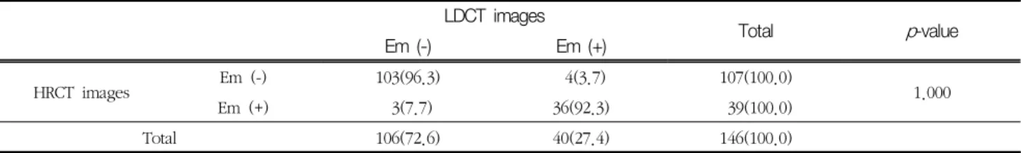
Emphysema was detected in 26.7% of participants on the HRCT images and in 27.4% of participants on the LDCT images (Table 2); there were no significant differences between the two methods. Among the 146 participants, 36 (24.7%) had emphysema on HRCT images as well as on LDCT images. Three (7.7%) out
LDCT images
Total p-value
Em (-) Em (+)
HRCT images Em (-) 103(96.3) 4(3.7) 107(100.0)
1.000
Em (+) 3(7.7) 36(92.3) 39(100.0)
Total 106(72.6) 40(27.4) 146(100.0)
Em, emphysema; HRCT, high-resolution CT; LDCT, low-dose CT. Data are expressed as the number of participants and as a percentage of the total number of participants. The k-value was 0.88 (standard deviation = 0.05; 95% CI = 0.79–0.97), calculated by unweighted kappa. The p-value was calculated with McNemar’s test.
Table 2 Emphysema diagnosis differences between HRCT and LDCT images
HRCT LDCT p-value
Radiation dose (mGy) 1.95
(0.54) 0.35
(0.10) 0.008
Image noise (HU) 40.1
(4.3) 99.6
(10.3) 0.021
HRCT, high-resolution CT; HU, Hounsfield units; LDCT, low-dose CT. Data are expressed as the mean (standard deviation). The p-value was calculated using a Mann–Whitney U-test.
Table 1 Comparisons of radiation dose and image noise between HRCT and LDCT
of 39 participants with emphysema on HRCT images were detected as negative for emphysema on LDCT images, and 4 (3.7%) out of 107 participants without emphysema on HRCT images were detected as positive on LDCT images. The diagnostic agreement for emphysema between HRCT and LDCT images was excellent, with a k-value of 0.88.
3. Diagnostic Agreements for Emphysema between HRCT and LDCT Images
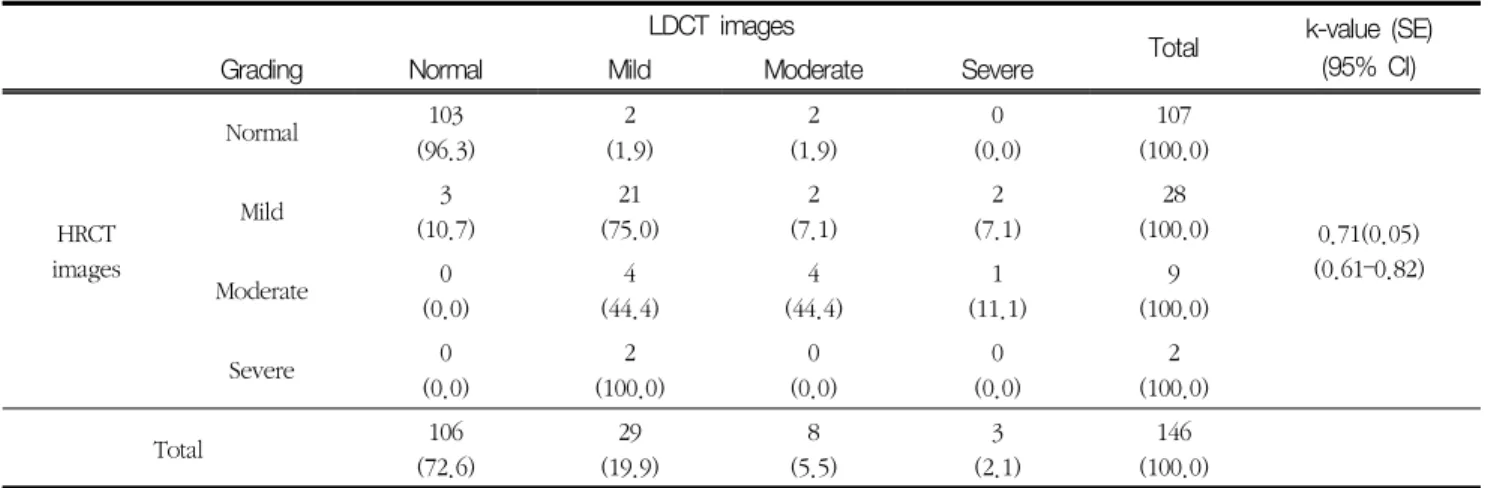
The k-value for the 4 emphysema grading categories on HRCT and LDCT images are listed in Table 3. The results showed good agreement, with a k-value of 0.71 (95% CI, 0.61–0.82). Emphysema detection was not significantly different between the two methods (McNemar–Bowker test, p=0.569). Of the 106 participants without emphysema on LDCT images, 3 participants were graded as having mild emphysema on HRCT images. Of the 29 participants with mild emphysema on LDCT images, the findings of 8 participants did not agree with that from the HRCT images, four and 2 participants were over-graded with moderate and severe emphysema on HRCT images, respectively. Of the 107 participants without emphysema on HRCT images, 4 participants were over-graded as having mild or moderate emphysema on LDCT images. Four
participants with mild emphysema on HRCT images were over-graded; 2 participants were graded as moderate and 2 as severe on LDCT images. One participant with moderate emphysema on HRCT images was graded as having severe emphysema on LDCT images.
4. Emphysema Grading Scores on HRCT and LDCT Images
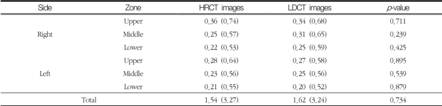
Emphysema grading scores were not significantly different between HRCT and LDCT images for all six lung zones (Table 4, Figure 1). The total emphysema grading score from LDCT images was higher than that from HRCT images; however, the difference was not statistically significant (p=0.734). The relationships between the emphysema grading scores from HRCT and LDCT images are shown in Figure 2. Emphysema grading scores from LDCT images were significantly correlated with increased scores on HRCT images; the correlation coefficient was 0.599 (p < 0.001) .
Ⅳ. DISCUSSION
CT is the best diagnostic modality to visualize structures in low-contrast regions such as the LDCT images
Total k-value (SE) (95% CI)
Grading Normal Mild Moderate Severe
imagesHRCT
Normal 103
(96.3) 2
(1.9) 2
(1.9) 0
(0.0) 107
(100.0)
0.71(0.05) (0.61–0.82)
Mild 3
(10.7) 21
(75.0) 2
(7.1) 2
(7.1) 28
(100.0)
Moderate 0
(0.0) 4
(44.4) 4
(44.4) 1
(11.1) 9
(100.0)
Severe 0
(0.0) 2
(100.0) 0
(0.0) 0
(0.0) 2
(100.0)
Total 106
(72.6) 29
(19.9) 8
(5.5) 3
(2.1) 146
(100.0)
HRCT, high-resolution CT; LDCT, low-dose CT. Data are expressed as the number of participants and as a percentage of the total number of participants. Emphysema was re-graded into 4 categories (normal, total score = 0; mild, total score = 1–6; moderate, total score = 7–12; and severe, total score = 13–18) from the total score summed after grading each of the 6 lung zones on a 4-point scale (normal = 0; mild = 1; moderate = 2; severe = 3). The k-value was calculated with linear weighting.
Table 3 Emphysema grading agreement for HRCT and LDCT images
lung parenchyma, which can undergo emphysematous changes. The diagnosis of emphysema is limited primarily by image noise, which is inversely related to radiation dose[19].
Radiation dose and image noise in CT generally depend on the choice of technique; parameters such as X-ray tube voltage and the current-time product (mAs) are the most important factors. The current-time product has been commonly used to reduce the radiation dose of CT images in clinical practice. In screening for emphysema, we have to consider reducing the radiation dose by using a lower current-time product. In this study, a reduction from 180 mAs to 30 mAs with a fixed tube voltage of 120 kVp resulted in decreasing the radiation dose of LDCT by up to one-sixth; however, the increase in image noise was 2.5-time larger than that of HRCT.
In our study, subjective emphysema grading was
performed according to the guidelines recommending that at least 6 slices with slice thicknesses of 1–2 mm be used in the evaluation of emphysema[5,6]. For screening purposes, 6–8 slices are taken distributed over all lung zones. Slice thicknesses were a bit different in HRCT and LDCT images in this study, which may have caused the noise to be increased in the thin slices used in HRCT[19].
LDCT images have been compared with HRCT and SDCT images for the diagnosis of emphysema[16-18]. The application of LDCT images to emphysema detection could be considered controversial, as image noise increases when using a lower mAs value[4,20]. There are two conflicting studies; A study by Horiuchi et al.[20] showed that HRCT images were more sensitive in detecting emphysema than LDCT images (75
Side Zone HRCT images LDCT images p-value
Right
Upper 0.36 (0.74) 0.34 (0.68) 0.711
Middle 0.25 (0.57) 0.31 (0.65) 0.239
Lower 0.22 (0.53) 0.25 (0.59) 0.425
Left
Upper 0.28 (0.64) 0.27 (0.58) 0.895
Middle 0.23 (0.56) 0.25 (0.56) 0.539
Lower 0.21 (0.55) 0.20 (0.52) 0.879
Total 1.54 (3.27) 1.62 (3.24) 0.734
HRCT, high-resolution CT; LDCT, low-dose CT. Data are expressed as the mean and standard deviation. The p-value was calculated using a paired t-test.
Table 4 Comparisons of emphysema grading scores between HRCT and LDCT images
Figure 1 High-resolution CT (HRCT, a) and low-dose CT (LDCT, b) images for lower zone of lung on same subject.
Emphysema grading scores between HRCT and LDCT were not difference in right(3 vs. 3) and left side(3 vs. 3).
Figure 2 A scatterplot shows the relationship between emphysema grading scores on high-resolution and low-dose CT images.
subjects vs. 54 subjects). On the other hand, another study found that measurements of the emphysema index, total lung volume, and mean lung density were not affected significantly by a decrease from 150 mAs to 25 mAs[4]. In our study, we did not find significant diagnostic differences in emphysema between the HRCT and LDCT images (39 participants vs. 40 participants), and concluded that emphysema detection was not affected by a lower mAs value. The total emphysema grading score was not significantly different between HRCT and LDCT images in this study, which was consistent with the results of previous studies reporting little difference between LDCT and SDCT images[17]. Therefore, for screening emphysema, evaluation of emphysema severity by LDCT images can be considered equivalent to HRCT images.
As shown in Figure 2, emphysema grading scores from HRCT images were significantly associated with those from LDCT images; such findings are in agreement with a study reported by Gierada et al.[16]
which showed that emphysema indices on LDCT and SDCT images were strongly correlated for all attenuation thresholds.
Emphysema has been quantified more objectively with the assistance of computer software[20]. A study by Gierada et al.[16] using pulmonary analysis software demonstrated that the LDCT technique has a minimal effect on the CT quantification of emphysema, in that CT measurements were not affected by lower mAs values, and their study showed no significant differences between mean and median lung attenuation on LDCT and SDCT images. However, emphysema quantification using software is time-consuming and increases the examination cost; therefore, subjective visual grading is typically used in clinical practice.
Subjective emphysema grading does not evaluate all image slices through the lungs; only 5–6 images are used[3,5,6]. However, objective measurement for emphysema quantification uses all image slices collected in spiral scanning mode to obtain volumetric data[16-18].
Emphysema could be overestimated by subjective grading[4] as well as by LDCT images[16]. In a previous study using macroscopic morphometric measurements as a gold standard in thin-section CT[4], subjective emphysema grading showed less agreement with macroscopic reference standard results (r=0.439-0.505, p < 0.05) than with objective CT densitometric results (r=0.555-0.623, p <0.001). Overestimation of emphysema grading on LDCT images can be avoided by using adaptive iterative dose reduction 3D processing for noise reduction in LDCT, as there are no remarkable differences between LDCT and SDCT images for emphysema quantification[17]. In our study, the subjective emphysema grading scores on LDCT images tended to be slightly overestimated compared with those on HRCT images. These results may be explained by the reduction of radiation dose resulting in an increase of noise[19] with subjective emphysema grading[4] affecting emphysema quantification, and because a software program was not used for noise reduction.
The results of our study are similar (data are not expressed) to the results of a study by Bankier et al.[4]
in which emphysema was more frequently observed in the upper zone of the lungs than in the middle or lower zones.
This study had a limitation that noise is strongly affected by the reconstruction filter utilized[19], and the use of filters was not investigated in this study.
To summarize, LDCT is effective in terms of reducing radiation dose, although image noise is greater than in HRCT. There was excellent agreement between the HRCT and LDCT images, and the imaging modalities did not show a difference in ability to diagnose emphysema. Therefore, considering the tradeoff between radiation dose and image noise, LDCT could be used as the gold standard method instead of HRCT for emphysema detection and grading.
REFERENCES
1. Takahashi M, Fukuoka J, Nitta N, et al: Imaging of pulmonary emphysema: a pictorial review, Int J Chron Obstruct Pulmon Dis, 3(2), 193-204, 2008 2. MMller NL, Staples CA, Miller RR, Abboud RT:
“Density mask”. An objective method to quantitate emphysema using computed tomography, Chest, 94(4), 782-787, 1988
3. Kuwano K, Matsuba K, Ikeda T, et al: The diagnosis of mild emphysema. Correlation of computed tomog- raphy and pathology scores, Am Rev Respir Dis, 141(1), 169-178, 1990
4. Bankier AA, De Maertelaer V, Keyzer C, Gevenois PA: Pulmonary emphysema: subjective visual grad- ing versus objective quantification with macroscopic morphometry and thin-section CT densitometry, Radiology, 211(3), 851-858, 1999
5. Kusaka Y, Hering KG, Parker JE, eds: International Classification of HRCT for Occupational and Environmental Respiratory Diseases, Tokyo, Springer-Verlag, 2005
6. Suganuma N, Kusaka Y, Hering KG, et al: Reliability of the proposed international classification of high-resolution computed tomography for occupa- tional and environmental respiratory diseases, J Occup Health, 51(3), 210-222, 2009
7. Blum T, Kollmeier J, Ott S, Serke M, SchMnfeld N, Bauer T: Computed tomography for diagnosis and grading of dust-induced occupational lung disease, Curr Opin Pulm Med, 14(2), 135-140, 2008
8. Majurin ML, Varpula M, Kurki T, Pakkala L:
High-resolution CT of the lung in asbestos-exposed subjects. Comparison of low-dose and high-dose HRCT, Acta Radiol, 35(5), 473-477, 1994
9. Huda W: Radiation doses and risks in chest computed tomography examinations, Proc Am Thorac Soc, 4(4), 316-320, 2007
10. Smith-Bindman R, Lipson J, Marcus R, et al:
Radiation dose associated with common computed tomography examinations and the associated life- time attributable risk of cancer, Arch Intern Med,
169(22), 2078-2086, 2009
11. Rumack CM: 2010 ACR presidential address, pa- tient-focused radiology: Taking charge of radiation dose, J Am Coll Radiol, 7(11), 837-844, 2010 12. Naidich DP, Marshall CH, Gribbin C, Arams RS,
McCauley DI: Low-dose CT of the lungs: preliminary observations, Radiology, 175(3), 729-731, 1990 13. Takahashi M, Maguire WM, Ashtari M, et al:
Low-Dose Spiral Computed Tomography of the Thorax Comparison with the Standard-Dose Technique, Invest Radiol, 33(2), 68-73, 1998 14. Fasola G, Belvedere O, Aita M, et al: Low-dose com-
puted tomography screening for lung cancer and pleural mesothelioma in an asbestos-exposed pop- ulation: baseline results of a prospective, non- randomized feasibility trial--an Alpe-adria Thoracic Oncology Multidisciplinary Group Study (ATOM 002), Oncologist, 12(10), 1215-1224, 2007 15. Won-Jeong Lee, Jong-Ryul Seon, Bong-Seon Ahn,
Young-Sun Park. Findings on Chest Low-Dose CT images of Group Exposed to Inorganic Dusts.
Journal of Korean Society of Radiological Technology, 34(4), 305-314, 2011
16. Gierada DS, Pilgram TK, Whiting BR, et al:
Comparison of standard- and low-radiation-dose CT for quantification of emphysema, AJR Am J Roentgenol, 188(1), 42-47, 2007
17. Nishio M, Matsumoto S, Ohno Y, et al: Emphysema quantification by low-dose CT: potential impact of adaptive iterative dose reduction using 3D process- ing, AJR Am J Roentgenol, 199(3), 595-601, 2012 18. Sverzellati N, Cademartiri F, Bravi F, et al:
Relationship and prognostic value of modified coro- nary artery calcium score, FEV1, and emphysema in lung cancer screening population: the MILD trial, Radiology, 262(2), 460-467, 2012
19. Goldman LW: Principles of CT: radiation dose and image quality, J Nucl Med Technol, 35(4), 213-225, 2007
20. Horiuchi N, Fujita J, Suemitsu I, Yamasaki Y, Higa F, Tateyama M: Low-dose multislice CT and high-resolution CT assessment of pulmonary em- physema in public school teachers, Lung, 185(1),
25-30, 2007
21. Gevenois PA, Zanen J, de Maertelaer V, De Vuyst P, Dumortier P, Yernault JC: Macroscopic assess-
ment of pulmonary emphysema by image analysis, J Clin Pathol, 48(4), 318-322, 1995
∙국문초록
폐기종에 대한 저선량 CT의 유용성 평가 : 고해상도 CT와 비교
이원정
대전보건대학교 방사선과
본 연구에서는 폐기종에 대해 고해상도 CT와 비교한 저선량 CT 의 유용성에 대해 평가하였다. 고해상도 CT와 저선량 CT 노출조건에서 선량과 영상 잡음을 3회 반복 측정하였다. 비슷한 노출조건에서 획득한 146명 의 고해상도 CT와 저선량 CT 영상에 대해 2명의 흉부영상의학과전문의 합의 판독결과에서 폐기종 소견 만을 본 연구에 사용하였다. SPSS ver. 19.0 프로그램 사용하여 고해상도 CT와 저선량 CT 간에 폐기종에 대한 진 단 차이는 McNemar’s tests, 일치도는 unweighted kappa tests, 선량과 잡음 차이는 Mann–Whitney U-test 로 분석하였다.
선량은 고해상도 CT가 저선량 CT 보다 높았지만(1.95 mGy vs. 0.35 mGy, p=0.008), 잡음은 낮았다(40.1 HU vs. 99.6 HU, p=0.021). 폐기종 진단에 대해서는 두 영상 간에 높은 일치도를 보였다(k-value=0.88).
폐기종 점수는 두 영상 간에 통계적인 유의한 차이를 보이지 않았고, 높은 상관성을 보였다(r=0.599, p < 0.001).
선량과 잡음을 고려했을 때, 저선량 CT는 폐기종 진단에 표준 진단 방법 인 고해상도 CT를 대신하여 사용 할 수 있을 것으로 사료된다.
중심 단어 : 고해상도 CT, 저선량 CT, 폐기종, 방사선 선량, 영상 잡음