Address for correspondence;
Dong Joo Yun, M.D.
Department of Neurology, Eulji University Hospital, Eulji University School of Medicine, 1306 Dunsan 2-dong, Seo-gu, Daejeon 302-779, Korea
Tel: +82-42-611-3431 Fax: +82-42-611-3858 E-mail: [email protected]
Case Report
거짓 장막힘과 감각신경세포병증으로 발현된 소세포폐암 1예
을지대학교 의과대학 을지대학병원 신경과
이현정․최영철․윤동주․고영채․장상현․윤수진․오건세․이수주
Gastrointestinal Pseudoobstruction and Sensory Neuronopathy in Small Cell Lung Cancer
Hyun Jeong Lee, M.D., Young Chul Choi, M.D., Dong Joo Yun, M.D., Youngchai Ko, M.D., Sang Hyun Jang, M.D., Soo Jin Yoon, M.D., Gun-Sei Oh, M.D., Soo Joo Lee, M.D.
Department of Neurology, Eulji University Hospital, Eulji University School of Medicine, Daejeon, Korea
Received 16 September 2011; received in revised form 8 November 2011; accepted 29 November 2011.
Subacute sensory neuronopathy and gastrointestinal pseudoobstruction are considered classical paraneoplastic neurological syndromes. We report a 56-year-old male who presented with typical symptoms of subacute sensory neuronopathy and autonomic neuropathy with gastrointestinal pseudoobstruction. The biopsy of the palpable supraclavicular lymph node revealed a small cell lung cancer. To our knowledge, intestinal pseudoobstruction and sensory neuronopathy in a small cell lung cancer have not been reported in Korea.
Key Words: Paraneoplastic polyneuropathy, Intestinal pseudoobstruction, Small cell lung carcinoma
Paraneoplastic neurological syndromes (PNS) are the remote neurological effects of cancer. Subacute sensory neuronopathy usually occurs as a paraneoplastic syndrome associated with the presence of anti-Hu antibodies in the serum in small cell lung cancer.1 Gastrointestinal pseudoobstruction is also considered classical PNS. However, this might be underreported because initially it was thought to be unrelated to the neurologic disorder. We report a case of subacute sensory neuronopathy
and autonomic neuropathy with intestinal pseudoobstruction.
Biopsy of the palpable supraclavicular lymph node revealed a small cell lung cancer.
Case Report
A 56-year-old man developed numbness and tingling sense in his feet and hands 10 days after a minor upper respiratory tract infection. He also complained of recurrent dizziness on standing, urinary difficulty, constipation, and recurrent abdominal pain. Numbness and paresthesia extended to both elbows and knees over the 4 weeks. Subsequently the symptoms spread to the neck and he had a severe unsteady gait within 2 months after the onset. There was no double vision or swallowing difficulty. He lost 10 kilograms over 2 months. He smoked
Table 1. Results of nerve conduction study
Motor nerve Terminal latency (ms)
Right/Left Amplitude (mV)
Right/Left NCV (m/s)
Right/Left F-latency (ms) Right/Left
Median nerve 30.0 (<29)
Wrist 3.35 (<3.6) 11.4 (>5)
Elbow 11.4 47.7 (>50)
Axilla 10.5 57.1 (>56)
Ulnar nerve 31.2 (<29)
Wrist 2.85 (<2.5) 17.3 (>5)
Below elbow 16.3 48.0 (>50)
Above elbow 16.0 53.5 (>52)
Peroneal nerve 56.1/56.5 (<48)
Ankle 4.5/4.5 (<4.7) 5.0/4.2 (>4)
Fibular head 4.1/4.1 39.4/36.5 (>41)
Knee 4.1/4.0 37.6/36.4 (>39)
Tibial nerve 53.0/56.3 (<53)
Ankle 4.5/4.1 (<5.1) 11.4/9.1 (>5)
Knee 8.7/7.0 38.3/39.0 (>40)
H-reflex (ms) NR (<30)
Sensory nerve Latency (ms) Amplitude (μV) NCV (m/s)
Median nerve 5.1
Finger-wrist 2.4 (>10) 21.7 (>41)
Wrist-elbow 11.6 45.1 (>49)
Elbow-axilla 17.5 50.2 (>53)
Ulnar nerve 3.7
Finger-wrist 1.2 (>10) 28.1 (>39)
Wrist-elbow 3.2 46.9 (>47)
Elbow-axilla 11.2 47.0 (>48)
Sural nerve
Calf 4.7/4.5 1.3/2.5 (>6) 29.8/31.1 (>34)
Superficial peroneal
Lateral leg NR/4.1 NR/1.0 (>4) NR/33.7 (>40)
NCV; nerve conduction velocity, NR; no response.
Reference value of normal limit within parentheses.
for 40 years. He had been diagnosed with atrial fibrillation and hypertension. Upon physical examination, multiple enlarged lymph nodes were palpable in his right supraclavicular fossa.
He had distended abdomen and the bowel sound was decreased. He was alert and cranial nerve functions were intact. Neurologic examinations revealed profoundly impaired vibratory sensation and joint positional sensation over all
extremities with a positive Romberg test. Sensations to pinprick and temperature were mildly impaired. He had minimal weakness (Medical Research Council grade 4) in the proximal limb muscles, but all tendon reflexes were absent. Despite the relative preservation of muscle strength, he could only lie in bed because of severe sensory ataxia and proprioceptive loss.
He exhibited neither clonus nor a Babinski sign.
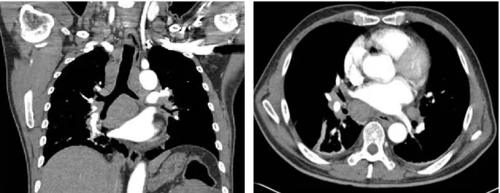
Figure 1. Contrast enhanced computed tomography of the chest showing markedly enlarged lymph nodes in bilateral supra- clavicular fossa, mediastinum, hilar and interlobar areas. Diffuse wall thinkening of proximal lobar and segmental bronchi in both lungs with multifocal linear atelectasis is revealed, probably due to conglomerated lymph nodes along bronchovas- cular bundle.
On diagnostic studies, levels of vitamin B1, B6, B12, folate were within normal range and antibodies to human immuno- deficiency virus, hepatitis surface antigen, cryoglobulin, rheumatoid factor, autoantibodies (antinuclear, anti-double stranded DNA, anti-Ro, anti-La, anti-neutrophil cytoplasmic antibodies) were all negative. Chest X-ray was unremarkable.
The abdominal X-ray showed multiple distended bowel loops with air-fluid levels. Cerebrospinal fluid (CSF) was clear and acellular with an elevated protein concentration of 110 mg/dL.
Both serum and CSF protein immunoelectrophoresis revealed no monoclonal gammopathy. Serum antiganglioside antibodies were examined by ELISA. Anti-GD1b IgM and IgG antibody were mildly increased to 48.9%, 40.17% respectively (normal
<30%). All the other tested anti-ganglioside antibodies (anti- GQ1b, anti-GM1 antibodies) and antibody to myelin-associated glycoprotein (MAG) were negative. The results of electro- physiology were summarized in Table 1. Nerve conduction studies showed markedly diminished amplitude of sensory action potentials with slightly reduced motor conduction velocities in all limbs. On autonomic function test, sympathetic skin response was absent in all limbs. We could not check heart rate variability due to atrial fibrillation. He had postural hypotension. Sensory neuronopathy with autonomic neuropathy was diagnosed by clinical findings and electrophysiologic studies. Tumor markers and anti-onconeuronal antibodies (anti-Hu, anti-Ri, anti-Yo antibodies) were negative. Chest CT
showed multiple enlarged lymph nodes in bilateral suprac- lavicular fossa, mediastinum, hilar and interlobar areas (Figure 1). Biopsy of the supraclavicular lymph node revealed a small cell lung cancer (Figure 2). We treated the patient with intravenous gamma globulin based on the autoimmune nature of sensory neuronopathy, but his sensory ataxia and autonomic dysfunction have not responded. After the first cycle of chemotherapy for small cell lung cancer, he could not continue treatment due to low blood pressure and progressive deterioration of general condition. The patient has been treated conservatively without any improvement of his neurological deficits.
Discussion
The electrodiagnostic studies of this patient identified diffuse sensory abnormalities and evidence of dysautonomia as characterized by absent sympathetic skin response and paralysis of the digestive tract. These findings initially were thought to be suggestive of nonmalignant inflammatory sensory neuronopathy or sensory demyelinating polyneuropathy. However, sensory neuronopathy with gastrointestinal pseudoobstruction is highly suggestive of paraneoplastic syndromes, and thought not to occur as part of immune mediated sensory neuronopathy.2 In addition, electrophysiological findings in the subset of patients reported with sensory demyelinating
Figure 2. The pathologic biopsy findings. Histopathologic findings of the biopsy specimen taken from the supraclavicular lymph node revealed a small cellcarcinoma (A: hematoxylin-eosin, ×40, B: ×200). (C) Immunohistochemical studies showing positive staining diffusely for CD56. (D) Neuroendocrine markers of chromogranin are identified by the brown staining in the cytoplasm of the neoplastic cells (immunohistochemistry, ×200).
polyneuropathy typically have evidence of demyelination of motor fibers.
Subacute sensory neuronopathy usually occurs as a paraneo- plastic syndrome associated with the presence of anti-Hu antibodies in the serum in small cell lung cancer. Many patients with paraneoplastic syndromes have antibodies in their serum and CSF that react with both the nervous system and the underlying cancer. Antibodies directed against neural antigens expressed by the tumor are called onconeural antibodies, which suggests that an autoimmune process underlies PNS.3 The antibodies highly specific for paraneoplastic sensory neuronopathy are anti-Hu, anti-CRMP5 (anti-CV2), and ANNA-3 antibodies. Most patients have anti-Hu antibodies,
which have 99% specificity and 82% sensitivity for the diagnosis of cancer in patients with subacute sensory neuronopathy.3 However, the absence of anti-Hu antibodies in a patient with sensory neuropathy of unexplained cause involving proprioceptive and kinesthetic sensation does not completely rule out the possibility of an underlying cancer.4 The diagnosis of PNS of the peripheral nervous system should be considered if a classical neurological syndrome, such as subacute sensory neuronopathy or gastrointestinal pseudoobstruction, and cancer that develops within five years of the diagnosis of the neurological disorder are present regardless of the presence of onconeural antibodies.5
The neurological evaluation in paraneoplastic sensory
A B
C D
neuronopathy may demonstrate involvement of the motor nerves, peripheral autonomic nervous system, or different areas of the brain.6 Enteric neuronopathy occurs most often with small cell lung canser. The pathological changes are a loss of myenteric plexus neurons, secondary axonal degeneration, and a lymphocytic inflammatory cell infiltrate.7 Circulating IgG antibodies reactive with neurons of myenteric and submucosal plexuses were found in paraneo- plastic pseudoobstruction with small cell lung cancer.
Gangliosides, a complex family of sialylated glycos- phingolipids, are components of the cell membrane that are particularly concentrated in the peripheral nervous system where several of them may be target antigens in immune mediated neuropathies.8 GD1b is expressed on human dorsal root ganglion neurons and may be a target molecule for autoantibody in some patients with sensory ataxic neu- ropathy.9 The role for gangliosides as onconeural antigens was supported by the expression of GM1 and GD1a at high levels in lung cancer and activation of T-cell dependent production of anti-ganglioside antibodies, which significantly decreased after cancer treatment.10 Anti-GD1b IgM and IgG antibody were mildly increased during the acute phase of illness in this patient. These findings suggest that the ectopic expression of gangliosides on neoplastic cells might elicit autoimmune responses, which may target the nervous system resulting in PNS. Whether PNS in this case resulted from chance association or immunological mechanisms induced by the tumor is still unknown. Although antigan- glioside antibodies were probably not responsible for the neuropathies in this patient, their presence may provide indirect support for an argument in favor of an autoimmune mechanism directed towards as yet unknown onconeural antigens in patients with suspected seronegative PNS.
Further researches for pathogenesis of sensory neuronopathy with anti-gangioside antibodies are needed to identify target molecules of cancer related neuropathies.
In conclusion, we report a case with severe disabling manifestations of sensory and autonomic neuropathy in small cell lung cancer. Paraneoplastic intestinal pseudoobstruction should be considered in any patient with a subacute course of gastroparesis associated with sensory neuronopathy regardless of the presence of onconeural antibodies. Early identification of this syndrome is important, since treatment of the primary cancer may halt the progression.
REFERENCES
1. Shin JH, Jo HJ, Kim DS, Jung DS, Park KH, Lee MK, et al.
A case of anti-Hu associated paraneoplastic subacute sensory neuronopathy. J Korean Neurol Assoc 2002;20:89-92.
2. Smith BE, Windebank AJ. Dorsal root ganglion disorders. In:
Katirji B, Kaminski HJ, Preston DC, Ruff RL, Shapiro BE.
Neuromuscular disorders in clinical practice. Boston: Butterworth- Heinemann. 2002;478-500.
3. Antoine J-C, Camdessanché J-P. Peripheral nervous system in- volvement in patients with cancer. Lancet Neurol 2007;6:75-86.
4. Molinuevo JL, Graus F, Serrano C, Rene R, Guerrero A, Illa I. Utility of anti Hu antibodies in the diagnosis of paraneo- plastic sensory neuropathy. Ann Neurol 1998;44:976-980.
5. Graus F, Delattre J, Antoine J, Dalmau J, Giometto B, Grisold W, et al. Recommended diagnostic criteria for paraneoplastic neurological syndromes. J Neurol Neurosurg Psychiatry 2004;
75:1135-1140.
6. Henson RA, Urich H. Part III. Paraneoplastic disorders. In:
Henson RA, Urich H. Cancer and the nervous system. Oxford:
Blackwell Scientific Publications, 1982;311-451.
7. Lennon V, Sas D, Busk M, Scheithauer B, Malagelada JR, Camilleri M, et al. Enteric neuronal autoantibodies in pseu- doobstruction with small-cell lung carcinoma. Gastroenterology 1991;100:137-142.
8. Gong Y, Tagawa Y, Lunn M, Laroy W, Heffer Lauc M, Li C, et al. Localization of major gangliosides in the PNS:
implications for immune neuropathies. Brain 2002;125:2491-2506.
9. Pan CL, Yuki N, Koga M, Chiang MC, Hsieh ST. Acute sensory ataxic neuropathy associated with monospecific anti-GD1b IgG antibody. Neurology 2001;57:1316-1318.
10. De Toni L, Marconi S, Nardelli E, Alberti D, Borsellino G, Fracasso G, et al. Gangliosides act as onconeural antigens in paraneoplastic neuropathies. J Neuroimmunol 2004;156:178-187.