Introduction
뼈는 고도로 석회화된 결합조직으로 파괴와 형성을 계속 반복하는 재 형성을 하며, 이러한 뼈의 재형성은 파골세포에 의한 골흡수와 조골세 포에 의한 골형성이 정교하게 균형을 맞춘 작용을 통해 동적으로 조절 된다[1]. 뼈의 재형성은 정상적인 골격형성, 골격의 기능, 무기물질의 항상성을 담당하고 있으며, 이러한 재형성의 균형이 깨진 경우 류마티 스관절염, 파제트 병, 치주질환 및 다발성 골수종과 같은 뼈와 관련된 질환을 야기한다[2]. 골 소실과 골질 손상으로 인한 뼈 질환은 고령화 인구의 주요한 건강문제이며, 사망률과 연관이 있다.
염증성 뼈파괴 질환의 극복을 위해 진행되는 연구는 주로 병적 뼈파 괴가 일어나는 동안 중요한 역할을 하는 파골세포의 생성 및 골흡수 정 도를 측정하거나[3,4], 관련 세포에서 신호전달과정 중 nuclear factor kappa-κB (NF-κB) 및 mitogen-activated protein kinase (MAPK) 등의 발현 및 활성을 측정하기도 한다[5,6]. 또한 류마티스관절염, 골관 절염 및 치주질환 등을 유도한 동물 모델을 통해 염증 매개물질을 측정
하거나, 결합조직분해 효소의 양을 측정하고, 혈청 내 골대사 표지 인자 를 측정하기도 한다[7-14].
류마티스관절염, 골관절염 및 치주질환은 뼈파괴 질환이라는 공통점 을 가지고 있고, 원인이 염증에 의한 경우가 대부분이다. 이러한 염증성 뼈파괴 질환의 치료목적으로 다양한 연구가 진행되고 있으며, 그 중 식 물유래 천연화합물이 염증 및 뼈파괴 완화에 도움이 된다는 연구도 다 수 진행되고 있다. 이 논문은 발표된 논문 중 polyphenol 또는 flavo- noid와 염증성 뼈파괴 질환 관련 검색어를 사용하였으며, 검색된 논문 에서 뼈 건강을 유지하는데 사용되는 다양한 식물유래 천연화합물을 구 조적으로 분류하였고, 이들 성분의 생물학적 효과와 관련 메커니즘을 정리하였다.
식물유래 천연화합물의 분류 및 종류
Phytochemical은 다양한 식물에서 유래하며, 항염증, 항산화 및 항 바이러스 등의 다양한 생물 활성을 가진 polyphenol 화합물로 알려져
Int J Oral Biol 44:130-143, 2019
pISSN: 1226-7155 • eISSN: 2287-6618 https://doi.org/10.11620/IJOB.2019.44.4.130
Effects of plant-derived natural products on inflammatory bone destructive disease
Seon-Yle Ko*
Department of Oral biochemistry and Institute of Dental Science, College of Dentistry, Dankook University, Cheonan 31116, Republic of Korea
Rheumatoid arthritis, osteoarthritis, and periodontal disease are bone destructive diseases mainly caused by inflammation. Various studies are being conducted to develop treatments for inflammatory bone destructive diseases. Many of these studies involve plant-derived natural compounds. In these studies, cell differentiation, signal transduction pathways, and bone resorption were measured at the cellular level. In disease-induced animal models, the amount of inflammatory mediators or matrix destructive enzymes and serum metabolic markers were measured.
This study examined the effects of plant-derived natural compounds, such as flavonoids, on inflammatory bone destructive diseases. In addition, we structurally classified various substances used to maintain bone health and summarized the biological effects and related mechanisms of the components.
Keywords: Polyphenol, Flavonoid, Arthritis, rheumatoid, Osteoarthritis, Periodontal diseases
Received November 21, 2019; Revised December 16, 2019; Accepted December 18, 2019
*Correspondence to: Seon-Yle Ko, E-mail: [email protected] https://orcid.org/0000-0003-2541-9556 Copyright © The Korean Academy of Oral Biology
CC
This is an open-access article distributed under the terms of the Creative Commons Attribution Non-Commercial License (http://creativecommons.org/licenses/by- nc/4.0/), which permits unrestricted non-commercial use, distribution, and reproduction in any medium, provided the original work is properly cited.
Invited Review IJOB
있다[15-17]. Phytochemical 구성성분인 polyphenol은 넓은 의미로 사용되며, flavonoid와 nonflavonoid로 분류될 수 있다. Flavonoid에 는 flavone, flavonol, isoflavone 등이 포함되는 반면, nonflavonoid 는 stilbene과 페놀산과 같은 다양한 종류의 polyphenol이 포함된다 [18].
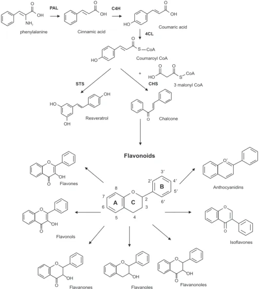
Flavonoid의 연구는 1937년 Albert Szent-Györgyi가 생물 활성을 밝히는 연구로 시작되었으며[19], 그 후 4,000여 종 이상의 flavonoid 가 밝혀졌다[20]. Flavonoid는 두 개의 벤젠고리 사이에 pyran 고리로 연결된, 15개의 탄소 골격 구조를 가지며(Fig. 1), phenylpropanoid 대사경로를 통해 전구물질인 phenylalanine과 malonyl-coenzyme A (CoA)로부터 생합성되는 저분자량 페놀 화합물이다(Fig. 2). Flavo- noid는 합성된 후 식물의 다양한 세포 내 소기관 및 세포벽 등으로 이동 하여 기능한다. Flavonoid는 인체에서 합성되지 않으므로 과일, 채소, 견과류, 씨앗, 허브 등을 통해 섭취할 수 있다[15-17].
Flavonoid는 Fig. 1의 flavonoid 기본골격 구조에서 C 고리의 4번 탄소의 산화 수준의 차이와 3번 탄소의 수산화 패턴의 차이에 따라 분 류한다. 즉 heterocyclic C 고리의 산화도 및 포화에 따라 flavones,
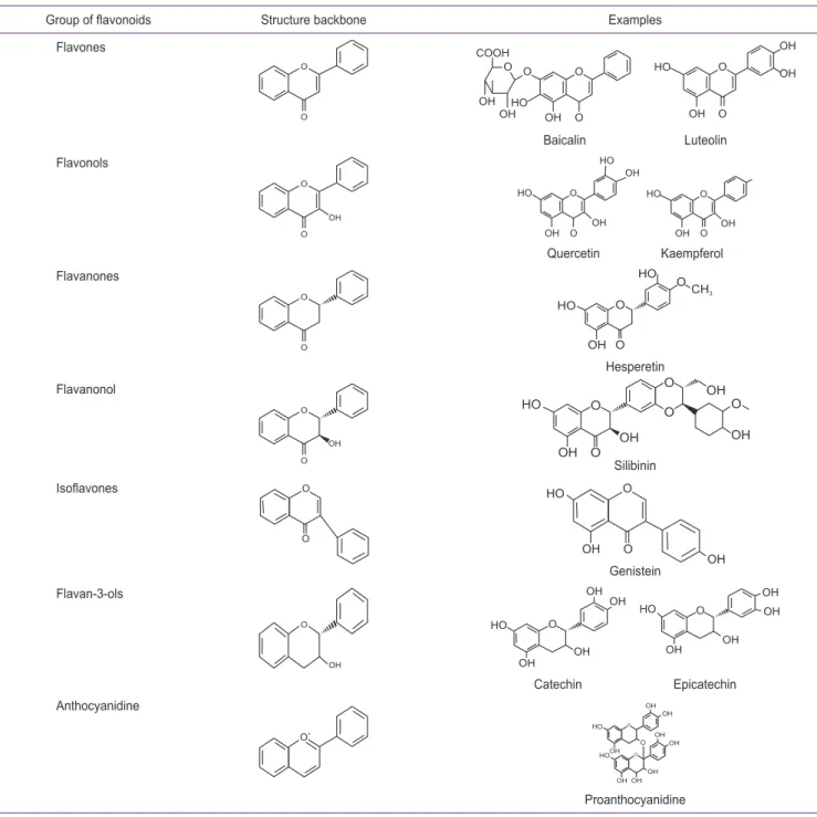
flavonols, flavanones, flavanonols, isoflavones, flavonols 및 an- thocyanidins으로 분류된다[7]. 분류에 따른 일반적인 구조는 Table 1 에 분류마다 포함된 예와 함께 구조를 표기하였다. 같은 분류 내의 개별
1
2
3
4 5
6 7
8 1 '
2 '
3 '
4 '
5 '
6 '
A C
B
O
Fig. 1. Basic flavonoid structure.
2 3
5 4 6 7
8
2 ' 3 '
4 ' 5 ' 6 '
A C
B
O
Flavonoids
O
+Anthocyanidins
O
O
Isoflavones
O
OH O
OH O
OH O
O OH
Flavonols O
O OH
Flavones
O Chalcone Resveratrol
HO
OH
OH
STS CHS
HO S
CoA
3 malonyl CoA O
O + HO
O
S CoA Coumaroyl CoA HO
O
Coumaric acid OH
4CL O
Cinnamic acid OH PAL C4H
phenylalanine O
OH NH
2Fig. 2. Biosynthesis of flavonoids and nonfla- vonoids via phenylalanine metabolic pathway and classification of flavonoids.
PAL, phenylalanine ammonia lyase; C4H,
cinnamate-4-hydroxylase; CoA, coenzyme
A; 4CL, coumaroyl-CoA ligase; STS, stilbene
화합물은 A 및 B 고리의 치환 패턴이 다른 구조를 가진다[21].
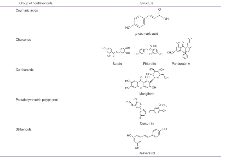
또한 polyphenol 중 flavonoid 구조 이외의 nonflavonoid는 flavo- noid 합성과정 중 중간산물인 coumaric acid, chalcone 및 p-cou- maroyl CoA로부터 stilbene synthase에 의해 합성된 resveratrol 등 을 포함하며, 구조는 Table 2에 포함된 예를 표기하였다.
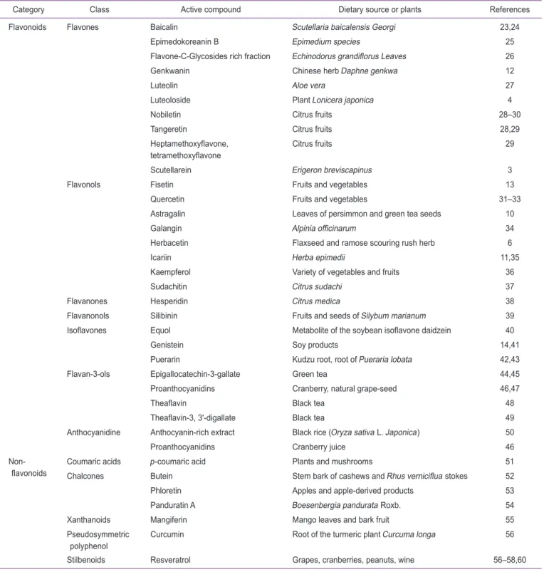
식물의 polyphenol 화합물은 모든 식물세포에 분포하는 물질이며, 여러 부류의 식물 유래 polyphenol 화합물과 화합물을 함유하는 공급 원을 Table 3에 기술하였다. 일반적으로 식품의 flavonoid는 색, 맛,
지방산화방지 등의 효과를 가지며, 식품에서 가장 풍부한 flavonoid는 flavonol의 종류이다[22-60].
류마티스관절염(rheumatoid arthritis)에서 식 물유래 천연화합물의 효과
류마티스관절염은 흔한 염증성 자가면역 질환으로, 활액조직(syno- vial tissue)에 림프구와 형질세포의 침윤 및 관절의 활막에 염증(활막
Table 1. Structure of flavonoids
Group of flavonoids Structure backbone Examples
Flavones
O
O
O O O
O OH OH
COOH
OH HO
O
O HO
OH
OH OH
Baicalin Luteolin Flavonols
O
O OH
O
O OH HO
HO OH
OH
O
O HO
OH OH
Quercetin Kaempferol Flavanones
O
O
O
O OH HO
CH
3HO O
Hesperetin Flavanonol
O
O OH
O
O OH
OH O
OH OH
HO
O
O
Silibinin
Isoflavones O
O
O
O OH HO
Genistein OH Flavan-3-ols
O
OH
HO
OH OH
OH OH
HO
OH
OH OH OH
O
O
Catechin Epicatechin Anthocyanidine
O
+O
O O
OH HO
OH OH
OH OH
OH
OH OH HO
Proanthocyanidine
염, synovitis)이 발생한다. 또한 류마티스관절염은 관절 연골 및 뼈의 파괴가 진행되어 기능손상과 비가역적인 강직 등의 변형을 초래한다 [59].
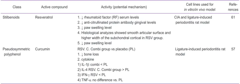
Polyphenol은 다양한 활성을 통해 류마티스관절염을 완화하는 것 으로 알려졌으며, polyphenol의 효과는 관절염을 유도한 동물 모델에 서 관절염 지수, 혈청 내 proinflammatory cytokine의 농도 변화, 활 막(synovium)의 반응과 활막 내 섬유아세포-유사 활막세포의 반응 및 다양한 신호전달과정의 변화를 통해 관찰되었으며, 이를 Table 4에 기 술하였다. 이러한 효과는 cytokine의 조절 및 synovial joint의 항상 성 파괴로 인한 뼈 침식을 조절하는 것에 기인한 것으로 조사되었다. 몇 몇 연구는 류마티스관절염의 병적 소견을 재현하는 adjuvant-유도 관 절염(adjuvant-induced arthritis, AIA) 및 collagen-유도 관절염 (collagen-induced arthritis, CIA) 동물 모델을 사용하였다.
Table 4에 기술된 내용 중 몇 종류의 효과를 보면, polyphenol 중 astragalin, galangin, genkwanin, genistein, theaflavin-3, 3’-di- gallate 및 p-coumaric acid 등은 동물 모델에서 proinflammatory
cytokine이나 matrix metalloproteinase (MMP)의 생성을 억제하는 등[10,12,34,41,49,51]의 효과를 나타냈다. 또한 resveratrol은 혈청 내 류마티스 지수를 억제하거나, 관절염이 유도된 동물의 발 붓기를 감 소시키는 등[60] 의 효과를 나타냈다. 이와 같이 polyphenol은 기질분 해효소의 발현 억제, 염증성 매개물질 생성 억제 등의 효과를 가지므로 염증반응을 개선할 수 있는 물질로 사용될 수 있다.
골관절염(osteoarthritis)에서 식물유래 천연화 합물의 효과
골관절염은 흔한 관절질환으로 연골의 구조 변형으로 통증과 관절 기 능 상실 등의 특징을 가진다. 염증성 질환에 포함되어 있지는 않지만, 염증이 골관절염의 병인에 기인한다는 증거가 증가하고 있다[61]. 연골 세포는 관절연골을 구성하고 다양한 기질 성분의 항상성을 유지하는 기 능을 하므로, 골관절염이 진행되는 동안 연골세포의 역할이 중요하다 [61].
Table 2. Structure of nonflavonoid polyphenol compounds
Group of nonflavonoids Structure
Coumaric acids O
OH HO
p-coumaric acid Chalcones
HO
OH
OH OH O
OH
HO OH HO
O
CH O
3OH OH O
Butein Phloretin Panduratin A Xanthanoids
HO
O O HO
HO OH
OH OH
OH O
HO
Mangiferin Pseudosymmetric polyphenol
OH HO
O O
O CH
3H C
3O
Curcumin Stilbenoids
HO
OH
OH
Resveratrol
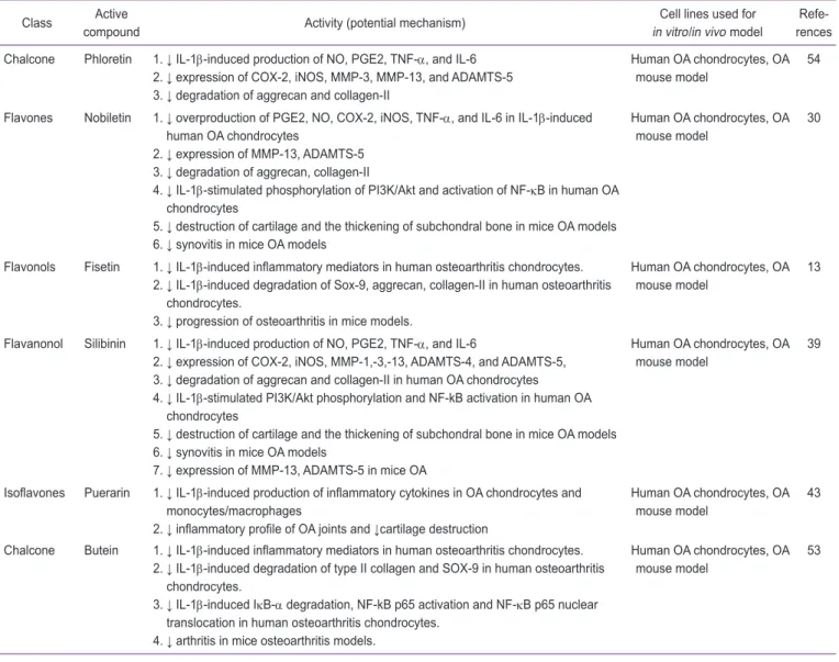
골관절염에서 polyphenol의 역할은 자극에 따른 기질성분 분해 감소 의 측면에서 연골보호효과를 주로 관찰하고 있으며, 이를 Table 5에 기 술하였다. 몇몇 연구는 골관절염을 재현하기 위해 골관절염을 유도한 동물모델을 사용하였다.
Table 5에 기술된 내용 중 몇 종류의 효과를 보면, polyphenol 중
phloretin, nobiletin 및 silibinin 등은 골관절염 모델에서 MMP-3, MMP-13 등의 기질분해효소 발현 감소 및 aggrecan과 2형 교원섬 유 등의 기질 성분 파괴를 억제하는 효과를 나타냈다[30,39,53]. 또한 nobiletin과 puerarin 등은 골관절염 모델에서 proinflammatory 매개 물질들의 생성을 억제하였다[30,43]. 이와 같이 polyphenol은 골관절
Table 3. Classification and food sources of some dietary phytochemical
Category Class Active compound Dietary source or plants References
Flavonoids Flavones Baicalin Scutellaria baicalensis Georgi 23,24
Epimedokoreanin B Epimedium species 25
Flavone-C-Glycosides rich fraction Echinodorus grandiflorus Leaves 26
Genkwanin Chinese herb Daphne genkwa 12
Luteolin Aloe vera 27
Luteoloside Plant Lonicera japonica 4
Nobiletin Citrus fruits 28–30
Tangeretin Citrus fruits 28,29
Heptamethoxyflavone, tetramethoxyflavone
Citrus fruits 29
Scutellarein Erigeron breviscapinus 3
Flavonols Fisetin Fruits and vegetables 13
Quercetin Fruits and vegetables 31–33
Astragalin Leaves of persimmon and green tea seeds 10
Galangin Alpinia officinarum 34
Herbacetin Flaxseed and ramose scouring rush herb 6
Icariin Herba epimedii 11,35
Kaempferol Variety of vegetables and fruits 36
Sudachitin Citrus sudachi 37
Flavanones Hesperidin Citrus medica 38
Flavanonols Silibinin Fruits and seeds of Silybum marianum 39
Isoflavones Equol Metabolite of the soybean isoflavone daidzein 40
Genistein Soy products 14,41
Puerarin Kudzu root, root of Pueraria lobata 42,43
Flavan-3-ols Epigallocatechin-3-gallate Green tea 44,45
Proanthocyanidins Cranberry, natural grape-seed 46,47
Theaflavin Black tea 48
Theaflavin-3, 3'-digallate Black tea 49
Anthocyanidine Anthocyanin-rich extract Black rice (Oryza sativa L. Japonica) 50
Proanthocyanidins Cranberry juice 46
Non- flavonoids
Coumaric acids p-coumaric acid Plants and mushrooms 51
Chalcones Butein Stem bark of cashews and Rhus verniciflua stokes 52
Phloretin Apples and apple-derived products 53
Panduratin A Boesenbergia pandurata Roxb. 54
Xanthanoids Mangiferin Mango leaves and bark fruit 55
Pseudosymmetric polyphenol
Curcumin Root of the turmeric plant Curcuma longa 56
Stilbenoids Resveratrol Grapes, cranberries, peanuts, wine 56–58,60
Table 4. Main polyphenolic compounds investigatad in rheumatoid arthritis and their effects demonstrated in the different cellular or animal model
Class Active compound Activity (potential mechanism) Cell lines used for
in vitro/in vivo model Refe- rences Flavones Flavone-C-
Glycosides rich fraction
1. ↓ neutrophil recruitment to the joint cavity and in periarticular tissue 2. ↓ chemokine (C-X-Cmotif) ligand 1, TNF-α, and IL-1β
3. ↓ joint inflammation and ↓ cartilage and bone destruction
AIA mouse model 26
genkwanin 1. ↓ paw swelling and arthritis index scores 2. ↓ serum TNF-α, IL-6, and NO
3. ↓ activation of JAK/STAT and NF-κB
AIA rat model 12
Flavonols Astragalin (kaempferol 3-glucoside)
1. ↓ pro-inflammatory cytokines (TNF-α, IL-1β, IL-6, and IL-8) production 2. ↓ MMPs (MMP-1,-3,-13) expression in chondrocytes and synovial cells 3. ↓ MMPs production in the TNF-α-induced MH7A cells
4. ↓ activation of p38, JNK, and the activation of c-Jun/AP-1 in the TNF-α- induced MH7A cells
CIA mouse model 10
Icariin 1. ↓ serum RANKL and the RANKL/OPG ratio
2. ↓ hyperplasia of joint synovium, infiltration of inflammatory cells, and the degree of articular cartilage destruction
CIA rat model 35
Kaempferol 1. ↓ IL-1β-stimulated, RANKL-mediated osteoclastogenesis
2 ↓ IL-1β-stimulated, RANKL-mediated phosphorylation of ERK1/2, p38, JNK MAPK
3. ↓ IL-1β-stimulated, RANKL-mediated expressions of c-Fos and NFATc1
Mouse bone marrow cells 36
Quercetin 1. ↓ IL-17-stimulated RANKL production in RA-FLS
2. ↓ Th17 differentiation RA fibroblasts-like
synoviocytes, Th17 cells 31 Galangin 1. ↓ arthritis clinical score, edema, and severity of disease
2. ↓ extensive cartilage, bone erosive changes
3. ↓ synovial inflammation, synovial hyperplasia, and pannus formation 4. ↓ IL-1β, TNF-α, and IL-17
5. ↓ osteoclastogenic factors and osteoclastogenesis in co-cultured cells 6. ↓ RANKL-induced phosphorylation of NF-κB, phospho-IκBα, and
inflammatory cytokines
7. ↓ osteoclastic bone destruction in CIA mice
CIA mouse model, bone marrow-derived macrophages, and osteoblast co-cultured cells
34
Flavanones Hesperidin 1. ↓ paw erythema
2. ↓ articular elastase activity 3. ↓ in glutathione levels 4. ↓ nitrite content
CIA rat model 38
Isoflavones Genistein 1. ↓ expressions of IL-1β, IL-6, and TNF-α in the serum 2. ↓ bone degradation (radiological results)
3. ↓ degree of inflammation 4. ↓ TRAP+ cells in the cartilage area
5. ↓ joint adhered and structures destroyed (micro-CT 3D images) 6. ↓ vascular endothelial growth factor expression
CIA mouse model 41
Equol 1. ↓ arthritis symptoms
2. ↓ CIA-induced BMD depression
3. ↓ IL-6 and receptor expression in inflammatory region 4. ↓ sclerostin, matrix extracellular phosphoglycoprotein 5. ↓ cathepsin K, Fos
6. ↓ secreted Spp1, integrin-binding sialoprotein 7. ↓ MMP-13, ADAMTS5
CIA mouse model 40
Flavan-3-ols Theaflavin-3, 3'-digallate (TFDG)
1. ↓ arthritis score
2. ↓ incidence in the CIA mouse model 3. ↓ expression of IL-1β, TNF-α, and IL-6 4. ↓ levels of MMP-1,-2,-3 in the synovium
5. ↓ activation of NF-kB and phosphorylation of p38, JNK2, and ERK
CIA mouse model 49
염에서 발생하는 퇴행성 과정을 감소시키는데 중요한 역할을 할 수 있 다.
치주질환(periodontitis)에서 식물유래 천연화 합물의 효과
치주질환은 구강 내 세균에 의해 일어나는 만성 염증성 질환으로 치 주인대, 결합조직, 치조골 등 치아주위 조직의 점진적 파괴를 특징으로 나타내며 치료하지 않는 경우 치아상실을 야기한다[62,63]
치주조직의 파괴는 biofilm을 형성하는 병원성 세균과 숙주의 면역 반응 사이의 복잡한 상호작용의 결과로 나타난다[64]. 치과병원성세균 biofilm은 300가지가 넘는 세균종으로 구성되며, 치주질환은 주로 치 주낭의 Porphyromonas gingivalis와 Prevotella intermedia 및 Ag- gregatibacter actinomycetemcomitans 등과 같은 세균에 의해 일 어나며[65], 이들 세균의 세포벽 성분 및 다양한 독성물질은 숙주 반응 을 유발하고, 치주조직의 파괴를 유도한다. 세균과 면역반응 사이의 반 응은 호중구, 비만세포, 대식세포 및 림프구를 포함한 숙주세포에 의해
분비된 cytokine 및 단백질 분해효소를 포함한 매개체에 의해 매개된다 [64].
치주질환의 치료는 치주낭의 세균을 감소시키기 위해 스케일링 등을 시행하고[66], 치료보조제로 항생제, 비스테로이드 항염증제 등을 사용 하는 경우가 있으며, 이에 대한 연구가 진행되었으나 치료보조제의 임 상적 효과는 불명확하다고 알려져 있다[67].
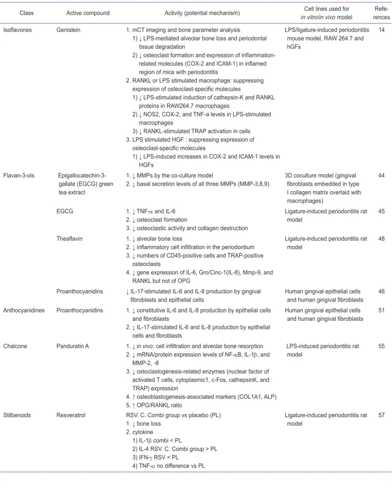
최근 치주질환을 포함한 염증성 질환의 치료목적으로 flavonoids와 proanthocyanidins를 포함하는 polyphenol의 약물 효과에 대한 연구 가 보고되었으며, 관련된 여러 연구에서 치조골 소실억제 및 면역반응 과 관련한 다양한 cytokine의 변화를 관찰하였으며, 이를 Table 6에 기 술하였다. 몇몇 연구에서는 치주결찰 또는 lipopolysaccharide (LPS) 를 이용하여 치주질환을 유도한 동물모델을 사용하여 연구하였다.
Table 6에 기술된 내용 중 몇 종류의 효과를 보면, polyphenol 중 baicalin, luteolin 및 puerarin 등은 결찰에 의해 유도된 치주질환에서 골흡수 억제 효과를 나타냈다[7,24,27]. 또한 genistein은 LPS에 자극 을 받은 대식세포에서 COX
2
, TNF-α 분비를 억제하였고[14], pandu- ratin A는 MMP-2, MMP-8의 발현을 억제하였다[46]. 여러 종류의Table 4. Continued
Class Active compound Activity (potential mechanism) Cell lines used for
in vitro/in vivo model Refe- rences Flavan-3-ols Proanthocyanidins 1. ↓ osteoclastogenesis and osteoclast activity
2. ↓ differentiation of mature osteoblasts
3. ↓ RANKL expression in fibroblasts from RA patients
4. ↓ human peripheral blood mononuclear cell-derived osteoclastogenesis
CIA mouse model 47
Coumaric acid p-coumaric acid 1. ↓ paw edema, body weight loss 2. ↓ TNF-α, IL-1β, IL-6, and MCP-1 3. ↓ RANKL, TRAP
4. ↓ TNF-α, IL-1β, IL-6, and IL-17 5. ↓ iNOS and COX-2 in arthritic rats 6. ↑ OPG expression
7. ↓ NF-κB-p65, and p-NF-κB-p65, NFATc-1, and c-Fos and MAP kinases expression in arthritic rats.
8. ↓ RANKL-induced NFATc-1 and c-Fos expression in vitro
9. Radiological (CT and DEXA scan) and histological assessments:↓TRAP, bone destruction and cartilage degradation in association with enhanced BMD
AIA rat model 52
Xanthanoids Mangiferin 1. exhibits strong pro-apoptotic effect in synoviocytes
2.↓ levels of matrix metalloproteinases in chondrocytes synoviocytes in human
synovia 56
Stilbenoids Resveratrol 1. ↓ rheumatoid factor (RF) serum levels
2. ↓ anti-citrullinated protein antibody (ACCPA) gingival levels 3. ↓ paw swelling level
4. Histological analyzes showed smooth articular surface and higher width of the subchondral cortical in RSV group.
5. ↓ paw swelling level
CIA and ligature-induced
periodontitis rat model 61
Stilbenoids Resveratrol ↓ tissue mRNA expressions of Wnt5a, MAPK3, Src kinase, and STAT3 CIA rat model 58 TNF, tumor necrosis factor; IL, interleukin; NO, nitric oxide; AIA, adjuvant-induced arthritis; NF-κB, nuclear factor kappa-B; MMP, matrix metalloproteinase;
CIA, collagen-induced arthritis; JNK, c-Jun N-terminal kinases; RANKL, receptor activator of nuclear factor kappa-Β ligand; OPG, osteoprotegerin; NFAT,
nuclear factor of activated T-cells; TRAP, tartrate-resistant acid phosphatase; BMD, bone mineral density; ERK, extracellular signal-regulated kinase; RA,
rheumatoid arthritis; MAPK, mitogen-activated protein kinase; STAT, signal transducer and activator of transcription.
polyphenol은 치조골 파괴의 중요한 역할을 담당하는 파골세포의 분화 를 억제하거나, 염증반응의 매개물질인 cytokine을 억제하고, 치주조직 을 이루는 결체조직 파괴에 관여하는 기질분해효소를 억제하는 효과를 가지므로, 치주질환 치료 전략의 하나로 사용될 수 있다.
Conclusions
식물유래 천연화합물은 류마티스관절염, 골관절염 및 치주질환과 같 은 뼈파괴 질환에서 통상적인 치료제의 부작용을 극복할 수 있는 대안 이 될 수 있으며, 이 논문은 flavonoid를 포함하는 polyphenol이 류마 티스관절염, 골관절염 및 치주질환의 치료목적으로 효과를 가지고 있음 을 정리하였다.
전달수준에서 MAPK를 비롯한 표적 경로의 물질의 발현 수준 조절, 관 련된 여러 염증성 매개물질의 발현 및 분비 조절뿐만 아니라 각 질환을 유도한 동물모델에서 치료목적의 효과를 가지는 것으로 보고되었다. 따 라서 이 논문은 다양한 식물유래 천연물을 뼈파괴 질환 극복에 이용할 수 있음을 보여주고 있다. 그러나 실제로 임상 환경에서 유익한지 확인 이 필요하므로, 추가 동물연구 및 사람을 대상으로 하는 임상 연구가 필 요하다.
Acknowledgements
이 논문은 2010년도 정부(교육과학기술부)의 재원으로 한국연구재단 의 지원을 받아 수행된 기초연구사업임(No. 2010-0023679).
Table 5. Main polyphenolic compounds investigated in osteoarthritis and their effects demonstrated in the different cellular or animal model Class Active
compound Activity (potential mechanism) Cell lines used for
in vitro/in vivo model Refe- rences Chalcone Phloretin 1. ↓ IL-1β-induced production of NO, PGE2, TNF-α, and IL-6
2. ↓ expression of COX-2, iNOS, MMP-3, MMP-13, and ADAMTS-5 3. ↓ degradation of aggrecan and collagen-II
Human OA chondrocytes, OA
mouse model 54
Flavones Nobiletin 1. ↓ overproduction of PGE2, NO, COX-2, iNOS, TNF-α, and IL-6 in IL-1β-induced human OA chondrocytes
2. ↓ expression of MMP-13, ADAMTS-5 3. ↓ degradation of aggrecan, collagen-II
4. ↓ IL-1β-stimulated phosphorylation of PI3K/Akt and activation of NF-κB in human OA chondrocytes
5. ↓ destruction of cartilage and the thickening of subchondral bone in mice OA models 6. ↓ synovitis in mice OA models
Human OA chondrocytes, OA mouse model
30
Flavonols Fisetin 1. ↓ IL-1β-induced inflammatory mediators in human osteoarthritis chondrocytes.
2. ↓ IL-1β-induced degradation of Sox-9, aggrecan, collagen-II in human osteoarthritis chondrocytes.
3. ↓ progression of osteoarthritis in mice models.
Human OA chondrocytes, OA mouse model
13
Flavanonol Silibinin 1. ↓ IL-1β-induced production of NO, PGE2, TNF-α, and IL-6
2. ↓ expression of COX-2, iNOS, MMP-1,-3,-13, ADAMTS-4, and ADAMTS-5, 3. ↓ degradation of aggrecan and collagen-II in human OA chondrocytes 4. ↓ IL-1β-stimulated PI3K/Akt phosphorylation and NF-kB activation in human OA
chondrocytes
5. ↓ destruction of cartilage and the thickening of subchondral bone in mice OA models 6. ↓ synovitis in mice OA models
7. ↓ expression of MMP-13, ADAMTS-5 in mice OA
Human OA chondrocytes, OA mouse model
39
Isoflavones Puerarin 1. ↓ IL-1β-induced production of inflammatory cytokines in OA chondrocytes and monocytes/macrophages
2. ↓ inflammatory profile of OA joints and ↓cartilage destruction
Human OA chondrocytes, OA mouse model
43
Chalcone Butein 1. ↓ IL-1β-induced inflammatory mediators in human osteoarthritis chondrocytes.
2. ↓ IL-1β-induced degradation of type II collagen and SOX-9 in human osteoarthritis chondrocytes.
3. ↓ IL-1β-induced IκB-α degradation, NF-kB p65 activation and NF-κB p65 nuclear translocation in human osteoarthritis chondrocytes.
4. ↓ arthritis in mice osteoarthritis models.
Human OA chondrocytes, OA
mouse model 53
NO, nitric oxide; PGE2, prostaglandin E2; TNF, tumor necrosis factor; IL, interleukin; MMP, matrix metalloproteinase; NF-κB, nuclear factor kappa-B; OA,
osteoarthritis.
Table 6. Main polyphenolic compounds investigated in periodontal diseases and their effects demonstrated in the different cellular or animal model
Class Active compound Activity (potential mechanism) Cell lines used for
in vitro/in vivo model Refe- rences
Flavones Baicalin 1. ↓ alveolar bone loss
2. ↓ levels of HMGB1, TNF-α, IL-1β, and MPO expression 3. ↓ numbers of inflammatory infiltrates in the gingival tissue 4. ↓ TLR2 and TLR4/MyD88/p38MAPK/NF-κB signaling
Ligature induced periodontitis, Porphyromonas gingivalis induced periodontitis rat model
24
Epimedokoreanin B 1. ↓ gingipain-induced hemostasis disorder in the P. gingivalis- diseased periodontal tissue
2. ↓ hemagglutinating activity of gingipains
Degradation of type I collagen 25
Luteolin 1. ↓ bone loss
2. ↑ osteoblast cell number
3. ↓ osteoclast and inflammatory cell numbers 4. ↑ BMP-2 expressions
5. ↑ TIMP-1 and BMP-2 expressions 6. ↓ MMP-8 and iNOS levels 7. ↑ OPG and ↓ RANKL levels
Ligature induced periodontitis rat model
27
Nobiletin and tangeretin ↓ LPS induced bone resorption RAW 264.7 cells 28
Nobiletin, tangeretin, heptamethoxyflavone (HMF), and tetramethoxyflavone (TMF)
↓ IL-1-induced osteoclast differentiation and bone resorption Co-Cultures, organ culture of mouse calvaria, OVX mouse model
29
Flavonols Sudachitin 1. ↓ c-fos, NFATc1, cathepsin K, DC-STAMP, Atp6v0d2 expression
2. ↓ OC generation
3. ↓ MAPKs activity (Erk and JNK) 4. ↓ ROS generation
LPS-induced calvarial bone destruction mouse model, co- culture
37
Quercetin 1. ↓ Aggregatibacter actinomycetemcomitans-induced bone loss
2. ↓ A. actinomycetemcomitans-induced IL-1β, TNF-α, IL-17, RANKL, and ICAM-1 production in the gingival tissue
A. actinomycetemcomitans induced periodontitis mouse model
33
Quercitrin 1. ↓ release of PGE2 and partially re-established the impaired collagen metabolism induced by IL-1β in human gingival fibroblasts (HGFs)
2. ↑ alkaline phosphatase (ALP) activity and mineralization in human mesenchymal stem cells
HGFs and mesenchymal stem cells
32
Isoflavones Puerarin 1. ↓ alveolar bone loss
2. ↓ collagen destruction and inflammatory cell infiltration 3. ↓ ratio of RANKL/osteoprotegerin and osteoclast activity 4. ↓ activation of NFkBp65 (lower IL-1β and TNF-α
production)
5. ↓ glycosylation of extracellular matrix metalloproteinase inducer
6. ↓ MMP-2 and MMP-9
7. ↓ production of RANKL, IL-1β, TNF-α, MMP-2, and MMP-9
Ligature-induced periodontitis rat
model 42
Table 6. Continued
Class Active compound Activity (potential mechanism) Cell lines used for
in vitro/in vivo model Refe- rences Isoflavones Genistein 1. mCT imaging and bone parameter analysis
1) ↓ LPS-mediated alveolar bone loss and periodontal tissue degradation
2) ↓ osteoclast formation and expression of inflammation- related molecules (COX-2 and ICAM-1) in inflamed region of mice with periodontitis
2. RANKL or LPS stimulated macrophage: suppressing expression of osteoclast-specific molecules
1) ↓ LPS-stimulated induction of cathepsin-K and RANKL proteins in RAW264.7 macrophages
2) ↓ NOS2, COX-2, and TNF-a levels in LPS-stimulated macrophages
3) ↓ RANKL-stimulated TRAP activation in cells 3. LPS stimulated HGF : suppressing expression of
osteoclast-specific molecules
1) ↓ LPS-induced increases in COX-2 and ICAM-1 levels in HGFs
LPS/ligature-induced periodontitis mouse model, RAW 264.7 and hGFs
14
Flavan-3-ols Epigallocatechin-3- gallate (EGCG) green tea extract
1. ↓ MMPs by the co-culture model
2. ↓ basal secretion levels of all three MMPs (MMP-3,8,9)
3D coculture model (gingival fibroblasts embedded in type I collagen matrix overlaid with macrophages)
44
EGCG 1. ↓ TNF-α and IL-6
2. ↓ osteoclast formation
3. ↓ osteoclastic activity and collagen destruction
Ligature-induced periodontitis rat model
45
Theaflavin 1. ↓ alveolar bone loss
2. ↓ inflammatory cell infiltration in the periodontium 3. ↓ numbers of CD45-positive cells and TRAP-positive
osteoclasts
4. ↓ gene expression of IL-6, Gro/Cinc-1(IL-8), Mmp-9, and RANKL but not of OPG
Ligature-induced periodontitis rat
model 48
Proanthocyanidins ↓ IL-17-stimulated IL-6 and IL-8 production by gingival
fibroblasts and epithelial cells Human gingival epithelial cells and human gingival fibroblasts 46 Anthocyanidines Proanthocyanidins 1. ↓ constitutive IL-6 and IL-8 production by epithelial cells
and fibroblasts
2. ↓ IL-17-stimulated IL-6 and IL-8 production by epithelial cells and fibroblasts
Human gingival epithelial cells and human gingival fibroblasts 51
Chalcone Panduratin A 1. ↓ in vivo: cell infiltration and alveolar bone resorption 2. ↓ mRNA/protein expression levels of NF-κB, IL-1β, and
MMP-2, -8
3. ↓ ostoclastogenesis-related enzymes (nuclear factor of activated T cells, cytoplasmic1, c-Fos, cathepsinK, and TRAP) expression
4. ↑ osteoblastogenesis-associated markers (COL1A1, ALP) 5. ↑ OPG/RANKL ratio
LPS-induced periodontitis rat
model 55
Stilbenoids Resveratrol RSV. C. Combi group vs placebo (PL) 1. ↓ bone loss
2. cytokine
1) IL-1β combi < PL
2) IL-4 RSV. C. Combi group > PL 3) IFN-γ RSV < PL
4) TNF-α no difference vs PL
Ligature-induced periodontitis rat
model 57
Conflicts of Interest
No potential conflict of interest relevant to this article was
reported.