I. Introduction
For the successful treatment of cleft lip and palate patients, close cooperation in treatment plan between the different specialties of medicine and dentistry is needed. Among the series of treatment plan, dental rehabilitation of residual alveolar cleft has been the main obstacle to the patients.
An important goal in the treatment of cleft patients is to normalize the anatomy of the cleft alveolar process. And one of the treatment procedures used to achieve this goal is bone grafting the defects in the alveolar process. Successful grafting improves the stability of maxillary segments, increases bony support for teeth close to the cleft from an adjacent area and support for alar
치조열환자에서 치조골 이식술의 예후
이수연 명훈 서병무 황순정 이종호 김명진 최진영, , , , , , *
서울대학교 치과대학 구강악안면외과학교실
ABSTRACT
Prognosis of Alveolar Bone Graft Alveolus in Cleft Patients : the preliminary report
Su-Yeon Lee, Hoon Myoung, Byoung-Moo Seo, Soon-Jung Hwang, Jong-Ho Lee, Pill-Hoon Choung , Myung-Jin Kim, Jin-Young Choi*
Department of Oral and Maxillofacial Surgery, College of Dentistry, Seoul National University
The case records of 24 patients in Seoul National University Hospital who had bone grafting of 29 alveolar clefts between 2001 and 2004 were examined. Details were recorded of age, sex, preoperative orthodontictreatment, the time of bone grafting, the type of donor site, cleft width, functional load applicationand the success of grafting as established by lowest marginal bone levels. Using this results and review of literature, we concluded that secondary bone graft with iliac bone before canine eruption with root development of 1/2 to 1/3 provide more favorable results and the functional load introduced to the grafted bone lower the resorption rates.
Key words : Alveolar cleft, Bone graft
구순구개 9:71~78, 2006
base. It eliminates oronasal fistulas. In addition, there is enhancement of alveolus and facial appearance
1). But the recommendation has been varied about the timing of operation and type of graft material. Pre-surgical cleft width, post-surgical functional load application, they are also known to have an effect on prognosis of resorption rate of grafted alveolus.
The purpose of this study was to evaluate the effect of the time of bone grafting, the type of donor site, cleft width, and functional loading at grafted site on the success of alveolar bone grafting with review of literatures.
II. Patients and Methods
Total 42 patients with cleft lip or cleft lip and palate had alveolar bone grafting at Seoul National University Dental Hospital in Korea from 2001 through 2004. Bone grafts were done by three operators. Only patients
who had complete records and radiographs taken before the bone graft and 1 year after surgical procedure available for evaluation were included. This was a retrospective study using records of 24 CLP patients. These consist of 5 patients with bilateral cleft lip and palate, and 19 patients with unilateral cleft lip and palate (all 29 cleft sites). There were 18 males and 6 females (Table 1). The age range at the time of surgery was 9 to 12, with a mean age of 13 years. We investigated the timing of bone graft, type of graft materials, cleft width and functional load application after bone graft.
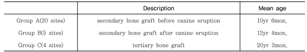
The Patients were divided into three groups with regard to eruption stage of the canine and age at the time of operation (Table 2). Group A represents the secondary bone grafting before canine eruption, Group B the secondary bone grafting after canine eruption, and Group C tertiary bone grafting.
The panoramic radiographs of patients were
Table 1. Distribution of the patients
UCLP BCLP
Male(n=18) 16 2
Female(n=6) 3 3
Total(n=24) 19 5
Table 2. Grouping according to operation time
Description Mean age
Group A(20 sites) secondary bone graft before canine eruption 10yr 6mon.
Group B(5 sites) secondary bone graft after canine eruption 12yr 4mon.
Group C(4 sites) tertiary bone graft 20yr 3mon.
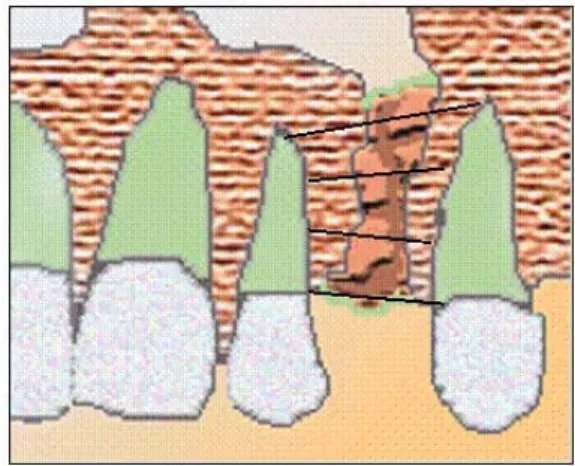
analyzed and bone resorption level was divided into four scores with the lowest
그림
marginal bone level ( 1) using the method described by Enemark et al.
2). Score 1 indicates the marginal bone level of normal interdental height, score 2 the bone level greater than 3/4 normal height, score 3 indicates the level less than 3/4 normal height, and score 4 indicates no bony bridge achieved. In this method, score 4 was considered fail. Cleft width was measured at its narrowest part near the cemento-enamel junction of adjacent teeth. According to the width of the cleft, it was divided into three groups and bone level was compared. Group I represent 3.1-5.9 mm of cleft width, Group II represent 6.0-8.9 mm of cleft width and Group III 9.0-12.5 mm of cleft width.
III. Results
1. Bone levels (General)
In the follow up of 29 secondary bone grafts, 23 (79.3%) showed a resorption of grade I, 4 (13.8%) a resorption of grade II, and 2 (6.9%) a resorption of grade III. All graft had a bone bridge in cleft site so no failure had occurred.
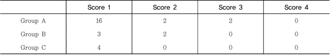
2. Bone levels according canine eruption At the time of this study, 20 of the 29 canines were still unerupted and 9 were fully erupted. Most were grafted by the age of 9-12 and 20 cleft sites were grafted before canine eruption. And bone resorption grade in each group were showed in Table 3. The outcome was favorable in all three groups.
3. Type of graft material
Type of graft material was all iliac cancellous bone.
4. Cleft width
A total of 29 cleft sites were observed.
The mean cleft width was 7.6 mm with a range of 3.1 mm to 12.5 mm. The bone level according to cleft width was shown in table 4. Regardless of the cleft width, all bone graft was successful.
5. Functional load
After 1 year follow-up, 12 cleft gap were closed by orthodontic movement of canine.
그림 1. Schematic representation of bone
resorption level : score 1-the marginal
bone level of normal interdental height,
score 2-the bone level greater than 3/4
normal height, score 3-the level less
than 3/4 normal height, and score 4-no
bony bridge achieved.
14 cleft gaps were just left waiting for appropriate timing of prosthodontic treatment.
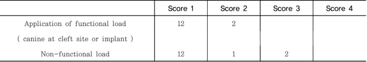
Implants were inserted in 2 cleft gap. And one cleft gap was closed by dental bridge work. We considered the cleft gap was under functional load when it was closed by implants or canine movement. There were no differences in resorption rate between the functional load and non- functional load group.
IV. Discussion
The objectives of alveolar bone grafting have evolved over time. Many reasons for this alveolar bone grafting include arch stability, tooth eruption, oronasal fistula closure, esthetic result, speech improvement.
Essential aim is the provision of bone to facilitate tooth eruption and orthodontic movement. But there are many factors affecting the prognosis of alveolar bone grafting.
With respect to the timing of the graft, it has been the subject of debate over many years and the recommended timing has varied. According to time of occurrence, the bone graft is divided into primary, secondary, or tertiary
3). When it is performed during the first stage of dentition, at the same time as the primary repair surgeries, bone graft is called primary. Primary grafting has become unpopular because of adverse effect on maxillary growth
4,5). Bone grafting is called secondary when performed at the end of the mixed dentition. It has become a well established procedure since the original work
of Boyne and Sand
6). It is ideally done between the age of 9 and 11
7,8). When a bone graft is performed in the permanent dentition, it is called tertiary. Tertiary graft are performed to enable prosthodontic and periodontal rehabilitation and to assist in the closure of persistent oronasal fistulae
9). But nowadays secondary bone graft is the prevailing consensus. After the Boyne et al.4) in 1972, many studies have supported this secondary alveolar bone grafting
10-13). Bergland et al. in 1986 suggested that the ideal time for grafting is between nine and 11 years before the eruption of canine and when canine root has 1/2 to 2/3 development.
And Enemark et al. demonstrated that significantly better results are achieved with secondary bone grafting if the treatment is performed before eruption of canine
9). In our study, the most of bone grafts were the secondary bone grafting and the tertiary bone grafting was performed only when patients visited after their growths. There were no differences in success between the groups according to the time of operation.
The secondary bone graft supports the eruption of canine and when the canine eventually erupts, it creates a periodontium of protection that usually maintains an interdental bone septum with good height.
Thus periodontal conditions are better than any other grafts
7). And it also have a minimal effect on subsequent vertical and antero-posterior development of maxilla
14).
A variety of donor sites have been
described for the alveolar bone grafting and these include the iliac crest, the calvarium, the rib, the mandibular symphysis, and the tibia. Historically the donor bone was usually obtained from the ilium or the cranium for the reconstruction of the bony clefts, while a few surgeons still recommend the use of others. Dracher first used tibial bone as a donor source in 1914. And a cranial bone graft was introduced in 1983 with the advances of craniofacial surgery and the advantages over iliac bone: it was easily harvested; the bone was membranous theoretically ideal replacement for the maxilla; and the donor site was much less painful. Boyne and Sands used the iliac bone and pointed out that the iliac bone is capable of responding physiologically to the orthodontic movement and migration of teeth. Thereafter, alveolar bone grafting using cancellous bone chips from the iliac
crest was used successfully by a number of centers, bringing it into common usages in the alveolar clefts. Now, the iliac crest is considered as the ‘gold standard’ for the alveolar grafting
15,16). In our cases, all donor sites were the iliac crest. So comparisons with other sites were unavailable.
Regarding the presurgical orthodontics, the role of it is to align and expand the maxillary arch to restore arch width and symmetry prior to bone grafting. But it also has the potential to increase the width of cleft which could be possible factor influencing the success of bone grafting. In this series three groups were divided according to the cleft width. They didn’t show any statistical differences of bone resorption level. Long et al.
1)suggested that the success of grafting is not related to the width of the cleft at the time of surgery.
Grafting the bone in a wide cleft is Table 3. Bone resorption according to the canine eruption group
Score 1 Score 2 Score 3 Score 4
Group A 16 2 2 0
Group B 3 2 0 0
Group C 4 0 0 0
Table 4. Bone resorption according to the cleft width
Score 1 Score 2 Score 3 Score 4
Group I (n=19) 15 4 0 0
Group II (n=9) 7 0 2 0
Group III (n=1) `1 0 0 0
* Table 4 : Group I represent 3.1-5.9 mm of cleft width, Group II represent 6.0-8.9 mm of cleft width and Group III 9.0-12.5 mm of cleft width
technically easier than in a narrow cleft.
After the bone graft, the closure of the gap in the upper dental arch can be possible by the help of orthodontic treatment, a dental bridge, or a dental implant. In our cases, the resorption rate between the functional load and non- functional load application group didnt have any differences (Table 5). ʼ However Dempf et al. contended that the best result can be achieved by aligning the cleft-adjacent teeth into the grafted area or installing the endosseous implant in grafted site. Because these can exert functional stimulation to the transplanted bone by mastication so a progressive resorption is prevented
17). Schultze-Mosgau et al. analyzed the bone resorption of secondary bone graft area after orthodontic gap closure and prosthodontic treatment and concluded that a more favorable result can be achieved with orthodontic correction of dental arch through gap closure by insertion of canine in the grafted bone than with a gap opening by prosthodontic treatment18). So the resorption of cleft site can be prevented by the functional stimulation of grafted bone. And the functional stimulation can be achieved by the orthodontic closure or an implant installation.
Traditionally, the successful grafting of alveolar cleft has been judged by following:
(1) the permanent closure of fistula, (2) the stability of the maxillary segment, (3) the radiographic evidence of bone across the alveolar cleft region, (4) the adequate support for the alar base, and (5) the absence of soft tissue recesses in the alveolus19). In a word, the most important thing that has been the consideration of success was the bone continuity of alveolar cleft area. However, functional load application prevents the resorption of grafted alveolar clefts and reported cases of implant installation on alveolar cleft area are increasing with the advancement of dental implantology. So, adequate bone volume and width for endosseous implant measured by dental CT can be added in this success criterion. Based on the review of the journals and our data, it is possible to conclude that the secondary bone graft with iliac bone before canine eruption with root development of 1/2 to 1/3 provides more favorable results and the functional load introduced to the grafted bone lowers the resorption rates. But the limitation of small study group and short term follow-up period, longer follow-up will be necessary.
Table 5. Bone resorption according functional loading
Score 1 Score 2 Score 3 Score 4
Application of functional load 12 2
( canine at cleft site or implant )
Non-functional load 12 1 2
REFERENCE
1. Long, R.E., Jr., B.E. Spangler, and M.
Yow, Cleft width and secondary alveolar bone graft success. Cleft Palate Craniofac J 1995;32:420-7.
2. Enemark, H., S. Sindet-Pedersen, and M. Bundgaard, Long-term results after secondary bone grafting of alveolar clefts.
J Oral Maxillofac Surg 1987;45:913-9.
3. Sindet-Pedersen, S. and H. Enemark, Comparative study of secondary and late secondary bone-grafting in patients with residual cleft defects. Short-term evaluation.
Int J Oral Surg 1985;14:389-98.
4. Friede, H. and B. Johanson, Adolescent facial morphology of early bone-grafted cleft lip and palate patients. Scand J Plast Reconstr Surg 1982;16:41-53.
5. Friede, H. and B. Johanson, A follow-up study of cleft children treated with primary bone grafting. 1. Orthodontic aspects. Scand J Plast Reconstr Surg 1974;8:88-103.
6. Boyne, P.J. and N.R. Sands, Secondary bone grafting of residual alveolar and palatal clefts. J Oral Surg 1972;30:87-92.
7. Amanat, N. and J.D. Langdon, Secondary alveolar bone grafting in clefts of the lip and palate. J Craniomaxillofac Surg 1991;19:7-14.
8. Newlands, L.C., Secondary alveolar bone grafting in cleft lip and palate patients.
Br J Oral Maxillofac Surg 2000;38:488-91.
9. da Silva Filho, O.G., et al., Secondary bone graft and eruption of the permanent canine in patients with alveolar clefts:
literature review and case report. Angle Orthod 2000;7:174-8.
10. Ames, J.R., D.E. Ryan, and K.A. Maki, The autogenous particulate cancellous bone marrow graft in alveolar clefts. A report of forty-one cases. Oral Surg Oral Med Oral Pathol 1981;51:588-91.
11. Hall, H.D. and J.C. Posnick, Early results of secondary bone grafts in 106 alveolar clefts. J Oral Maxillofac Surg 1983;41:289-94.
12. Bergland, O., G. Semb, and F.E.
Abyholm, Elimination of the residual alveolar cleft by secondary bone grafting and subsequent orthodontic treatment.
Cleft Palate J 1986;23:175-205.
13. Kortebein, M.J., C.L. Nelson, and A.M.
Sadove, Retrospective analysis of 135 secondary alveolar cleft grafts using iliac or calvarial bone. J Oral Maxillofac Surg 1991; 49:493-8.
14. Daskalogiannakis, J. and R.B. Ross, Effect of alveolar bone grafting in the mixed dentition on maxillary growth in complete unilateral cleft lip and palate patients. Cleft Palate Craniofac J 1997;34:455-8.
15. Cohen, M., et al., Iliac versus cranial
bone for secondary grafting of residual
alveolar clefts. Plast Reconstr Surg
1991;87:423-7; discussion 428.
16. LaRossa, D., et al., A comparison of iliac and cranial bone in secondary grafting of alveolar clefts. Plast Reconstr Surg 1995;96: 789-97; discussion 798-9.
17. Dempf, R., et al., Alveolar bone grafting in patients with complete clefts: a comparative study between secondary and tertiary bone grafting. Cleft Palate Craniofac J 2002;39: 18-25.
18. Schultze-Mosgau, S., et al., Analysis of bone resorption after secondary alveolar cleft bone grafts before and after canine eruption in connection with orthodontic gap closure or prosthodontic treatment. J Oral Maxillofac Surg 2003;61:1245-8.
19. Kearns, G., et al., Placement of endosseous implants in grafted alveolar clefts. Cleft Palate Craniofac J 1997;34:520-5.
교신 저자
최진영 서울대학교 치의학대학원 구강악안면외과학교실,
서울시 종로구 창경궁로62-1 110-749/전화02-2072-3992/ e-mail: [email protected]