150 This is an Open-Access article distributed under the terms of the Creative Commons Attribution Non-Commercial License (http://
creativecommons.org/licenses/by-nc/3.0) which permits unrestricted non-commercial use, distribution, and reproduction in any medium, provided the original work is properly cited.
J. Mushrooms 2021 September, 19(3):150-159 http://dx.doi.org/10.14480/JM.2021.19.3.150 Print ISSN 1738-0294, Online ISSN 2288-8853
© The Korean Society of Mushroom Science
Ki Nam Yoon(Associate professor), Tae Soo Lee (Emeritus professor)
*Corresponding author E-mail : [email protected]
Tel : +82-32-835-4617,Fax : +82-32-835-0763 Received August 31, 2021
Revised September 13, 2021 Accepted September 24, 2021
Melanin synthesis and skin wrinkle inhibitory effects of the medicinal mushroom Ganoderma applanatum
Ki Nam Yoon1 and Tae Soo Lee2*
1Department of Clinical Laboratory Science, Ansan University, Ansan Dae-hak-ro, Sangrok-gu, Ansan 15328, Korea
2Division of Life Sciences, Incheon National University, 119 Academy-ro, (Songdo-dong) Yeonsu-gu, Incheon 22012, Korea
ABSTRACT: Anti-melanogenesis and skin anti-wrinkle effects of methanol (ME) and hot water (HE) extracts from the fruiting bodies of Ganoderma applanatum were investigated in this study. The total phenolic contents of the ME and HE of the mushroom were 11.68 and 3.15µg GAEs/mg, respectively, whereas the total flavonoid contents of the ME and HE were 21.82 and 2.69µg QEs/mg, respectively. The survival rate of B16-F10 murine melanoma cells treated with 750 µg ME and HE were 83.46% and 85.54%, respectively, thereby suggesting that mushroom extracts were slightly cytotoxic at the tested concentration.
The in vitro tyrosinase inhibition by ME (83.15%) and HE (83.44%) was significantly lower than that of kojic acid (99.61%), the positive control, at 2.0 mg/mL. Although the inhibition of cellular melanin synthesis in B16-F10 melanoma cells by 2.0 mg/mL of ME (50.24%) and HE (51.24%) was lower than that of arbutin (64.84%), the inhibition by both ME and HE was higher than 50%.
Collagenase inhibition by HE was comparable to 2.0 mg/mL epigallocatechin (EGCG), the positive control; however, elastase inhibition by ME and HE was lower than that of EGCG at the concentration tested. The results showed that the fruiting bodies of G. applanatum had good anti-tyrosinase, good anti-collagenase, and moderate anti-elastase activities, which might be useful for developing novel skin-whitening and anti-wrinkle agents.
KEYWORDS: Anti-melanogenesis, Anti-wrinkle, Cytotoxicity, Ganoderma applanatum
INTRODUCTION
Tyrosinase is one of the key enzymes responsible for melanin biosynthesis, catalyzing the hydroxylation of L- tyrosine to 3,4-dihydroxyphenylalanine (DOPA) and converting DOPA to dopaquinone (Zawistowski et al., 1991). Melanin is a colored pigment, which plays a key role in protecting the skin from harmful ultraviolet (UV) light. Skin hyperpigmentation, resulting from increased melanin-synthesizing enzyme activity caused by excessive
exposure to UV radiation may be responsible for skin disorders, including age spots, freckles, melasma, and malignant melanoma (Robb, 1984). Recently, tyrosinase inhibitors have become important treatments to prevent hyperpigmentation and other melanin-related disorders.
Since light-colored skin is preferred by some people, the cosmetic industry has turned its attention to tyrosinase inhibitors to treat skin hyperpigmentation (Brenner and Hearing, 2008). To address these problems, natural compounds derived from plants and microorganisms, including kojic acid, arbutin, azelaic acid, gentisic acid, and others, have also been developed (Chang, 2009). The effectiveness of depigmentation agents is determined by mushroom tyrosinase inhibition in cell-free systems or by the inhibition of cellular tyrosinase and new tyrosine inhibitors from natural sources are continually being screened.
Skin aging is a complex biological process of living organisms influenced by both intrinsic and extrinsic factors that include hormonal changes, metabolic process, inheritance, cigarette smoking, and UV radiation. These factors can cause changes in skin thickness, loss of skin tone, and wrinkles (Zhang and Duan, 2018). Skin is composed of epidermal, dermal, and subcutaneous tissue
and the soft outermost part of the skin is an extracellular matrix (ECM) consisting of collagen, fibronectin, and elastin (Muiznieks and Keeley, 2013). Collagen contributes flexibility, strength, and elasticity to the skin. It also preserves the structural integrity of the skin and plays a key role in normal cellular functions during skin morphogenesis (Shoulders and Raines, 2009). Elastin is a crucial protein existing in the connective tissue of the ECM that contributes elasticity and three-dimensional integrity to the skin. Collagenase, a zinc-dependent endopeptidase, breaks down peptide bonds, specifically ECM type 1 collagen. Elastase, a proteolytic enzyme, which degrades collagen and elastin, also regulates the mechanical and structural integrity of the ECM connective tissue (Kim et al., 2004). Therefore, there is increasing interest in natural products that can halt and delay skin aging, skin sagging, and wrinkling.
Mushrooms have been used worldwide as a good source of food and folk medicines for thousands of years.
The fruiting bodies of mushrooms possess biologically beneficial metabolites, such as polysaccharides, polyphenols, flavonoids, terpenoids, and other compounds, with good medicinal qualities that contribute to immune potentiation, as well as antitumor, anti-hyperglycemic, anti-hyperlipidemic, and anti-inflammatory activities (Lindequist et al., 2005; Wasser and Weis, 1999). Wu et al. (2016) reported that many mushrooms have known to possess ingredients such as phenolics, flavonoids, terpenoids, polysaccharides, vitamins, and volatile organic compounds. These compounds are beneficial to anti-aging, anti-wrinkle, skin whitening, and good moisturizing effects, which make them good candidates for cosmetics materials. Lee et al. (2016) demonstrated that hot water extract from fruiting bodies of Tremella fuciformis exhibited the anti-melanogenesis effect by decreasing melanin contents and tyrosinase activity in B16-F10 melanoma cells. Furthermore, the extract showed anti-wrinkle effect by increasing the synthesis of type I procollagen and reduced mRNA expression of matrix metalloproteinase 1 (MMP-1) activity in the human dermal fibroblast, which suggested that hot water extract of T. fuciformis reduce the cellular melanogenesis expression and protect against wrinkles caused by ageing, and UV radiation damage.
G. applanatum, known as an artist's conk mushroom, belongs to the phylum Basidiomycota, class Agaricomycetes, order Polyporales, and family Ganodermataceae, and is distributed worldwide (Park and Lee, 2003). Several
mushroom species belonging to the genus Ganoderma have been studied for their potential medicinal antioxidant, anti- diabetic, anti-hyperlipidemic, anti-microbial, and antitumor activities (Ying et al., 1987). Although a few species of Ganoderma have been used in Asian countries for thousands of years for their medicinal properties, only G. lucidum has been cultivated on an industrial scale and utilized for medicinal purposes for human health. However, there are only a few published reports on the medicinal effects of G.
applanatum (Tsivileva et al., 2018).
Therefore, the aim of this study was to evaluate the anti-melanogenesis, and anti-wrinkle effects of methanol and hot water extracts from the fruiting body powder of G. applanatum.
MATERIALS AND METHODS
Chemicals and reagents
Dimethyl sulfoxide, (DMSO), tyrosinase, L-3,4- dihydroxyphenylalanine (DOPA), kojic acid, synthetic melanin, arbutin, collagenase, and porcine pancreatic elastase were purchased from Sigma-Aldrich, Co. (St. Louis, MO, USA). All other chemicals and solvents used for the study were of analytical grade.
Mushroom extract
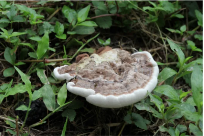
The fruiting bodies of G. applanatum were collected from Incheon City Grand Park and identified by Dr.
Kyung-Rim Lee, mycologist. The voucher specimen (IUM 3393) was deposited in the “Culture Collection of Mushrooms”, Incheon National University (Fig. 1). The fruiting bodies were air-dried (45oC for 48 h) and finely pulverized. To prepare the methanol extract (ME), 20 g of the powder and 400 mL of 80% methanol were
Fig. 1. Fruiting body of Ganoderma applanatum used in this study.
mixed in an orbital shaker (125 rpm) for 24 h at 25oC.
The solution was filtered through filter paper. To obtain the hot water extract (HE), the same amount of mushroom powder was boiled for 3 h in 400 mL of deionized distilled water and filtered. Then, the first ME and HE residues were extracted with 400 mL of 80%
methanol or hot water, respectively, twice, using the same method described above. The extracts were evaporated to dryness at 40oC and remaining water and solvent were removed with freeze-drier.
Total phenolics and flavonoid contents
The total phenolic contents of fruiting bodies of G.
applanatum were determined by the Folin–Ciocalteu method (Meda et al., 2005). Briefly, 0.1 g of methanol and hot water extracts of the mushroom dissolved in 1 mL deionized water. The solution (0.1 mL) was mixed with 2.8 mL of deionized water, 2 mL of 2% sodium carbonate (Na2CO3), and 0.1 mL of 50% Folin–Ciocalteau reagent. After incubation at room temperature for 30 min, the absorbance of the reaction mixture was measured at 750 nm against a deionized water blank on a spectrophotometer. Gallic acid (GA) was used as a positive control. the data were expressed as microgram (µg) gallic acid equivalents (GAE)/mg of lyophilized powder. The data were expressed as microgram (µg) gallic acid equivalents (GAE)/mg.
The total flavonoid content was determined by the aluminum chloride colorimetric method described by Chang et al. (2002) , Briefly, 0.1 g of methanol and hot water extracts were dissolved in 1 mL deionized water.
The solution (0.5 mL)was mixed with 1.5 mL of 95%
alcohol, 0.1 mL of 10% aluminum chloride hexahydrate (AlCl3), 0.1 mL of 1 M potassium acetate (CH3COOK), and 2.8 mL of deionized water. After incubation at room temperature for 40 min, the absorbance of reaction mixture was measured at 415 nm against deionized water blank on a spectrophotometer. Quercetin was used as a positive control.
Cytotoxicity assay Cell culture
B16-F10 murine melanoma cells were purchased from the Korean Cell Line Bank of the Korean Cell Line Research Foundation in Seoul, Korea. The B16-F10 melanoma cells were cultured in Dulbecco’s Modified Eagle’s Media (DMEM) supplemented with 10% heat- inactivated fetal bovine serum (FBS), penicillin (100 U/mL),
streptomycin (100 μg/mL), and 1% L-glutamine at 37oC with 5% atmospheric CO2 in a humidified incubator.
Cell viability test
The effect of ME and HE on B16-F10 melanoma cell viability was evaluated by the MTT assay (Mosmann, 1983). The cells (5 × 10⁴) were incubated in a 96-well plate for 24 h at 37oC. Then, the cells were treated with various concentrations of ME and HE (25-750 μg/mg) for the B16-F10 melanoma cells and incubated for 48 h in MTT solution (200 μL of 0.5 mg/mL) was added to each well and the plate was incubated for 4 h in the dark. The resulting colored formazan crystals were dissolved in 200 μL of dimethyl sulfoxide (DMSO) and the absorbance of the solution was measured at 570 nm with a microplate reader.
Inhibitory effect of melanin synthesis Inhibition of tyrosinase
Inhibition of tyrosinase by the mushroom extracts was determined using a published method (Kim and Ryu, 2012) with slight modification. Briefly, the reaction mixtures were prepared by the addition of 40 µL of 31 units/mL tyrosinase to 40 µL of varying concentrations of ME and HE (0.125-2.0 mg/mL), 40 µL of 1.5 mM L- tyrosine, and 80 µL of 0.1 M phosphate buffer (pH 6.8).
Then, the mixture was incubated for 10 min at 37oC.
The absorbance was measured with a microplate reader at 475 nm. Tyrosinase inhibition was calculated using the following formula:
Inhibition of tyrosinase (%) = [(Absc – Abss)/Absc] × 100
where Absc represents the absorbance of the vehicle control and Abss represents the absorbance of the test sample. BHT was used as a reference standard.
Inhibition of L-DOPA autooxidation
The inhibition of L-DOPA autooxidation by ME and HE was performed using a slight modification of a published method (Masuda et al., 2005). Briefly, the reaction mixtures were prepared by the addition of 40 µL of 31 units/mL tyrosinase to 40 µL of varying concentrations of ME and HE (0.125-2.0 mg/mL) and incubated for 10 min at 37oC. Then, the reaction was initiated by adding 40 µL of 2.5 mM L-DOPA solution in phosphate buffer and 80 µL of 0.1 M phosphate
buffer (pH 6.8) and incubated for 10 min at 37°C in the dark. The absorbance of the solution was measured at 475 nm with a microplate reader. The percentage of L- DOPA autooxidation inhibition was calculated using the following formula:
Inhibition of DOPA autooxidation (%) = [(Absc – Abss)/Absc] × 100
where Absc represents the absorbance of the vehicle control and Abss represents the absorbance of the test sample. Kojic acid was used as a reference standard.
Inhibition of cellular melanin synthesis
The melanin content of B16-F10 melanoma cells treated with varying concentrations of mushroom extracts was performed using the method published by Hosoi et al.
(1985). The cells (4 × 10⁴ cells/well) were cultured in a 96- well plate in DMEM. After 24 h of incubation, the cells were treated with ME and HE (25-500 µg/mL) for 72 h containing 0.4 µM melanocyte-stimulating hormone (MSH).
The cells were harvested by the addition of 300 µL of 1%
Triton X-100 (in PBS), centrifuged for 5 min at 3,000 rpm, and the supernatant removed. Then, 100 µL of 1N NaOH and 200 µL of 10% DMSO were added to the cell pellets and kept for 1 h at 60oC. The absorbance of the solution was measured using a microplate reader at 405 nm and compared to standard curves of synthetic melanin. Arbutin was used as a reference standard.
Skin anti-wrinkle effect Inhibitory effect of elastase
The elastase inhibitory effect of ME and HE was assessed by previously reported method (Kim et al., 2004). Porcine pancreatic elastase (PE-EC. 3.4.21.36) was dissolved in sterile water to make a 3.33 mg/mL stock solution. The substrate, N-Succinyl-Ala-Ala-Ala-p- nitroanilide (AAAPVN), was dissolved in 0.2 M Tris- HCl buffer (pH 8.0). The reaction was carried out in a 96-well microplate. Each well contained 120 µL of 0.2 M Tris-HCl buffer, 50 µL mushroom extract (0.125 – 2.0 mg/mL) in Tris-HCl buffer, 30 µL PE, and 50 µL of 1.6 mM AAAPVN. The extract was preincubated at 25oC for 15 min before the addition of the substrate.
The absorbance was measured with a microplate reader at 402 nm. The percent inhibition of elastase was calculated as follows:
Elastase inhibition (%) = [(Absc – Abss)/Absc] × 100 where Absc represents the absorbance of the vehicle control and Abss represents the absorbance of the test sample. EGCG was used as a reference standard and distilled water was used as a negative control.
Inhibitory effect of collagenase
The inhibition of collagenase by ME and HE was investigated using previously reported method (Van Wart and Steinbrink, 1981) with some modifications. Collagenase from Clostridium histolyticum (ChC - EC 3.4.24.3) was solubilized in the 50 mM of 1.44 units/mL Tricine buffer (pH 7.8). The substrate, 4-phenylazobenzyloxycarbonyl-Pro- Leu-Gly-Pro-D-Arg trifluoroacetate salt (PZ-peptide), was dissolved in 2.5 mM tricine buffer. Twenty-five microliters of mushroom extracts (0.125-2.0 mg/mL) were incubated with 20 µL of enzyme in 155 µL buffer for 15 min at 37oC before addition of 50 µL PZ-peptide to initiate the reaction and the mixture was incubated for 60 min at 37oC. The reaction was terminated by adding 1 mL of citric acid (3%).
Then, 5 mL of ethyl acetate was added and the solution was vigorously shaken and allowed to stand for 10 minutes. The absorbance of the supernatant was measured at 320 nm using a microplate reader. EGCG was used as a reference standard. The inhibition of collagenase was calculated using the following formula:
Collagenase inhibition (%) = [(Absc – Abss)/Absc] × 100
where Absc represents the absorbance of the vehicle control and Abss represents the absorbance of the test sample.
Statistical Analysis
All data were expressed as mean ± standard deviations (SD) and SPSS V.13 (SPSS Inc., Chiago, IL, USA) was used for statistical analysis. One-way analysis of variance followed by Tukey multiple comparisons were used to compare means between groups. Differences between means at the 5% (p ≤ 0.05) level were considered statistically significant.
RESULTS AND DISCUSSION
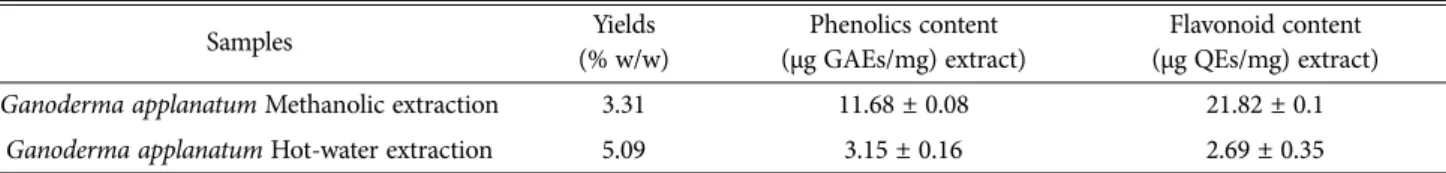
Total phenolic and flavonoid contents
The results showed that the total phenolic and flavonoid
contents in the metahnol and hot water extracts of G.
applanatum varied considerably. Total phenolic contents from methanol extract and hot water extract of G.
applanatum ranged from 11.68 ± 0.08 to 3.15 ± 0.16 µg GAE/mg, respectively (Table 1). This study showed that total phenolic contents in the methanol extract was higher than that of hot water extract.
The total flavonoid contents in the methanol and hot water extracts of G. applanatum ranged from 21.82 ± 0.10 to 2.69 ± 0.35 µg QEs/mg, respectively (Table 1), suggesting that extraction by methanol gave the higher phenolic and flavonoid contents as compared to the hot water extract.
Nguyen et al. (2013) reported that phenolic contents of methanol extract from fruiting bodies of Phellinus xeraticus was 16.37 µg GAE/g, suggesting that phenolic contents of fruiting bodies from P. xeranticus was higher compared to G. applanatum, whereas flavonoid content of P. xeranticus was lower (2.40 µg QEs/mg) than that of G. applanatum.
(3.15 µg QEs/mg).
Phenolics and flavonoids are well-known for possessing one or more aromatic rings with large numbers of hydroxyl groups, which benefit human
health by scavenging free radicals and inhibiting hydrolytic enzyme and tyrosinase activity (Walter and Marchesan, 2011; Yoon et al., 2011). Many researchers have demonstrated a positive relationship between the concentrations of phenolic compounds and in vitro tyrosinase inhibition and cellular melanin synthesis inhibition activities in B16 mouse melanoma cells (Sun et al., 2017).
Cell viability
To determine the extracts from G. applanatum fruiting bodies exhibited a cytotoxic effect on B16-F10 melanoma cells, the cells were treated with varying concentrations of ME and HE for 72 h and the cell viability was determined by the MTT assay. The cell viability of the B16-F10 melanoma cells treated with ME and HE ranged from 108.60 to 83.45% and from 111.80 to 88.54%, respectively, at 50 to 750 μg/mL (Fig. 2).
The cell viabilities of the B16-F10 melanoma cells decreased with increasing concentrations of the extracts.
Since the viabilities of B16-F10 melanoma cells treated with 750 μg/mL ME and HE demonstrated 83.46% and Table 1. Total phenolics and flavonoid contents of methanol and hot-water extract from fruiting bodies of Ganoderma applanatum
Samples Yields
(% w/w)
Phenolics content (μg GAEs/mg) extract)
Flavonoid content (μg QEs/mg) extract)
Ganoderma applanatum Methanolic extraction 3.31 11.68 ± 0.08 21.82 ± 0.1
Ganoderma applanatum Hot-water extraction 5.09 3.15 ± 0.16 2.69 ± 0.35
Results are expressed as means ± standard deviation (SD, n = 3) GAEs, gallic acid equivalents; QEs, quercetin evalents
Fig. 2. Cytotoxic effects of methanol and hot water extracts from fruiting bodies of Ganoderma applanatum on B16-F10 melanoma cells. The results are expressed as means ± SD (n = 3).
85.54% viabilities, respectively, suggesting that 750 μg/
mL ME and HE of G. applanatum were slghtly cytotoxic to the B16-F10 melanoma cells. Therefore, the inhibition of cellular tyrosinase and melanin synthesis in B16-F10 melanoma cells were analyzed at 50-750 μg/
mL mushroom extracts.
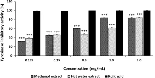
Inhibitory effect of melanogenesis Inhibition of tyrosinase
The tyrosinase inhibition by ME and HE from the fruiting bodies of G. applanatum ranged from 31.88% to 83.15% and from 38.88% to 83.44%, respectively, at 0.125-2.0 mg/mL (Fig. 3) and increased with increasing concentrations. However, kojic acid, the reference standard, demonstrated excellent tyrosinase inhibition of 98.74 to 99.61% at 0.125-2.0 mg/mL. These results suggest that ME and HE exhibited a moderate inhibitory effect at the concentrations tested. Alam et al. (2011) reported that the tyrosinase inhibition by methanol and hot water extracts from the fruiting bodies of P.
salmoneostramineus ranged from 20.90% to 63.29% and from 15.25% to 49.25%, respectively, at 0.125-1.0 mg/
mL, demonstrating greater tyrosinase inhibition by G.
applanatum than by P. salmoneostramineus.
Phenolic compounds are aromatic compounds containing phenolic rings and carboxylic acid. Flavonoids are a group of polyphenolic compounds found in many plant and mushroom species. Polyphenols are regarded as substrates for tyrosinase and are the largest group of tyrosinase inhibitors. Several polyphenols were shown to inhibit tyrosinase by binding to the active site of the enzyme,
resulting in irreversible inactivation of the enzyme by a substrate-like interaction (Wang et al, 2014). Antioxidant activity may also be one of the important mechanisms of tyrosinase inhibition. Some flavonoids like quercetin, naringin, and kaempferol also inhibit tyrosinase by chelating the copper ions in tyrosinase (Kim et al., 2006).
The inhibitory effect of ME and HE toward tyrosinase might be due to the polyphenolic compounds and flavonoids present in the mushroom extracts.
Inhibition of L-DOPA auto-oxidation
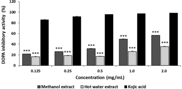
L-DOPA is one of the precursors of melanin during melanogenesis. L-DOPA is synthesized from L-tyrosinase by tyrosinase hydroxylase. Tyrosinase can also convert L-DOPA to dopaquinone, eventually leading to melanin. The in vitro inhibition of L-DOPA autooxidation by G. applanatum is summarized in Fig. 4. The inhibition of L-DOPA autooxidation by ME and HE ranged from 38.25% to 78.46% and from 29.68% to 44.77%, respectively, at 0.125 to 2.0 mg/mL, whereas kojic acid demonstrated excellent L- DOPA inhibition of 86.14 to 98.72% at the concentration tested. The results showed that ME and HE moderately inhibited L-DOPA autooxidation. The inhibition of L-DOPA autooxidation by ME and HE, however, was less when L- DOPA was used as a substrate than when L-tyrosine was used as a substrate of tyrosinase. Since tyrosinase uses two different binding sites for tyrosine hydroxylase and L-DOPA oxidase (Hwang and Lee, 2007), our experimental results suggest the selective effectiveness of tyrosinase for the two different substrates of the mushroom during melanogenesis.
Fig. 3. Tyrosinase inhibitory activities of methanol and hot water extracts from fruiting bodies of Ganoderma applanatum.
Results are expressed as means ± standard deviation (SD, n=4). ***p < 0.001 vs. Kojic acid group.
Inhibition of cellular melanin synthesis
To confirm whether the moderate cellular tyrosinase inhibition detected in the previous experiment was related to the inhibition of melanin synthesis in B16-F10 melanoma cells, melanin synthesis in the melanoma cells was further investigated. The inhibition of melanin production in the B16-F10 melanoma cells treated with ME and HE ranged from 18.74% to 50.24% and from 26.09% to 51.23%, respectively, at 25-500 µg/mL, whereas inhibition of melanin synthesis treated with arbutin on B16-F10 cells ranged from 26.5% to 64.84%
at the concentration tested (Fig. 5). The results indicated that the inhibition of melanin synthesis by arbutin was
greater than those of ME and HE. Although the inhibition of melanin synthesis in B16-F10 melanoma cells by ME and HE was significantly lower than that of arbutin, the inhibition of melanin synthesis by ME and HE at 500 µg/mL was greater than 50%. Increased concentrations of the mushroom extracts resulted in decresed cellular melanin synthesis in tthe B16-F10 melanoma cells.
Chen et al. (2021) reported that culture broth and mycelia extracted with 50% ethanol from medicinal mushroom Antrodia cinnamomea exhibited 32% reduction of melanin production in B16-F10 melanoma cells at 200 µg/mL, indicating that inhibition of melanin synthesis in Fig. 4. DOPA auto-oxidation inhibitory activities of methanol and hot water extracts from fruiting bodies of Ganoderma applanatum. Results are expressed as means ± standard deviation (SD, n=4). ***p < 0.001 vs. Kojic acid group.
Fig. 5. Melanin synthesis activities of methanol and hot water extracts from fruiting bodies of Ganoderma applanatum in B16- F10 melanoma cells. Results are expressed as means ± standard deviation (SD, n=4). ***p < 0.001, **p < 0.01 vs. Arbutin group.
the melanoma cells of the ME (49.58%) and HE (47.09%) was higher than that of the Antrodia cinnamomea ethanol extract at the same concentration tested. The results demonstrated that the mushroom extracts, not only inhibited in vitro tyrosinase activity and L-DOPA autooxidation, but also melanin synthesis in B16-F10 melanoma cells. Taken together, our findings suggest that the ME and HE from G. applanatum fruiting bodies may be regarded as potential novel anti-melanogenesis sources which can protect skin pigmentation from hazardous ultraviolet radiation.
Skin anti-wrinkle activity Inhibitory effect of elastase
Elastase, a protease, is responsible for the breakdown of elastin in the ECM. Elastases can break down elastin, as well as other substrates, including collagen, fibronectin, and ECM proteins. Under natural conditions, elastase activity is crucial for the degradation of foreign proteins delivered through wounds (Tyagi and Simonn, 1993). To slow down the visible effects of skin aging, such as wrinkles, sagging, and freckles, the degradation of ECM- related proteins must be blocked, thereby terminating the loss of skin elasticity. The inhibition of elastase by ME and HE from the fruiting bodies of G. applanatum ranged from 35.59% to 69.29% and from 32.81% to 62.82%, respectively, at 0.125-2.0 mg/mL, whereas EGCG demonstrated elastase inhibitions of 80.92 to 92.27% at the concentration tested (Fig. 6). Therefore, the elastase inhibition by ME and
HE might be considered moderate compared to EGCG.
Kim et al. (2014) reported that mycelial extract of Tricholoma matsutake demonstrated elastase inhibition of 81.4% at 100 μg/mL, indicating that the inhibitory effects of ME and HE from G. applanatum on elastase were lower than that of the T. matsutake extract. Choi and Lee (2016) demonstrated that the elastase inhibition by methanol and hot water extracts from the fruiting bodies of Coriolus versicolor ranged from 21.5% to 35.47% and from 17.57% to 25.86% at 1.0 to 3.0 mg/
mL, respectively, indicating that the elastase inhibition by ME and HE was greater than that of the C.
versicolor fruiting bodies. Thus, the results suggest that the mushroom extracts blocked elastin and collagen from degradation and may represent potential anti- wrinkle agents.
Inhibitory effect of collagenase
Collagenases are metalloproteinases capable of cleaving various molecules found within the cells and ECM, including collagen, elastin, fibronectin, gelatin, and laminin. The bacterial collagenase (Clostridium histolyticum, ChC) used in this experiment also degrades proteins in the ECM (Pruteanu et al., 2011). Therefore, the bacterial collagenase was used to test the anti- collagenase activity of the mushroom extracts. The collagenase inhibition by the ME and HE from the fruiting bodies of G. applanatum ranged from 19.62% to 47.20%
and from 18.53% to 45.56% at 0.125-2.0 mg/mL, Fig. 6. Collagenase inhibitory activities of methanol and hot water extracts from fruiting bodies of Ganoderma applanatum.
Results are expressed as means ± standard deviation (SD, n=4). EGCG, epigallocathechin-3-gallate. ***p < 0.001, **p < 0.01, *p <
0.05 vs. EGCG group.
respectively (Fig. 7), while EGCG inhibited collagenase 17.93 to 45.31% at the concentration tested. The results suggest that ME was an excellent inhibitor, whereas HE revealed good inhibitory effects comparable to EGCG at the concentrations tested. Cheon et al. (2008) reported that the collagenase inhibition by hot water, and ethanol extracts from fruiting bodies of Phellinus linteus ranged from 3.4% to 6.8% and from 33.4% to 89.6%, respectively, at 0.05-1.0 mg/mL, indicating that the collagenase inhibition by ME and HE from G. applanatum was higher than that of the water extract, whereas collagenase inhibition by fruiting body extracts of G. applanatum were lower than that of the ethanol extract from the Ph. linteu at 1.0 mg/mL. Since ME and HE exhibited good inhibitory effects on collagenase, the extracts may protect the ECM and collagen from degradation caused by photoaging due to UV light exposure. Although most elastase and collagenase inhibitory activities have been demonstrated in plants and microorganisms, including white tea, green tea, mulberry bark, ginkgo fruits, and mushrooms (Ghimeray et al., 2015; Melzig et al., 2001).
This is the first report of elastase and collagenase inhibition by the fruiting body extracts from G. applanatum and may represent the source of a new anti-wrinkle agent.
In conclusion, the fruiting body extracts from G.
applanatum exhibited good anti-tyrosinase, anti-melanin synthesis activities, as well as elastase and collagenase inhibition. The tyrosinase, and melanin synthesis inhibitory activities indicate that G. applanatum extracts might be new
natural sources for controlling inflammation and decreasing skin pigmentation. The inhibition of elastase and collagenase also suggest that the G. applanatum extracts might be good sources of compounds to treat skin wrinkle-related disorders.
Additional research is necessary to verify the components providing the anti-tyrosinase, anti-melanin synthesis, and anti-wrinkle effects.
REFERENCES
Alam N, Yoon KN, Cha YJ, Kim JH, Lee KR, Lee TS. 2011.
Appraisal of the antioxidant, phenolic compounds concentration, xanthine oxidase and tyrosinase inhibitory activities of Pleurotus salmoneostramineus. Afr. J. Agric. Res. 6: 1555–1563.
Brenner M, Hearing VJ. 2008. The protective role of melanin against UV damage in human skin. Photochem Photobiol.
84(3): 539–549. doi: 10.1111/j.1751-1097.2007.00226.x.
Chang CC, Yang MH, Wen HM, Chern JC. 2002. Estimation of Total Flavonoid Content in Propolis by Two Complementary Colorimetric Methods. J Food Drug Anal. 10(3): 178–182.
Chang TS. 2009. An updated review of tyrosinase inhibitors. Int J Mol Sci 10(6): 2440–2475. doi: 10.3390/ijms10062440.
Chen HY, Cheng KC, Wang HT, Hsieh CW, Lai YJ. 2021. Extracts of Antrodia cinnamomea mycelium as a highly potent tyrosinase inhibitor. J Cosmet Dermatol. 20(7): 2341–2349.
doi: 10.1111/jocd.13847.
Cheon SJ, Jang MJ, Jang YA, Choi EY, Jun DH, Kim YH, Cho WA, Jeong YS, Kwon HB, Kim TH, Choi KI, Do JR, Lee CE, Lee JT. 2008. Anti-wrinkle effect of Cambodian Phellinus linteus extracts. J Life Sci. 18(12): 1718–1721. doi:10.5352/
JLS.2008.18.12.1718.
Choi BY, Lee HH. 2016. Antioxidant and physiological activities of Coriolus versicolor fruit body crude extracts J Korea Acad Fig. 7. Elastase inhibitory activities of methanol and hot water extracts from fruiting bodies of Ganoderma applanatum. Results are expressed as means ± standard deviation (SD, n=4). EGCG, epigallocathechin-3-gallate. ***p < 0.001, **p < 0.01, *p < 0.05 vs.
EGCG group.
Ind Coop Soc.17(8): 415–422.
Ghimeray AK, Jung US, Lee HY, Kim YH, Ryu EK, Chang MS.
2015. In vitro antioxidant, collagenase inhibition, and in vivo anti-wrinkle effects of combined formulation containing Punica granatum, Ginkgo biloba, Ficus carica, and Morus alba fruits extract. Clin Cosmet Investig Dermatol. 16(8): 389–
396. doi: 10.2147/CCID.S80906.
Hosoi J, Abe E, Suda T, Kuroki T. 1985. Regulation of melanin synthesis of B16 mouse melanoma cells by 1 alpha, 25- dihydroxyvitamin D3 and retinoic acid. Cancer Res. 45(4):
1474–1478.
Hwang JH, Lee BM. 2007. Inhibitory effects of plant extracts on tyrosinase, L-DOPA oxidation, and melanin synthesis. J Toxicol Environ Health A. 70(5): 393–407. doi: 10.1080/
10937400600882871.
Kim D, Park J, Kim J, Han C, Yoon J, Kim N, Seo J, Lee C. 2006.
Flavonoids as mushroom tyrosinase inhibitors: a fluorescence quenching study. J Agric Food Chem. 54(3):935–941. doi:
10.1021/jf0521855.
Kim MJ, Ryu MJ. 2012. Inhibition of melanogenesis and anti-UV properties Reynoutria elliptica. Kor J Aesthet Cosmetol.
10(4):961–968.
Kim SY, Go KC, Song YS, Jeong YS, Kim EJ, Kim BJ. 2014.
Extract of the mycelium of T. matsutake inhibits elastase activity and TPA-induced MMP-1 expression in human fibroblasts. Int J Mol Med. 34(6): 1613–1621. doi: 10.3892/
ijmm.2014.1969.
Kim YJ, Uyama H, Kobayashi S. 2004. Inhibition effects of (+)- catechin-aldehyde polycondensates on proteinases causing proteolytic degradation of extracellular matrix. Biochem Biophys Res Commun. 320(1): 256–261. doi: 10.1016/j.bbrc.2004.05.163.
Lee KH, Park HS, Yoon IJ, Shin YB, Baik YC, Kooh DH, Kim SK, Jung HK, Sim MO, Cho HW, Jung WS, Kim MS. 2016.
Whitening and anti-wrinkle effects of Tremella fuciformis extracts. Korean J Med Crop Sci. 24(1): 38–46.
Lindequist U, Niedermeyer TH, Jülich WD. 2005. The pharmacological potential of mushrooms. Evid Based Complement Alternat Med. 2(3): 285–299.
Masuda T, Yamashita D, Takeda Y, Yonemori S. 2005. Screening for tyrosinase inhibitors among extracts of seashore plants and identification of potent inhibitors from Garcinia subelliptica. Biosci Biotechnol Biochem. 2005 69(1): 197–201.
doi: 10.1271/bbb.69.197.
Meda A, Lamien CE, Romito M, Millogo J, Nacoulma OG. 2005.
Determination of the total phenolic, flavonoid and proline contents in Burkina Fasan honey, as well as their radical scavenging activity. Food Chem. 91(3):571–577.
Melzig MF, Löser B, Ciesielski S. 2001. Inhibition of neutrophil elastase activity by phenolic compounds from plants.
Pharmazie. 56(12): 967–970.
Moreno MI, Isla MI, Sampietro AR, Vattuone MA. 2000.
Comparison of the free radical-scavenging activity of propolis from several regions of Argentina. J Ethnopharmacol. 71(1-2):
109–114. doi: 10.1016/s0378-8741(99)00189-0.
Mosmann T. 1983. Rapid colorimetric assay for cellular growth and survival: application to proliferation and cytotoxicity assays. J Immunol Methods. 65(1-2): 55–63. doi: 10.1016/
0022-1759(83)90303-4.
Muiznieks LD, Keeley FW. 2013. Molecular assembly and mechanical properties of the extracellular matrix: A fibrous
protein perspective. Biochim Biophys Acta. 1832(7): 866–875.
doi: 10.1016/j.bbadis.2012.11.022.
Nguyen TK, Shin DB, Lee KR, Shin PG, Cheong JC, Yoo YB, Lee MW, Jin GH, Kim HY, Im KH, Lee TS. 2013. Antioxidant, anti-inflammatory, and anti-AChE activities of the fruiting bodies of Phellinus xeranticus. The Korean Soc Mushroom Sci.
11(4): 278–286. https://doi.org/10.14480/JM.2013.11.4.278.
Park WH, Lee HD. 2003. Illustrated book of Korean medicinal mushrooms. 2nd ed. Seoul: Kyo-Hak Publishing Co. Ltd.
Pruteanu M, Hyland NP, Clarke DJ, Kiely B, Shanahan F. 2011.
Degradation of the extracellular matrix components by bacterial- derived metalloproteases: implications for inflammatory bowel diseases. Inflamm Bowel Dis. 17(5):1189–200. doi: 10.1002/
ibd.21475.
Robb DA. Tyrosinase. In: Lontie R, editor. 1984. Copper and copper enzymes Boca Raton: CRC Press, p. 207–240.
Shoulders MD, Raines RT. 2009. Collagen structure and stability.
Annu Rev Biochem. 78: 929–958.
Sun L, Guo Y, Zhang Y, Zhuang Y. 2017. Antioxidant and Anti- tyrosinase Activities of Phenolic Extracts from Rape Bee Pollen and Inhibitory Melanogenesis by cAMP/MITF/TYR Pathway in B16 Mouse Melanoma Cells. Front Pharmacol.
8:104. doi: 10.3389/fphar.2017.00104.
Tsivileva O, Pankratov A, Misin V, Zavyalov A, Volkov V, Tsymbal O, Yurasov N, Nikitina VE. 2018. Antioxidant properties of the Artist's Conk medicinal mushroom, Ganoderma applanatum (Agaricomycetes), upon cultivation with para-substituted phenolic compounds and tea leaf extracts. International journal of medicinal mushrooms. Int J Med Mushrooms 20: 549–560.
Tyagi SC, Simon SR. 1993. Regulation of neutrophil elastase activity by elastin-derived peptide. J Biol Chem 268: 16513–16518. https:/
/doi.org/10.1615/IntJMedMushrooms.2018026329.
Van Wart HE, Steinbrink DR. 1981. A continuous spectrophotometric assay for Clostridium histolyticum collagenase. Anal Biochem.
113(2): 356–365. doi: 10.1016/0003-2697(81)90089-0.
Walter M, Marchesan E. 2011. Phenolic compounds and antioxidant activity of rice. Braz Arch Biol Technol. 54: 371–
377.
Wang Y, Curtis-Long MJ, Lee BW, Yuk HJ, Kim DW, Tan XF, Park KH. 2014. Inhibition of tyrosinase activity by polyphenol compounds from Flemingia philippinensis roots. Bioorg Med Chem. 22(3): 1115–20. doi: 10.1016/j.bmc.2013.12.047.
Wasser SP, Weis AL. 1999. Therapeutic effects of substances occurring in higher Basidiomycetes mushrooms: a modern perspective. Crit Rev Immunol. 19(1): 65–96.
Wu YZ, Choi MH, Li JS, Yang HT, Shin HJ. 2016. Mushroom cosmetics: The present and future. Cosmetics. 3(3):22.
doi:10.3390/cosmetics3030022.
Ying CC, Wang YC, Tang H. 1987. Icons of medicinal fungi from China. Beijing: Science Press.
Yoon KN, Alam N, Lee KR, Shin PG, Cheong JC, Yoo YB, Lee TS. 2011. Antioxidant and antityrosinase activities of various extracts from the fruiting bodies of Lentinus lepideus.
Molecules. 16(3):2334–47. doi: 10.3390/molecules16032334.
Zawistowski J, Biliaderis CG, Eskin NA. 1991. Polyphenol oxidase. In: Robinson DS, Eskin NA, editors. Oxidative enzymes in foods. London: Elsevier. 217–273.
Zhang SB, Duan EK. 2018. Fighting against skin aging: The way from bench to bedside. Cell Transplant. 27(5): 729–738. doi:
10.1177/0963689717725755.