With Extended Life Span: In Vitro Model for Ocular Studies
Carolyn R. Kahn* Elaine Young,-\ Ihn Hwan Lee,% andjohng S. Rhim%
Purpose. To develop an in vitro model of human corneal epithelium that can be propagated in serum-free medium that is tissue specific, species specific, and continuously available.
Methods. Primary explant cultures from human cadaver donor corneas were generated and subsequently infected with Adeno 12-SV40 (Adl2-SV40) hybrid virus or transfected with plas- mid RSV-T.
Results. Several lines of human corneal epithelial cells with extended life span were developed and characterized. Propagation of both primary cultures and lines with extended life span, upon collagen membranes at an air-liquid interface, promoted multilayering, more closely approximating the morphology observed in situ.
Conclusions. In vitro models, using primary cultures of corneal epithelium and lines of corneal epithelial cells with extended life span, retain a variety of phenotypic characteristics and may be used as an adjunct to ocular toxicology studies and as a tool to investigate corneal epithelial cell biology. Invest Ophthalmol Vis Sci. 1993; 34:3429-3441.
JL he corneal epithelium has a unique function com- pared with dermal epithelium: Although it provides the characteristic epithelial barrier, it also maintains its transparency and helps to maintain the transpar- ency of the underlying corneal stroma. This function, which allows light penetration to the retina, is crucial to visual acuity. Injury of the corneal epithelium may result in the loss of sight. To protect against inadver- tent damage from commercial materials, United States government agencies require testing of products for ocular irritancy.1 The currently accepted method for testing ocular irritancy employs rabbits and involves placing a foreign substance directly into the conjuncti- val sac of the rabbit eye. This assay (Draize test) is simple to perform, provides a conservative model for human ocular safety testing, allows a quick economical result, and uses a laboratory animal that is easy to breed and maintain.2 Yet there are morphologic and biochemical differences between the rabbit eye and
From *Gille.Ue Medical Evaluation Laboratories, Gaithersburg, Maryland;
•{National Center for Research Resources, Boston, Massachusetts, and the ^National Cancer Institute, National Institutes of Health, Bethesda, Maryland; and the
%Department of Anatomy, Keimyung University, Korea.
Supported fry the Gillette Company, Gaithersburg, Maryland.
Submitted for publication January 21, 1993; accepted April 22, 1993.
Proprietary interest category: N.
Reprint requests: Dr. Carolyn R. Kahn, Gillette Medical Evaluation Laboratories, 401 Professional Drive, Gaithersburg, MD 20879.
the human eye that have led the animal model to be challenged almost since its inception.34
Alternatives to in vivo animal models have been proposed. Human corneal organ culture techniques have been developed56 whereas tissue culture of hu- man corneal epithelium has been used to model the ocular surface in vitro.7"9 These systems, whether maintained with fetal bovine serum, fibroblast feeder layers, or growth supplements became senescent after several passages in vitro. In addition to the restriction of finite life span, the availability of donor corneal ma- terial is uncertain.
Models that use continuous cell lines have been proposed for ocular toxicology studies. These models include the SIRC cell line (rabbit origin), which in fact has a fibroblast morphology,10 and the MDCK line (ca- nine origin), which is derived from kidney.11 Neither of these cell lines provide species and tissue specificity.
This study reports, to the best of our knowledge, the first species-specific, tissue-appropriate model of the ocular surface that can be propagated serially in vitro.
The availability of human corneal epithelial cell lines that retain their phenotypic traits may make it possible to model the human ocular surface in vitro.
Such a model may be useful as an adjunct in studying the mechanisms underlying human ocular toxiclty.
Corneal epithelial events, such as ulceration, wound
Investigative Ophthalmology & Visual Science, November 1993, Vol. 34, No. 12
Copyright © Association for Research in Vision and Ophthalmology 3429
healing, parasite infection, regulation of gene expres- sion, and radiation damage, may be investigated using this experimental paradigm because the epithelial contribution to these events can be analyzed in isola- tion.
MATERIALS AND METHODS Donor Material
Human donor corneas, previously found to be nonreactive to hepatitis B, hepatitis C, and HIV and stored in McCarey-Kaufman or Dexsol storage media at 4°C, were obtained from the Maryland Eye Bank (Baltimore, MD). No restrictions were placed on the age of the donor.
Cell Cultures
Donor corneas were placed epithelial-side up on a sterile surface and cut into 12 triangular-shaped wedges, using a single cut of the scalpel and avoiding any sawing motion. Careful handling of the cornea in this manner decreases damage to the collagen matrix of the stroma and prevents liberation of fibroblasts.
Each corneal segment was then turned epithelial-side down, and four segments were planted in each well of a six-well tray (precoated with rat tail collagen, type I, Biocoat, Collaborative Research, Bedford, MA). Each segment was gently pressed down with forceps to en- sure good contact between the tissue and the tissue culture surface. The tissue was allowed to dry for 20 minutes. One drop of antibiotic and serum-free me- dium (KGM, Clonetics, Irvine, CA) containing 0.15 niM calcium, human epidermal growth factor (0.1 ng/
ml), insulin (5 /ig/ml), hydrocortisone (0.5 jug/ml), and bovine pituitary extract (30 /ig/ml) was carefully placed upon each segment and the tissue was allowed to incubate overnight (37°C, 5% CO2). Although the donor corneas received from the eye bank were stored in antibiotic containing medium (either McCarey- Kaufman or Dexsol), all subsequent manipulations were performed under antibiotic-free conditions. The next day, 1 ml of medium was added to each well and, on day 5, the tissue segment was removed with forceps and 3 ml of medium was added. After this initial out- growth period, cultures were fed twice a week. At 70%
to 80% confluence, cells were rinsed in Dulbecco's phosphate-buffered saline (D-PBS) and released with trypsin-ethylenediaminetetraacetic acid (EDTA) (0.05% trypsin, 0.53 mM EDTA, Gibco, Grand Island, NY) for 4 minutes at 37°C. The reaction was stopped with 10% fetal bovine serum (FBS) in D-PBS. Cells were washed, resuspended, and plated at 1 X 10⁴ cells/cm2 onto tissue culture surfaces coated with a solution of commercially prepared fibronectin and collagen (FNC; Bethesda Research Faculty and Facil-
ity, Ijamsville, MD). This solution consists of fibronec- tin (10 mg/ml), collagen (35 /um/ml) with bovine serum albumin (BSA, 100 /urn/ml) added as a stabi- lizer. These cultures are denoted passage 1 (PI); by convention, passage number denotes the number of serial trypsinizations. One day after trypsinization and reseeding, culture medium was exchanged with fresh medium. Unless otherwise noted, serum-free medium was used throughout and all culture plastic ware was coated with FNC immediately before addition of cells.
All incubations occurred at 37°C, 95% air, 5% CO2. Morphologic Studies
Phase-contrast and bright-field microscopy were per- formed with a Zeiss (New York, NY) ICM 405 micro- scope equipped with a Nikon (Melville, NY) 35 mm camera and a Polaroid (Cambridge, MA) 4 X 5 format camera.
Hybrid Virus Infection
Adl2-SV40 virus was grown in African green monkey kidney (Vero, Rockville, MD) cells as described previ- ously.12 Primary cultures at Pj, obtained from a single donor, were grown to 60% confluence in four individ- ual T-25 flasks. Three flasks were inoculated with Adl2-SV40 hybrid virus at a multiplicity of infection of approximately 100. Each dilution of virus was pre- pared in 5 ml of medium. The control flask was inocu- lated with medium only. Cells were incubated over- night with virus at 37°C, and the medium was ex- changed the next day and twice weekly thereafter.
Cultures were passaged as they approached con- fluence, 2 to 5 days after inoculation.
Assay for Virus Production
To assess whether HCE lines were shedding virus, sam- ples of culture supernatant from each line at multiple passages were examined for their ability to infect Vero cells.12 Vero cells were plated in six well plates at 20%
confluence in Eagle's minimal essential medium con- taining 5% FBS. After 24 hours' incubation, medium was removed and 0.5 ml of "spent," filtered medium from each HCE culture was added along with 2.5 ml of fresh medium to a test well. Each plate contained one control well that received spent medium from a nonin- fected culture. Supernatants were tested immediately and also stored frozen for retesting at a later date.
Each HCE line was tested at several passages. Vero cultures were then fed with fresh media twice weekly for 21 days, at which time cytopathic effects (CPE) were scored. CPE was defined as any culture that ap- peared different from the control.
Transfection
Plasmid RSV-T (pRSV-T) (gift from Dr. J. Brady and Dr. B. Howard, National Cancer Institute) are SV40 ori-constructs containing the SV40 early region genes and the Rous sarcoma virus long-terminal repeat.
Plasmids were amplified and banded twice in cesium chloride by Lofstrand Labs (Gaithersburg, MD). Pri- mary or secondary cultures of corneal epithelial cells were transfected by lipofection in 25 cm2 flasks by the method of Feigner.13 Briefly, lipofectin (Gibco BRL, Grand Island, NY) (30 /xm/bO ix\ distilled water) was mixed 1:1 with plasmid DNA (10 /zm/50 jul distilled water) in polystyrene tubes. After gentle agitation, the mixture was allowed to stand at room temperature for 15 minutes. Five milliliters of medium was then added to the flask, and 0.1 ml of mixture was added by the drop to each T-25 flask of epithelial cells. Flasks were incubated overnight at 37°C, at which time the me- dium was exchanged. Cultures were fed twice weekly thereafter. Control cultures received lipofectin only.
Monoclonal Antibodies
Monoclonal anti-SV40 T antigen (Antibody 2, Onco- gene Science, Inc. Uniondale, NY) reacts with the 94 kD SV40 large T antigen and identifies SV40-infected cells.14 Monoclonal anti-cytokeratin hybridoma cul- ture supernatants (AE1, AE3, and AE5) were kindly provided by Dr. T. T. Sun (New York University School of Medicine, New York, NY). Culture superna- tant AE1 recognizes a group of acidic keratins includ- ing keratin 16, which is an acidic protein of 48 kD found in hyperproliferative epithelium. AE3 reacts with a number of basic keratins and is found in all epithelial cells, whereas AE5 is specific for keratin 3, a 64 kD protein found in differentiated corneal epithe- lial cells.15 Monoclonal anti-vimentin (Oncogene Science) stains intermediate filaments found in cells derived from the mesenchymal germ layer, migrating epithe- lial cells in vivo that are involved in wound repair, and other epithelia propagated in vitro, including corneal epithelium.16
Indirect Immunofluorescence
Cells were cultured for varying lengths of time in wells of FNC-coated glass toxoplasmosis slides (Roboz, Rockville, MD). Before staining, the medium was aspi- rated and the slides were rinsed in phosphate buffered saline (PBS) and fixed in ice cold acetone:methanol (1:1) for 5 minutes to precipitate protein and solubi- lize lipids. After rehydration in PBS and air drying, the slides were incubated with 20 jiil/well of antibody (1:50 in 3% BSA) or hybridoma culture supernatant in a humidified chamber for 60 minutes at 37°C. Control supernatant consisted of an anti-ELAM monoclonal antibody (provided by Dr. Walter Newman, Otsuka America, Rockville, MD) raised against an endothelial cell-specific receptor. After two 15-minute washes in 1 1 of PBS, the slides were dried for 2 to 3 minutes, and fluorescein isothiocyanate conjugated goat anti-mouse antibody (1:200 in 3% BSA) was added to each well.
Slides were incubated for 60 minutes at 37°C. After washing, slides were air dried, mounted using mount- ing medium for fluorescent microscopy (Kirkegaard and Perry, Gaithersburg, MD), and sealed under a cov- erslip with clear nail polish to preserve the slide for photomicroscopy. Cells were viewed with a Zeiss epi- fluorescence microscope (model ICM 405, 100-watt light source) using a 10X objective with a 40X lens (0.75 NA) and fluorescein filters. Fluorescence photo- micrographs were prepared using 100 ASA Kodak (Rochester, NY) color slide or print film with a 30-sec- ond exposure time.
Mycoplasma Testing
HCE cell culture supernatants were monitored for mycoplasma contamination using a commercial assay (Myco Test Kit, Gibco, Grand Island, NY) and 3T6 cells (American Type Culture Collection, Rockville, MD), as recommended by the manufacturer. All the HCE cultures were determined to be negative for my- coplasma contamination, as evidenced by the con- fluent lawns of 3T6 fibroblasts remaining after their coculture with supernatants derived from each of the HCE lines.
Saturation Density
Cultures were established at 1 X 10⁴ cells/cm2 and allowed to propagate, with regular media changes but without passage, for 11 to 14 days. This was done to determine the maximal growth that could be sup- ported in the submerged culture format. The postcon- fluent cultures were released with trypsin and counted using a Coulter (Hialeah, FL) counter, model ZM. Sat- uration density was calculated by dividing the total cell number by the area of the growth surface.
Kinetics of Growth
The kinetics of growth were determined for control cultures and for each HCE line. Cells from each line (1 X 10⁴ cells/cm2) were apportioned into one 24-well cluster plate coated with FNC. After 24 hours, me- dium was exchanged and cultures were fed twice weekly during the course of the experiment. Cells were harvested with trypsin and counted at 24-hour intervals using a Coulter counter, model ZM. Popula- tion doubling time was calculated from growth curves during log-phase growth, as described by Jakoby and Pastan.17
Soft Agar Cloning
Autoclaved agar (0.9%, Difco, Detroit, MI) was dis- solved in Dulbecco's minimal essential medium (DMEM, 20% FBS) aliquoted (5 ml) into sterile 60 mm Petri plates, and incubated at 37°C overnight. This base was overlaid with 1 X 10⁵ HCE or control cells in 0.36% agar (2.0 ml). Plates were incubated at 37°C for
4 weeks, at which time colony formation was scored.
Only colonies containing more than four cells were counted. Negative controls were nonvirus-exposed corneal epithelial cells derived from primary cultures of donor tissue, and positive controls were mos onco- gene transformed endothelial cells (kindly provided by Dr. Michael Seidman, Otsuka America, Rockville, MD).
Karyotype and Isozyme Analysis
Karyotypic and isozyme analyses were carried out by Dr. Ward D. Petersen, Jr., Children's Hospital of Mich- igan (Detroit, MI). For each cell strain, chromosomes were counted in 40 to 145 metaphases, and a mini- mum of seven Giemsa-banded karyotypes was exam- ined. The following seven enzymes were used for species identification; lactate dehydrogenase (LDH), glucose-6-phosphate dehydrogenase (G6PD), purine nucleoside phosphorylase (NP), malate dehydroge- nase (MDH), mannose phosphate isomerase (MPI), as- partate aminotransferase (AST), and peptidase B (PEPB). The seven enzymes used to calculate the phe- notype frequency product were G6PD, phosphoglu- comutase-1 (PGM1), phosphoglucomutase-3 (PGM3), esterase D (ESD), malic enzyme, mitochondrial (Me- 2), adenylate kinase (AK-1), and glyoxalase-1 (GLO-1).
Cryopreservation
Trypsin-dispersed cells were pelleted (60 g X 5 min- utes), resuspended in FBS containing 10% dimethyl- sulfoxide (DMSO, Sigma, St. Louis, MO), were appor- tioned and placed into cryotubes (1-2 X 10⁶ cells/
cryotube), and frozen at a rate of l°C/min using a controlled temperature freezing apparatus (Forma, Marietta, OH).
Three-Dimensional Tissue Constructs
Cells were seeded (2.4 X 10⁵ cells/cm2) upon collagen membranes fixed within plastic cylinders (Cellagen disks CD24, ICN, Irvine, CA). Each unit was placed in one well of a 24-well culture plate. Cells were propa- gated, submerged in 1.5 ml of serum-free medium, and exposed to an air-liquid interface on day 3 by re- moving the 1.5 ml of spent serum-free medium and refeeding from the basal aspect with 1 ml of a 1:1 mixture of DMEM and Ham's F12 (DMEM-F12, Gibco, Grand Island, NY) containing 2% FBS, or with serum-free medium supplemented with 1 mm CaCl.
When fed from below in this manner, the plastic cul- ture units float on the medium, exposing the apical surface to air and the basal surface to the culture me- dium. Alternatively, cells were cultured upon collagen discs impregnated with dermal fibroblasts (Organo- genesis, Cambridge, MA) according to the manufac- turer's instructions.
Transepithelial Permeability Measurements Measurements were made essentially as described by Tchao.18 Briefly, 0.2 ml of sterile PBS containing Na- fluorescein (0.02%) (Sigma) was added to the apical (air exposed) surface of the cells and incubated at 37°C for 30 minutes. One milliliter of culture fluid from the basal surface of the cells (medium in the well bathing the underside of the membrane) was diluted 1:1 in PBS and read at 490 nm in a spectrophotometer (Hewlett-Packard, Palo Alto, CA). Data are expressed as percent of dye retained above the membrane and were calculated by the following formula:
Dye diffusion across a membrane without cells is linear (data not shown).
Histology
Three-dimensional tissue constructs were fixed in for- malin (10%) and embedded in paraffin. Sections were stained with hemotoxylin and eosin.
RESULTS
The morphologic appearance of a primary culture was followed as shown in Figure 1. A primary cell culture of corneal epithelium was generated from an explant of a single cadaver donor cornea. Segments from each cornea were planted epithelial-side down (Fig. la).
During the initial 24-hour culture period, emigration of cells could be observed only from the limbal region of the cornea (Fig. lb). No cells were observed migrat- ing away from the central cornea or the sclera (Fig.
lc). The leading edge, or most peripheral cells, are stellate and highly migratory, but where the cultures are dense they form a cobblestone monolayer, as clearly seen by day 5 (Fig. Id). By using a serum-free medium low in calcium (0.15 mM), minimizing disrup- tion of the collagen matrix, and leaving donor tissue in vitro for no longer than 5 days, corneal fibroblast (ker- atocyte) outgrowth was minimized. After corneal tis- sue was removed, adherent cells continued to prolifer- ate, and, within 2 weeks from the time of establish- ment of the culture, confluent monolayers formed displaying the typical cobblestone morphology asso- ciated with epithelia (Fig. le). These cultures are de- noted Po. Approximately 6 X 10⁶ cells/cornea were generated from each Po culture. Primaries allowed to propagate past confluence stratified in discrete areas over the confluent monolayer (Fig. If).
Primary cultures were routinely passaged at 1 X 10⁴ cells/cm2 (5% confluence) and designated Pj.
Immediately after passage, cells appear more spindle shaped, are refractile, and are highly migratory, but within a week they develop into a cobblestone mono- layer; note the appearance of giant cells (the cultures
FIGURE l. Preparation of primary cultures from donor corneas, (a) Photograph of tissue that has been sliced and explanted epithelial-side down, (b) 24 hours: Initial emigration of cells is visible by phase-contrast photomicroscopy. (c) Emigration of cells away from the donor cor- neal slice is restricted to the corneal-scleral junction; corneal slice was underlined with marker, cells are stained with giemsa. (e) The margins of the donor corneal slice were marked with pen and the cells were stained (Giernsa). (d) 5 days: Tissue slice is removed and a mitotic population of homogeneous-appearing epithelial cells remains adherent to the culture sub- strate, (e) By 2 weeks, confluence is achieved and the cobblestone morphology is apparent, (f) Clumps of attached cells are supported by the confluent monolayer when the primary cul- tures are allowed to become postconfluent.
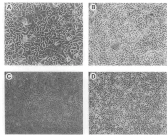
are not as uniform in appearance as the Po) (Fig. 2a).
When Pj cultures become 70% to 80% confluent they are passaged and designated P2. Early passage cultures derived from donor cornea continue to display a cobblestone morphology, and, if allowed to become postconfluent, the cultures retain the ability to stratify in discrete areas (Fig. 2b), but after P3 the ability to
stratify is diminished, In vitro transformed HCE lines also appear stellate and highly migratory when plated at 1 X 10⁴ cells/cm2, and they also develop into con- fluent monolayers (2c). Similar to the later passage control cultures, they no longer exhibit the ability to stratify when propagated while submerged. When al- lowed to become highly postconfluent, the cells be-
FIGURE 2. Passaged primary cultures are compared to HCE lines, (a) Control corneal epithe- lial cultures, serially passaged once, exhibit the typical cobblestone morphology; note that several giant cells are visible, (b) Postconfluent twice passaged control cultures support areas of multilayered growth, (c) HCE line with extended life span forms a confluent, cobblestone- like monolayer, and (d) postconfluent HCE line will not support multilayered growth when propagated submerged on plastic.
come very tightly packed on the surface of the plastic substrate (2d).
Although control cultures could be expanded un- til P5t (approximately 9 to 10 population doublings), most of the proliferation occurs between passages 1 and 3 (Fig. 3a). Approximately 125 X 106 cells/donor cornea can be generated, yielding a 20-fold amplifica- tion in cell number. Senescence always ensued by P5 in control cultures. In contrast, HCE have a constant rate of passage (3b) over the 20 passages thus far char- acterized, and a 1 X 10⁹ amplification of the starting population can be achieved. Because the starting pop- ulation of cells is generally several million, significant amplification of HCE can be generated in this manner.
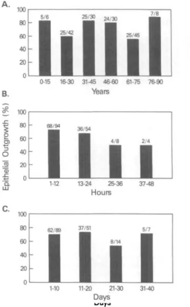
Characteristics such as age of the donor, time be- tween death of the donor and storage of the cornea, or length of storage in the eye bank, did not appear to significantly affect the ability to generate a primary outgrowth (Fig. 4). There was no statistically signifi- cant relationship between the age of the donor and its ability to generate a primary outgrowth. Increasing
age did not correlate with decreased ability to propa- gate in vitro. Of the donor tissue processed, 111/161, or 69% of the corneas explanted, yielded confluent primary cultures.
Infection and Transfection
Foci of growing cells were detected by phase-contrast photomicroscopy and appeared in the inoculated flasks after 4 to 6 weeks (Fig. 5a). Optimal results were generally obtained when cultures were infected with Adl2-SV40 at a multiplicity of 1:10. Actively growing cultures were subcultured by trypsinization. Control cultures always became senescent by the fifth passage (Fig. 5b), whereas Adl2-SV40 infected cultures exhib- ited extended life span, as previously illustrated in Fig- ure 4a. Several independent lines developed by trans- fection with plasmids and containing the early region SV40 genes exhibited large T antigen immunoreactiv- ity and extended life span. Morphologically, these lines were indistinguishable from the virally infected lines (data not shown).
Cells Generated
10 15 20
B Population Doublings per Week
0 10 15
Passage #
20
FIGURE 3. Growth kinetics of control (- • -) and HCE (- • -) lines, (a) Cells generated from one control donor cornea versus one HCE line, (b) Population doublings per week are used as a means of comparing control and HCE line growth potential.
All lines were stored frozen in liquid nitrogen and are recoverable as viable cells with a 70% to 80% seed- ing efficiency, identical to that of trypsinized cells that have not experienced cryopreservation. Cryopre- served cells require about 2 days to resume normal growth rates.
Viral Shedding/Transmissible Virus
The production of infectious virus was examined by lysis of Vero cells. There was no correlation between the dose of virus to which the cells were exposed (mul- tiplicity of infection) and the time at which viral shed- ding ceased. Shedding of whole virus into the culture supernatant ceased as early as P3 in HCE line 54 and persisted at least until P27 in HCE line 5.099.1 A. Six of the 22 HCE lines generated by hybrid virus infection continued to shed virus and were not used in this study (Table 1).
Morphology
Phase-contrast photomicroscopy demonstrated that early passage (Pi~P3) control cultures and HCE (P2- P20) develop into confluent monolayers when propa- gated submerged on plastic substrates (Figs. 2a, 2c).
Stratification can be detected in early passage, post- confluent, submerged, control cultures as raised areas of cell growth on a cobblestone monolayer (Fig. 2b). A three-dimensional, tissue-like morphology develops
A.
B.
i 1
&2 o
lial
' a*-»
LU
c.
\W
80 60 40 20 0
100 80 60 40 20 0
1UU
80 60 40 20 0
5/6 26/30 K/JO 1L
- m • • I
- I 1 I I £S I
1 1 1 1 1 1 1 1 1 1 1 1
• • m m m m0-15 16-30 31-45 46-60 61-75 76-90Years
" ^ J 36/54
^M ^M 4/8 2/4
- • I • •
1 H ^1 ^1
^^1 ^^H ^^H ^^B^H ^H 1H ^M1-12 13-24 25-36 37-48
Hours
62/89 3 7 / 5 1
5/7
• | - I
• I I I
" 1 1 1 1 m • • m
11-20 21-30 1-10
FIGURE 4. History of donor corneas and its relationship to the establishment of primaiy outgrowth from the explain.
In the top panel donor age (years) is shown to be indepen- dent of the ability for the development of a healthy out- growth. In the middle panel, the time (hours) between the death of the donor and the preservation of the cornea is shown to be a limiting factor, and in the bottom panel the length of time (days) on storage in the eye bank does not seem to impact on subsequent success of the primaiy cul- tures.
FIGURE 5. Five weeks after infection with Ad 12-SV40 hybrid virus, (a) Transformed foci are visible, (b) Control culture contains only senescent cells.
when cultures are propagated upon collagen mem- branes (Cellagen, 1CN, Irvine, CA) at air-liquid inter- faces. Both control and HCE lines appear to be tightly packed and more uniformly stratified than when culti- vated on plastic, more closely approximating in vivo morphology. HCE grown on collagen membranes at air-liquid interfaces can be seen to develop 3 to 5 cell layers when viewed in cross-section (Fig. 6a) and ap- proximately 6 cell layers when grown on collagen gels impregnated with dermal fibroblasts (Genesis, Cam- bridge, MA) (Fig. 6b).
Growth in Soft Agar
Control cultures do not form colonies in soft agar, indicating that substrate attachment is prerequisite to growth (data not shown). Nine of the HCE lines tested
did not form colonies in semi-solid agar, but two lines (50.Bl and 50.C2) did express the ability to develop small, disperse colonies. The colonies developed in 50.Bl and 50.C2 ranged from 0.05-0.08 mm in diame- ter, indicating a moderate degree of anchorage inde- pendence. A mos transformed line, which served as the positive control, developed 40-fold more colonies, with each colony being fourfold greater in diameter, reflecting its highly anchorage-independent trans- formed phenotype.
Karyotype and Isozyme Analysis
Karyotypes of two of the HCE lines (50.B1 and 50.C2), generated from the same donor, were analyzed. Al- though they both contained the Y chromosome of the donor, different marker chromosomes were detected in the two cultured lines, indicating that they had be- come genetically different from one another during propagation in vitro. The karyotypes of the control cultures were near-diploid, whereas the karyotypes of the HCE were heteroploid (Figs. 7a, 7b), as is typical of virally immortalized lines.
TABLE l. Virus Production by HCE Lines as Detected by Cytopathic Effects in Vero Cells
46*
5t 6t 7 8 9 10 11 50.B2
8t
9 11
72 4f 5t
89.B3
8t9 12
50 7t iot
50.B3 11 13 15
73 5t
8
89.C1 7 10
* HCE line names.
t CPE is observed .n 50.Al
7
8
9 12
15
50.B4
l i t 12t13 15
73.CJ 6 8
99 4f
5t
iliai passage 50.A2
7 9 10
50.C2
7 11 12
88 5t 8
5.099.1 A
17t 22t 27f
50.A3 7t 8t l i t
54 2f 3 4 5 8 9 89. A1
6t
87t
9
50.B1 9 11 12 15
62 4f fit7t 8f
89.B1 8
B
FIGURE 6. Photomicrographs of hemotoxylin and eosin stained HCE. (a) HCE on an optically clear collagen mem- brane (ICN). (b) HCE upon collagen disc impregnated with epidermal fibroblasts, 320X magnification.
The human origin of the lines examined was con- firmed by analysis of seven enzymes, all of which were found to be human (data not shown). Isozyme pheno- type frequencies were used for calculating the pheno- typic frequency product, which was determined to be 0.00116, indicating that less than 1% of cell lines might be expected to have an isozyme phenotypic pro- file identical to this.
Cell Size
Although the HCE are aneuploid and control cells are diploid, the diameter of control cells was not different from the diameter of the HCE cells (Fig. 8a), as deter- mined by Coulter counter, ZM analysis.
Kinetics and Population Doubling Time Seeding efficiency is approximately 85% for both con- trol and HCE lines. Log-phase growth generally oc-
curs between days 2 and 6, and by day 7 growth begins to plateau. Population doubling times (Fig. 8b) deter- mined on plastic growth surfaces during log-phase growth reveal no difference between control cultures and lines. Doubling times of control cultures ranged from 24 to 26 hours compared to 25 to 30 hours for the HCE lines.
Saturation Density
Saturation density determinations, or the number of cells packed within a defined area, reflected differ- ences among the lines. Control cultures have an aver- age saturation density of 1.7 X 10⁵ cells/cm2, whereas each HCE line has a characteristic saturation density ranging from 1.7 to 3.7 X 10⁵ cells/cm2 (Fig 8c).
Immunofluorescence
The pattern of reactivity of HCE was developed with antibodies specific for certain differentiated cell popu-
un
£6S 181
MI* Iisw -i
••» i n n
H I ••-
Mt r
1*1
B
)J )! |H lin
il Ml HC inl ii ntu fMII
Itl si»t III
i n ««« i
l« »4M I I I Hi rain
FIGURE 7. Karyotypes of two HCE lines derived from the same donor demonstrate the heteroploid nature of the lines, (a) 50.B1, (b) 50.B2.
Cell Diameter
30
20~ Control 5.099.1A 50.A1 50.A2 50.B1 D.91.139
linn
Cell Line
B Population Doubling Time
50
Control 5.099.1A 50.A1 50.A2 50.B1 D.91.139
Cell Line
Saturation Density
« 5
~ 3
1 1
Eiiiiii
Control 5.099.1A 50.A1 50.A2 50.B1 D.91.139Cell Line FIGURE 8. Comparisons of control cultures and various HCE lines, (a) Cell diameter, (b) Population doubling times, (c) Saturation density. Determinations were each made from four replicate wells within an experiment, and each experi- ment was done on aL least three different days.
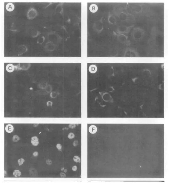
lations, as shown in Figure 9. When propagated on tissue culture plastic in the absence of fibroblast feeder cells, all control cultures and all but one HCE line that was examined reacted with AE1, AE3, and AE5 (Fig. 9a to 9c, respectively). This indicated the epithelial (AE3), hyperproliferative (AE1), and cor- neal (AE5) nature of the cells. Cytokeratin staining was concentrated in the cytoplasm with a distinct network clearly visible. Both control (not shown) and HCE (Fig.
9d) when propagated submerged reacted with anti-vi- mentin antibody, as is typical of corneal epithelium
undergoing wound healing in situ. When propagated on collagen membranes at an air-liquid interface, vi- mentin immunoreactivity disappeared (data not shown). Characteristic T antigen staining, typified by an immunoreactive nucleus containing nonimmunore- active nucleoli, was seen only in the HCE (Fig. 9e). The SIRC cell line served as a negative control for AE3, AE1, and AE5 immunoreactivity.
Functional Integrity of Synthetic Membranes Corneal control cells and HCE lines cultured on colla- gen membranes at the air-liquid interface retarded the diffusion of Na-fluorescein by 83% to 97%. Control cultures, at passages one through five, reflected dif- ferent degrees of functional integrity. P! control cul- tures could retain 83% of the Na-fluorescein. This level of retention did not increase but was stable for 1 week. Control cultures that had been passaged two to three times (P2 and P3) retained 97% and 96% of the fluorescein. These levels were maintained for a mini- mum of 1 week and a maximum of 2 weeks. P4 and P5 control cultures retained 86% and 87% of the fluores- cein, but these levels were maintained for only 1 day.
In contrast, a typical HCE line retained 94% of the Na-fluorescein and maintained this level for a mini- mum of 1 week and a maximum of 2 weeks. In addi- tion, the HCE lines exhibited a constant level of Na- fluorescein retention over 20 passages in vitro. Each HCE line has a characteristic level of Na-fluorescein retention associated with it. SIRC cells retarded diffu- sion approximately 69% and maintained this level for 1 week while corneal fibroblasts were not able to re- tard the flow of the ionic dye marker beyond 20% (Fig- ure 10).
DISCUSSION
In vitro transformation using Adi 2-SV40 hybrid virus causes normal epithelial cells to overcome cellular se- nescence and confers "immortality" without leading to tumorigenicity.19 Culture techniques for epithelial cells, such as serum-free media and raft systems that provide an air-liquid interface, allow maintenance of phenotypic differentiation and response to growth stimuli during propagation in vitro,20"22 although lev- els of expression of specific gene products may differ between primary cultures and immortalized lines.23 In- terestingly, cells expressing SV40 large T antigen that develop within transgenic mice achieve a normal phe- notype,24 suggesting that the ability to respond to ex- ternal growth stimuli is not necessarily lost as the re- sult of viral immortalization and may in fact be due to inadequate culture conditions.
The ability to maintain the differentiated pheno- type does not seem to be infinite.22 Nevertheless, enor-
FIGURE 9. Immunofluorescent analysis of cytokeratin expression in an HCE line, (a) AE1, (b) AE3, <c) AE5, (d)Vimentin, (e) T Antigen, (f) negative control (400X magnification).
~ 100 80 60 40 20
Control HCE SIRC Fibroblasts FIGURE 10. Na-fluorescein retention as measured across membranes formed by human corneal epithelial cells de- rived from twice-passaged control corneal cells and an HCE line are compared to SIRC cells and human corneal fibro- blasts.
mous numbers of phenotypically specific cells can be generated during extended life span while new lines are not difficult to generate once the laboratory has gone through the procedure and established evalua- tion criteria for substantiating phenotypic specificity.
We have established culture techniques for the ex- pansion of normal human corneal cadaver donor tis- sue. We have shown that donors should not be re- jected upon the basis of age. Homogeneous cultures of epithelial cells were generated from approximately 70% of all the donor corneas processed.
It is important to note that the finite life span of serially passaged cultures, five passages in vitro, lim- ited the number of experiments that could be per- formed. Therefore, several HCE lines with extended life span have been generated to overcome some of the problems associated with using primary cultures. Both whole virus infection and origin viral transfection was
employed to generate HCE lines with extended life span. No differences in morphology or function were noted between lines generated by these two tech- niques. All new lines are currently being generated in the laboratory using transfection techniques to avoid the necessity of testing for whole virus shedding.
One of the well-studied phenotypic traits of cor- neal epithelium is its synthesis of corneal specific cyto- keratins. Cytokeratins are 10 nm intermediate fila- ments that form a cytoskeletal network thought to provide mechanical integrity to the cell in context of its tissue.25 The keratins exist in a 1:1 ratio of type I (acidic) and type II (basic) keratins, which form hetero- dimers. The types of keratins synthesized are specific to both the developmental stage and the phenotype of the cell.26 In cornea, AE1 is found in hyperprolifera- tive epithelial cells, AE3 immunoreactivity is seen in all epithelial cells, and AE5 immunoreactivity is observed in all but the basal cells of the limbus.15 Our studies using immunofluorescence to detect keratin produc- tion indicate that the corneal phenotype of HCE can be preserved in vitro. Qualitatively, cytokeratin immu- nofluorescence is brightest in situ. Likewise, cytokera- tin staining is brighter in serially passaged corneal epi- thelium than it is in HCE with extended life span.
In addition to traditional submerged monolayer cultures, a raft system was developed to promote strati- fication in vitro. Data indicate that membranes formed by HCE lines can inhibit the flow of ionic material such as Na-fluorescein across their surfaces. Modulation of barrier function by external agents (liquids or solids) can thus be measured using a spectrophotometer. In addition, the effects of irradiation upon the cornea, such as that generated during laser surgery, might be effectively monitored in the three-dimensional format.
In vitro models must satisfy several stringent crite- ria to be informative.27 They must be species and tissue specific so that key biochemical and tissue specific mechanisms may be studied. Because of the limited availability of donor cornea tissue and short life span (up to five passages) of cultures generated from this tissue, the availability of HCE lines with extended life span (up to 25 passages) should facilitate investigation of basic cell biologic mechanisms.
By our calculations, the use of lines over a period of 20 passages can generate a 1 X 10⁹-fold amplifica- tion in terms of cell number. The number of cells that can be generated during their extended life span is significant and can provide many researchers with bio- logic material.
Key Words
human corneal epithelium, immortalized epithelial cell lines, in vitro model, serum-free medium, transepithelial per- meability
Acknowledgments
The authors thank Bernard Madison and Dawn Duffy of the Maryland Eye Bank. Sincerest thanks are extended to Tracey L. Walker, who provided expert technical skills in propagat- ing the cell cultures, and to Mary Alice Crawford for prepar- ing histologic materials. We are grateful to Louis C. DiPas- quale for stimulating discussions.
References
1. Federal Food Drug and Cosmetics Act per Food and Drug Administration (FFDCA) 21 CFR, Chapter I, Part 312, subpart B, Section 312.23. Federal Insecti- cide, Fungicide and Rodenticide Act per Environmen- tal Protection Agency, 40 CFR, Chapter I, Part 158, Subpart D, Section 158,202. Federal Hazardous Sub- stances Act for Consumer Product Safety Commission (FHSA), 16 CFR Chapter II, Part 1500, Section 1500.42. Toxic Substances Control Act per Environ- mental Protection Agency, 40 CFR, Chapter I, Part 798, subpart B, Section 798.4500.
2. Draize JH, Woodard G, Calvery HO. Methods for the study of irritation and toxicity of substances applied topically to the skin and mucus membranes,/ Pharma- col Exp Ther. 1944;82:377-390.
3. Frazier JM, Gad SC, Goldberg AM, McCulley JP. A Critical Evaluation of Alternatives to Acute Ocular Irrita- tion Testing. Vol. 4. New York: Mary Ann Liebert, Inc;
1987:21-22.
4. Bruner LH. In Vitro Toxicity Testing: Applications to Safety Evaluation. New York: Marcel Dekker, Inc;
1992:160.
5. Doughman DJ. Prolonged donor cornea preservation in organ culture long-term clinical evaluation. Trans Am Ophthalmol Soc. 1980;CLXXVIII:567-628.
6. Richard NR, Anderson JA, Weiss JL, Binder PS. Air/
liquid corneal organ culture: A light microscopic study. Current Eye Res. 1991; 10:739-749.
7. Sun T-T, Green H. Cultured epithelial cells of cornea, conjunctiva and skin: Absence of marked intrinsic di- vergence of their differentiated states. Nature. 1977;
269:489-493.
8. Ebato B, Friend J, Thoft RA. Comparison of central and peripheral human cornealepithelium in tissue cul- ture. Invest Ophthalmol Vis Sci. 1987; 28:1450-1456.
9. Hainsworth S. Modified culture method for human corneal epithelial cells./ Tiss Culture Meth. 1991; 13:
45-48.
10. Neiderkorn JY, Meyer DR, Ubelaker JE, Martin JH.
Ultrastructural and immunohistological characteriza- tion of the SIRC corneal cell line. In Vitro Cell Dev Biol. 1990; 26:923-930.
11. Madin S, Darby NB. American Type Culture Collection:
Catalog of Strains. 6th ed. 1988:21.
12. Rhim JS, Trimmer R, Arnstein P, Huebner RJ. Neo- plastic transformation of chimpanzee cells induced by adenovirus type 12 simian virus 40 hybrid virus (Adl2-SV40). Proc Nat Acad Sci USA. 1981;78:313- 317.
13. Feigner PL, Gadek TR, Holm M, et al. Lipofection: A highly efficient, lipid-mediated, DNA-transfection
procedure. Proc Nat Acad Sci USA. 1987;84:7413- 7417.
14. Harlow E, Crawford LV, Pirn DC, Williamson NM.
Monoclonal antibodies specific for simian virus 40 tu- mor antigens./ Virol. 1981;39:861-869.
15. Schermer A, Glavin S, Sun T-T. Differentiation-re- lated expression of a major 64K corneal keratin in vivo and in culture suggests limbal location of corneal epithelial stem cells./ Cell Biol. 1986; 103:49-62.
16. SunderRaj N, Rizzo JD, Anderson SC, Gesiotto JP.
Expression of vimentin by rabbit corneal epithelial cells during wound repair. Cell Tissue Res. 1992; 267:
347-356.
17. Jakoby WB, Pastan IH. Methods in Enzymology: Cell Culture. San Diego: Academic Press, Inc; 1979; 58:
150.
18. Tchao R. Trans-epithelial permeability of fluorescein in vitro as an assay to determine eye irritants. In: Gold- berg AM, ed. Alternative Methods of Toxicology. New York: Mary Ann Liebert, Inc; 1988;6:27l-283.
19. Rhim JS, Jay G, Arnstein P, Price FM, Sanford K, Aar- onson SA. Neoplastic transformation of human epi- dermal keratinocytes by AD12-SV40 and Kirsten Sar- coma viruses. Science. 1985;227:1250-1252.
20. Stoner GD, Kaighn ME, Reddel RR, et al. Establish- ment and characterization of SV40 T-antigen immor- talized human esophageal epithelial cells. Cancer Res 1991;51:365-371.
21. Chopra DP, Taylor GT, Mathieu PA, Hukku B, Rhim
JS. Immortalization of human tracheal gland epithe- lial cells by Adenovirus 12-SV40 hybrid virus. In Vitro Cell Dev Biol. 1991;27A:763-765.
22. Lechner MS, Laimins LA. Human epithelial cells im- mortalized by SV40 retain differentiation capabilities in an in vitro raft system and maintain viral DNA ex- trachromasomally. Virology. 1991; 185:563-571.
23. Sheibani N, Rhim JS, Allen-Hoffmann BL. Malignant human papillomavirus type 16-transformed human keratinocytes exhibit altered expression of extracellu- lar matrix glycoproteins. Cancer Res. 1991; 51:5967—
5975.
24. Hauft SM, Kim SH, Schmidt GH, et al. Expression of SV-40 T antigen in the small intestinal epithelium of transgenic mice results in proliferative changes in the crypt and reentry of villus-associated enterocytes into the cell cycle but has no apparent effect on cellular differentiation programs and does not cause neoplas- tic transformation./ Cell Biol. 1992; 117:825-839.
25. Coulombe PA, Hutton ME, Vassar R, Fuchs E. A function for keratins and a common thread among different types of epidermolysis bullosa simplex dis- eases./ Cell Biol. 1991; 115:1661-1674.
26. Steinert PM, Roop DR. Molecular and cellular biology of intermediate filaments. Ann Rev Biochem. 1988; 57:
593-625.
27. Flint OP. In vitro test validation: A house built on sand. A Item To Lab Animals. 1992; 20:196-198.