D o s e - D e p e n de n t In h ib it ory E f f e c t o f N it ric O x ide o n E m b ry o D e v e lo p m e n t
S e a - H ee P ark , Bo- Su n J oo
*, H w a - S ook M oon
*, Yoon - Y eon Kim
* *, J ae - Don g Ch o
* *, H o- Sun g K an g , an d H an - Do Kim
Dep art m ent of M olecular Biolog y , Colleg e of N at ur al S cien ce , P u s an N ation al Un iv er s ity , P u s an 609- 735,
*
Infertility Clin ic , M oon H w a H ospit al, P u s an 601- 062,
* *
Ch o J ae Don g OB/ GY , Ch an g - W on 641- 530, K or ea
N it ric O x ide 의 농 도 의 존 적 인 배 발 생 억 제 효 과
박세희 , 주보선
*, 문화숙
*, 김윤연
* *, 조재동
* *, 강호성 , 김한도
부산대학교 분자생물학과, 문화병원 불임클리닉
*, 조재동 산부인과의원
* *주소 : 경상남도 창원시 소답동 233- 7 조재동 산부인과 의원 T el : 0551- 253- 2111
F ax : 0551- 253- 2114
INTRODUCTION
Approximately 14-40% of infertile couples have infertility caused by immunologic disorders which result from the production of antisperm antibody in humoral immune response
1and the soluble products of activated lymphocytes and macrophages in cellular immune response
2 ,3.
Especially, endometriosis which is inflammatory state characterized by the presence and growth of ectopic endometrial tissue outside the uterine cavity, occurs at 20-40%
of high incidence in infertile women
4. As peritoneal fluid is able to go into the oviductal cavity, changes of cellular and hormonal components within peritoneal fluid may influence the early reproductive processes such as fertilization and early embryo development by modulating the microenvironment that surrounds the embryo
5. Peritoneal fluid in women with endometriosis has been reported to increase the number of activated macrophages
4and the production of cytokines such as interleukin-1 (IL-1)
6, interleukin-6 (IL-6)
7and tumor necrosis factor- (TNF- )
8 ,9. These cytokines are known to have adverse effects on sperm cell function, fertilization and embryo development
10. However, the correct mechanism for this inhibition is unclear.
It is an established fact that these cytokines stimulate macrophages to produce nitric oxide (NO) during conversion of L-arginine to L-citrulline by NO synthase
1 1. It has been reported that NO plays important roles in various physiological systems. The physiologic actions of NO are regulating smooth muscle relaxation, platelet aggregation and adhesion, cell growth, neurotransmission, apoptosis and immune response
12. Since the cells of these systems are integral parts of the reproductive organs, NO is thought to play an important role in reproduction. Indeed, many studies have demonstrated that NO regulates pulsatile hypothalamic GnRH secretion
13, human granulosa-luteal cell steroidogenesis
14, ovulation
1 5and penile erection
16. Therefore, it is suggested that the increased production of NO from the activated macrophage by endometriosis or certain other causes may affect directly or indirectly fertilization and embryo development causing infertilty.
NO induces cell death via apoptosis or necrosis. This effects of NO were thought
to be solely mediated via activation of 3',5'-cyclic guanosine monophosphate
(c-GMP)
17. However, recent studies have demonstrated that NO can also induce biological effects via non-cGMP dependent pathways
1 8 , 19.
Therefore, this study was performed to understand the effect of NO on embryo development using sodium nitroprusside (SNP), stable NO donor and peritoneal fluids collected from women undergoing laparoscopic operation. NO concentration in PF was measured to determine whether NO level within PF correlates with the embryo development. We also investigated the occurrence of apoptosis following by treatment of SNP and the effect of cGMP analogue on embryo development to understand the mechanism and the pathway for the action of NO.
MATERIALS AND METHODS
1. Materials
The original stock of C57BL/6 × CBA/Ca hybrid F 1 female mice was purchased from Korean Laboratory Animal Center (Korea). PM SG (pregnant mare's serum gonadotropin), hCG (human chorionic gonadotropin), hyaluronidase, mineral oil, sodium nitroprusside, N-(1-naphthyl)-ethylendiamine (NED), sulphanilamide, H
3PO
4 ,NaNO
2 ,8-bromo-cGMP, trypan blue, Hoechst 33342 and sodium citrate were purchased from Sigma Chemical Company (St. Louis, MO). Synthetic serum substitute (SSS) was purchased from Irvine Scientific (Santa Ana, CA). Culture dish (60mm) was purchased from Falcon Inc. (Becton Dickinson, USA). SNP and 8-bromo-cGMP was prepared as a concentrated stock in serum free modified human tubal fluid (m-HTF) and stored at -20℃ until use.
2. Zygote embryo collection and culture
Zygote embryos were obtained from superovulated hybrid F 1 female mice (4-6
weeks old). Superovulation was induced by intraperitoneal inj ections of 5 IU of
PMSG followed by 5 IU of hCG after 48 hours. Mating was confirmed by the
appearance of a vaginal plug in the next morning. Zygotes were collected from
excised oviducts 18 to 20 hours after hCG inj ection and cultured in m-HTF medium
supplemented with 10% SSS at a 37℃ incubator in 5% CO
2for 4 days. Adherent
cumulus cells were removed by micropippeting in serum free modified human tubal medium (m-HTF) containing 0.1% hyaluronidase.
3. Treatment of SNP and cGMP-analogue
Zygotes were treated with the various concentration of SNP (0, 100 nM, 10 M, 50 M, 100 M, 200 M, 1mM) or 8-bromo-cGMP, an analogue of cGMP (0, 50 M, 100 M, 500 M, 1mM). And 200 M SNP was treated at 2-cell, 4-cell and 8-cell embryonic stage to examine the effect of SNP is dependent on SNP treatment stages.
Experiments were repeated three or more times and 10-20 embryos were usually used in each experiment.
4. Peritoneal fluid processing
Peritoneal fluid (PF) was obtained from 42 women undergoing laparoscopy for various causes (e.g. endometriosis, myoma) and was centrifuged at 800g for 10 minutes within 30 minutes after collection. The supernatant was divided into two parts. One part was inactivated complement proteins in a water bath at 56℃ for 30 minutes and stored at -20℃ to use for embryo culture. The other was immediately stored to assay NO concentration. Before use in culture, an aliquot of PF was filtered (0.22㎛ filter, Millex GS, Millipore Corporation, Bedford, MA) and 1ml of 10% PF in m-HTF medium was provided for in vitro zygote embryo culture. The groups of control were cultured in m-HTF medium containing 10% SSS. Experiments was repeated three times for each PF sample and 10-20 zygote embryos were used in each experiment.
5. Measurement of NO
NO concentration in PF was measured using Griess method
2 0. Griess assay reagents
〔Nitroprusside 0.2% N-(1-naphthyl)-ethylendiamine (NED) and 2% sulphanilamide in
5% concentrated H
3PO
4〕were premixed and then incubated with a peritoneal fluid
sample (ratio 1:1) to form a purple azo dye, and absorption was determined at a
wavelength of 540 nm by ELISA Reader. NaNO
2was used to generate standard
curve, and NO concentration was calculated from standard curve.
6. Detection of A poptosis in embryos
To observe the occurrence of apoptosis, normal developing 2-cell embryos, which was not treated with SNP, and 2-cell embryos arrested by treatment of 200 M SNP were stained with trypan blue and incubated with Hoechst 33342 working solution for 3 min. Hoechst 33342 (Sigma) dye was prepared as stock solution (1mg/ml) and added to 2.3% sodium citrate in ethanol (10 l/ml) for working solution. The stained samples were mounted and apoptosis was examined under a fluorescence microscope.
7. Statistical analysis
Statistical analysis was performed by the Student's t-test and a p value of <0.05 was considered significant.
RESULTS
1. Inhibition of mouse embryo development by SNP treatment
To investigate the effect of nitric oxide on mouse embryo development, SNP was treated to mouse zygote culture media with various concentration (0 to 1mM). The rate of embryo development to blastocyst in the different concentration of SNP is shown in Fig. 1. The addition of SNP inhibited mouse embryo development in a dose-dependent manner. The rates of embryo development were 71% in control, 53%
in 0.1 M, 48% in 10 M, 27% in 50 M, 8% in 100 M, 0% in 200 M and 1mM.
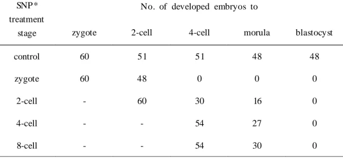
Especially above 200 M of SNP mouse embryo development was completely arrested and degenerated since 2-cell embryonic stage. To determine whether this effect of SNP was treatment stage-specific, 200 M SNP was treated at 2-cell, 4-cell and 8-cell embryonic stage. As showed in Table 1, mouse embryo did not develop to blastocyst whenever SNP was treated.
2. The relationships betw een the level of N O in PF and em bry o development
The addition of PF in mouse embryo culture media resulted in the different
development rate. We measured the concentration of NO in PF to determine whether
this different rate was caused by changes of cellular component within PF. NO concentration was different from each PF sample ranged from 0.79 M to 4.4 M. PF was divided into 6 groups according to its NO level and the rates of embryo development were represented in Table 2. The rate of embryo development was significantly lower in group V (26.5± 16.6%) and group VI (29.2± 11.0%) compared with control (62.8±6.1%), which was cultured in medium with 10% SSS (p<0.05).
However, the rate of embryo development in 2.5 M or less (group I to IV) was similar compared with that of control.
3. Inhibitory effect of NO on the mouse embryo development via cGMP-dependent or independent pathway
To determine whether this effect of NO on mouse embryo development was mediated by the elevating level of cGMP, 8-bromo-cGMP was added to embryo culture media. As noted in Fig.2, embryo development was not influenced by 8-bromo-cGMP regardless of the treated concentrations (50 M to 1mM). This result suggests that NO may inhibit mouse embryo development through cGMP-independent pathway.
4. D etection of apoptosis in embryo arrested by SN P treatment
To investigate whether the arrest of embryo development following SNP treatment resulted from cell death via NO-mediated apoptosis, we examined the nucleus fragmentation in 200 M SNP treated 2-cell embryos. Compared with normal developing embryos, the morphological changes of nucleus were not detected in embryos treated with SNP and the size of nucleus is equal to that of control (Fig.
3).
DISCUSSION
NO can pass easily into the intracellular area and have many different effects on
cell metabolism in the biology, physiology, and pathophysiology of reproduction
12.
Because NO is a free radical, it is degraded easily and has very short life span. Thus
most studies used a more stable nitric oxide donor to elucidate the role of NO in vitro. SIN-1, sodium nitroprusside (SNP), SNAP are representative nitric oxide donor.
Among them, SNP released only NO in vitro cell culture system, while other two donors released superoxide anion or/and hydrogen peroxide as well as NO
2 1, and SNP concentration in cell is known to be in proportion to production of NO
2 2. Weinberg et al.
2 3reported that SNP reduced sperm motility and this inhibition was blocked by the NO quencher hemoglobin. This suggests that SNP is more suitable to study the effect of only NO compared with other NO donors.
Our present study showed that mouse embryo development to blastocyst was inhibited dose-dependently by SNP, especially reduced at high concentration of 100μ M or more. However, this SNP-induced inhibitory effect on embryo development was not stage-specific because embryo development was inhibited regardless of SNP treatment stages. We also found that embryo development was severely arrested in media containing PF with the higher concentration of NO than in media containing qualified controlled serum. It has been recognized that PF is able to go into the oviductal cavity and may influence the early reproductive processes by modulating the microenvironment that fertilization and early embryo development occur. These results imply that high concentration of NO may adversely affect embryo development, and that changes of component within PF especially such as increased concentration of NO can be closely associated with embryo development.
Previously, many studies have observed that the level of macrophage, cytokines and other cellular components were increased and activated with the extent of pelvic disease and inflammation
5 , 8 ,9. PF in women with endometriosis adversely affected the cleavage of mouse two-cell embryos
2 4and contained high concentration of cytokines such as TNF-α and IL-1
10. However, the correct mechanism for interaction between these cytokines in PF and embryo toxicity has not been elucidated yet. A common pathway of the action of TNF-α on various cell systems is the induction of an inducible nitric oxide synthase (iNOS), which results in the generation of NO. Thus it is possible that a certain disease such as endometriosis or other factors stimulate the production of cytokines, which subsequently induce the production of NO thereby directly affecting embryo development.
Generally the effects of NO were known to be mediated via activation of soluble
guanylate cyclase and guanosine 3',5'-cGMP, which inhibits Ca
2 +influx and activates its efflux, resulting in a lower intracellular [Ca
2 +]. Decreased intracellular [Ca
2 +] can inactivate PKC, and thus inhibit PKC-dependent cell proliferation
2 5. But, the inhibitory effect of NO on mouse embryo development may be not mediated by a cGMP-dependent pathway, since the addition of 8-bromo-cGMP, agent which raises cellular cGMP, did not affect embryo development regardless of treated concentration.
This result is supported by recent studies, which suggest that NO can induce its biological effects via non-cGMP-dependent pathways by forming high-affinity-nitroso complexes with a variety of metal compound or binding the heme-containing proteins and iron-sulphur-containing proteins
14 , 1 8 , 19.
The effects of NO via these pathway may be dependent on dose and the cell type.
One hundred micromole of SNP was shown to inhibit sperm motility
2 6, while lower concentration of SNP (100nM or less) was beneficial to the maintenance of post-thaw sperm motion, viability and motility
2 7. And NO has the double-edged role in inducing apoptosis. High level of NO induced by pathophysiolocal conditions stimulates apoptosis while the continuous release of endothelial NO inhibits apoptosis
2 8. We investigated whether the inhibition of embryo development by higher concentration of SNP caused by inducing apoptosis of embryo, but apoptotic characteristics did not found in embryos arrested by treatment of SNP.
Another possibility we can consider for this inhibitory effect is the inhibition of embryonic-stage specific protein synthesis by SNP. A lot of stage-specific proteins are necessary for normal embryo development. NO is capable to posttranslationally interfere with heme proteins and enzymes containing iron-sulfur cluster or to modulate enzyme activity by S-nitrosylation
2 9. Thus, the further study is needed to prove the correct mechanism for the inhibition of embryo development.
In conclusion, high concentration of SNP and PF containing high amounts of NO
inhibited early embryo development. This result suggests that in vivo, if there is
increase of NO level in reproductive systems such as peritoneum and tubal fluid by
certain causes, this may correlate with infertility by causing damage to embryo.
REFEREN CES
1. Naz RK, Menge AC: Antisperm antibodies. Origin, regulation, and sperm reactivity in human infertility. Fertil Steril 1994; 61: 1001-13.
2. Hill JA, Cohen J, Anderson DJ. The effects of lymphokines and monocytes on human sperm fertilizing ability in the zona-free hamster egg penetration test. Am J Obstet Gynecol 1989; 160: 1154-59.
3. Jung BJ, Lee SH, Hur M. The efficacy on the immunotherapy with paternal lymphocytes in unexplained infertility. Korean J Fertil Steril 1997; 24: 293-300.
4. Ramey JW, Archer DF. Peritoneal fluid: its relevance to the development of endometriosis. Fertil Steril 1993; 60: 1-14.
5. Badawy SZA, Cuenca V, Marshall L, Munchback R, Rinas AC, Coble DA.
Cellular components in peritoneal fluid in infertile patients with and without endometriosis. Fertil Steril 1984; 42: 704-8.
6. Fakih H, Baggett B, Holtz G, Tsanh KY, Lee JC, Williamson HO.
Interleukin-1: a possible role in the infertility associated with endometriosis. Fertil Steril 1987; 47: 2 13-7.
7. Harada T, Yoshioka H, Yoshida S, Iwabe T, Onohara Y, Tanikawa M, Terakawa N . Increased interleukine-6 levels in peritoneal fluid of infertile patients with active endometriosis. Am J Obstet Gynecol 1997; 176: 593-7.
8. Halme J, Hammond MG, Hulka JK. Retrograde menstruation in healthy women and in patients with endometriosis. Obstet Gynecol 1984; 64: 151-4.
9. Eisermann J, Michael JG, Jorge P, Odem RR, Collins JL. Tumor necrosis factor in peritoneal fluid of women undergoing laparoscopic surgery. Fertil Steril
1988; 50: 573-9.
10. Taketani Y, Kuo TM, Mizuno M . Comparison of cytokine levels and embryo toxicity in peritoneal fluid in infertile women with untreated or treated endometriosis. Am J Obstet Gynecol 1992; 167: 265-70.
11. Bredt DS, Snyder SH. Nitric oxide: A physiologic messenger molecule. Annu
Rev Biochem 1994; 63: 175-95.
12. Rosselli M, Keller PJ, Dubey RK. Role of nitric oxide in the biology, physiology and pathophysiology of reproduction. Human Reprod 1998; 4: 3-24.
13. Moretto M, Lopez FJ, Negro-Vilar A. Nitric oxide regulates luteinizing hormone-releasing hormone secretion. Endocrinology 1993; 133: 2399-2402.
14. Van Voorhis BJ, Dunn M S, Snyde GD, Weiner CP. Nitric oxide: An autocrine regulator of human granulosa-luteal cell steroidogenesis. Endocrinology
1994; 135: 1799-1806.
15. Hwsla JS, Preutthipan S, Maguire MP, Chang TS, Wallach EE, Dharmaraj an AM . Nitric oxide modulates human chorionic gonadotropin-induced ovulation in the rabbit. Fertil Steril 1997; 67: 548-52.
16. Holmquist F, Stief CG, Joncs U . Effect of the nitric oxide synthase inhibitor NG-nitro-L-arginine on the erectile response to cavernous nerve stimulation in the rabbit. Acta Physiol Scand 1991; 143: 299-304.
17. Murad F. Regulation of cytosolic guanylyl cyclase by nitric oxide: the NO-cyclic GMP signal transduction system. Adv Phamocol 1994; 26: 19-33.
18. Brunswig-Spickenheier B, Mukhopadhyay AK. Stimulation of nitric oxide-cyclic guanosine monophosphate pathway in bovine ovarian theca cells by tumor necrosis factor- (TNF- ). Is this pathway implicated in the TNF- -induced inhibition of utenizing hormone-stimulated prorenin production. Biol Reprod 1997;
57: 700-6.
19. Kroncke KD, Fehsel K, Kolb-Bachofen V. Nitric oxide: cytotoxicity versus cytoprotection ; How, Why, When, and Where? NITRIC OXIDE : Biol Chem
1997; 1: 107-20.
20. Marzinzig M, Nussler AK, Stadler J, Marzinzig E, Barthlen W. Improved methods to measure end products of nitric oxide in biological fluids: Nitrite, Nitrate, and S-Nitrosothiols. NITRIC OXIDE : Biol Chem 1997; 1: 177-89.
2 1. Ioannidis I, Groot H. Cytotoxicity of nitric oxide in Fu 5 rat hepatoma cells;
evidence for co-operative action with hydrogen peroxide. Biochem J 1993; 296:
34 1-5.
22. Kowulak EA, Seth P, Fung HL. Metabolic activation of sodium nitroprusside in vascular smooth muscle. J Pharmacol Exp Ther 1992; 262: 916-22.
23. Weinberg JB, Doty E, Bonaventura J, Haney AF. Nitric oxide inhibition of human sperm motility. fertil steril 1995; 64: 408-13.
24. Morcus RN, Gibbons WE, Findley WE. Effects of peritoneal fluid on in vitro cleavage of 2-cell mouse embryos: possible role in infertility associated with endometriosis. Fertil Steril 1985; 44: 678-83.
25. Khatib AM, Siegfrid G, Quintero M, Mitrovic DR. The mechanism of inhibition of DNA synthesis in articular chondrocytes from young and old rates by nitric oxide. NITRIC OXIDE : Biol Chem 1997; 1: 2 18-25.
26. Rosselli M, Dubey RK, Imthurn B. Effect of nitric oxide on human spermatozoa: evidence that nitric oxide decreases sperm motility and induces sperm toxicity. Human Reprod 1995; 10: 1786-90.
27. Hellstrom WJG, Bell M, Wang R . Effect of sodium nitroprusside on sperm motility, viability, and lipid peroxidation. fertil steril 1994; 61: 1123-8.
28. Dimmeler S, Zeiher AM . Nitric oxide and apoptosis: Another paradigm for the double-edged role of nitric oxide. NITRIC OXIDE :Biol Chem 1997; 1: 275-81.
29. Stamler JS, Jaraki O, Osborne J. Nitric oxide circulates in mammalian plasma primarily as a S-nitroso adduct of serum albumin. Proc Natl Acad Sci
1992; 89: 7674-6.
Figure Legends
Figure 1. Effect of SNP on mouse embryo development.
SNP was treated with various concentrations (control, 100 nM, 10 M, 50 M, 100 M, 200 M, 1 mM) to embryo culture medium. Embryos were cultured from zygote to blastocyst and embryonic development was examined. Development rate was expressed with % of developing to blastocysts from zygotes. Data was mean of three experiments. Values in vertical bar are the number of embroys reached to blastocyst/the total number of embryos provided for experiment. *p<0.05
Figure 2. Effect of cGMP-analogue (8-bromo-cGMP) on mouse embryo development. 8-bromo-cGMP was treated with concentrations of 0 to 1 mM to embryo culture medium. Embryos were cultured from zygote to blastocyst and embryonic development was examined and development rate was expressed with % of developing to blastocysts from zygotes. Data was mean of three experiments. Values in vertical bar are the number of embryos reached to blastocyst/the total number of embryos provided for experiment. *p 0.05
Figure 3. Fluorescence micrographs of 2-cell embryos stained with Hoechst 33342
for detection of apoptotic bodies. Zygote embryos were culture in m-HTF medium
without (Fig.3a) or with (Fig.3b) 200 M SNP. After 24 hours, 2-cell embryos were
stained with Hoechst 33342 and observed at ×400 by fluorescence microscope.
Table 1. Effect of SNP on mouse embryo development depending on the SNP treatment stages.
SNP*
treatment stage
No. of developed embryos to
zygote 2-cell 4-cell morula blastocyst
control 60 51 51 48 48
zygote 60 48 0 0 0
2-cell - 60 30 16 0
4-cell - - 54 27 0
8-cell - - 54 30 0
* 200 M SNP treated to 2-cell, 4-cell and 8-cell embryonic stage during zygote
embryos were cultured to blastocyst stage.
Table 2. Effect of peritoneal fluid on mouse embryo development according to the level of NO in peritoneal fluid
Group ( n
*)
NO level ( M ) ( Mean±SEM )
No. of embryos
% of developed embryos
†to
2-cell 4-cell Morula Blastocyst control 120 87.4±2.9 85.2±3.3 76.9±4.5 62.8±6.1
I (9 ) ≥ 1.0 (0.84±0.05) 90 96.6±2.1 92.7±3.0 84.9±2.9 68.3±8.6
II (7 ) 1.0~ 1.5 (1.20±0.07) 70 100±0.0 98.2± 1.7 91.9±4.0 70.0±8.0
III (5 ) 1.5~2.0 (1.81±0.07) 50 95.5±4.4 95.8±4.1 93.0±4.1 91.9±5.2
aIV (8 ) 2.0~2.5 (2.15±0.03) 80 90.2±3.6 89.9±4.6 82.7±4.9 71.2±5.2
V (5 ) 2.5~3.0 (2.68±0.04) 50 85.5± 11.9 70.5±23.6 40.3±24.9
a26.5± 16.6
aVI (8 ) <3.0 (4.36±0.05) 80 89.0±3.3 69.8± 12.3 42.8±23.6
a29.2± 11.0
aNote : Peritoneal fluid was obtained from 42 women with endometriosis of other causes by laparoscopy
*
; n = number of peritoneal fluid sample
†
; Mean ± SEM
a
; p<0.05 (versus control)
= 국문초록 =
목 적 : 일산화질소 (nitric ox ide ; NO)는 생식계를 비롯한 여러 생체내 기관에서 다양 하고도 중요한 작용을 하는 것으로 알려져 있으며, 복강액은 난관내강과 연결되어 복강액 내의 세포 성분의 변화는 난관의 미세환경을 변화시켜 수정과 초기 배아 발 생에 영향을 줄 수 있다 . 본 연구는 배아 발생에 있어서 일산화질소의 역할을 이해 하고 복강액내의 NO 농도 변화가 배아 발생에 미치는 역할을 조사하기 위해 수행되 었다 .
방 법 : 과배란시킨 1세대 잡종 암컷 생쥐 (C57BL×CBA/ Ca )로부터 1세포기 배아를 얻 어 10% synthetic serum substitute가 첨가된 m odified hum an tubal fluid 배양액에서 4 일 동안 체외배양하였다 (대조군 ). 실험을 위해 이러한 배양조건에 sodium nit r opru s side (S NP )를 0~ 1m M 의 다양한 농도로 배양초기부터 첨가하거나, 200 M S N P 를 2- , 4- , 8- 세포기의 각기 다른 배아 시기에 첨가하였으며, 복강경수술을 받는 42명의 여성으로부터 채취한 복강액을 S S S 대신 단백질원으로 사용하여 포배아까지 의 배아 발달율을 관찰하였으며 . 복강액 내의 NO 농도를 Gries s 방법에 의해 측정하 였다 . 배아의 apopt otic b ody는 H 33342 염색법으로 조사하였으며 배아 발달율은 3회 이상 반복 실험한 결과의 m ean±SEM 으로 나타내었다 .
결 과 : S NP는 농도에 의존적으로 배발생을 억제하였으나 배아 단계에 대한 특이성은 관찰할 수 없었으며, 특히 100 M 이상의 고농도의 SNP는 2- 세포기 단계에서 배아 발생을 정지시켰다 . 또한 단백질원으로 복강액 이용시 배발생율은 복강액 내의 NO 농도에 따라 현저한 차이가 발견되었으며, 2.5 M 이상의 NO를 함유한 복강액에서
배양한 배아의 발생율은 현저하게 감소하였다 . cGM P an alog u e인 8- brom o- cGM P를 배양액에 첨가시 배아 발생에는 변화가 없었으며, SNP에 의해 배발생이 정지된 2- 세포기 배아에서 apopt otic b ody를 발견할 수 없었다 .
결 론 : 이상의 결과로 보아 NO는 고농도에서 배아 발생을 저해하며, 복강액내의 NO 와 같은 성분의 변화는 배아 발생에 유해한 효과를 유발할 것으로 사료된다 . 이러한 N O의 배아 발생 억제효과는 cGM P 로 중재되는 경로나 apopt osis 유발과는 관계가 없는 것 같다 .