INTRODUCTI ON4 )
Follicular dendritic cells (FDC s) are maj or m icro- environm ental components of secondary lymphoid follicles, and are known to play critical roles in the survival, proliferation , selection , and differentiation of germinal center (GC) B cells ( 1) . FDC s display distinct m orphological characteristics and retain unprocessed antigen s in the form of immune complexes for a long tim e (2) . Tran sfer experim ents with bone marrow cells have shown that FDC s are not from the bone m arrow (3) . An experim ent designed to investigate whether FDC s
Correspondence: Jongseon Choe, Department of Microbiology, Kangwon National University College of Medicine, Chunchon, Kangwon 200-70 1 Korea
Tel: 033-250-8862, Fax: 033-242-757 1 Email: jchoe @kangwon.ac.kr
are derived from haem atopoietic cells or the surrounding stromal components, dem on strated the host origin of FD Cs induced in the spleen s of SCID mice after the tran sfer of allogeneic lymphocytes (4) . We recently suggested that antigen -activated B cells stimulate the m aturation of FD C precur sors, whose identity is unclear, via lymphotoxin -α and that m ature FD Cs, in turn, m ay provide potent proliferation signals to centroblasts (5) . Therefore, the ontogeny of FDC is unknown .
We reported previou sly upon the establishm ent of an FD C line, HK , from hum an ton sils to overcom e the practical difficulty in isolating pure FDCs and to mim ic the GC reaction in vitro (6) . HK cells indeed have the functional features of FDC s by delaying apoptosis, and by stimulating growth and differentiation of GC B cells.
HK cells bind and prevent the apoptosis of GC B cells preferentially and have co-stimulatory effects on the proliferation of CD40-stimulated GC B cells (7,8) .
= Abs t r ac t =
Background : Follicular dendritic cells (FDCs) play key roles during T cell-dependent humoral immune responses by allowing antigen-specific B cells to survive, proliferate, and differentiate within the FDC networks of secondary follicles, i.e., germinal centers (GC). M ethods: A novel monoclonal antibody , 3C8, was generated by immunizing with an FDC line HK, in order to understand the molecular signals involved in the FDC-B cell interactions in the microenvironment of the GC. Results: The 3C8 antibody did not bind to mononuclear cells, including T cells, B cells, and monocytes. Murine L 929 and human skin fibroblasts exhibited no or little reactivity to 3C8. However, 3C8 specifically recognized HK cells by flowcytometry . Furthermore, the antigen recognized by 3C8 was restricted to the GC of the human tonsil. Dendritic networks of the GC were intensely stained by 3C8, but cells outside the GC were not . Conclu sion : Our results suggest that the antigen 3C8 may play some unique role on FDCs during the GC reactions.
Key W ords: follicular dendritic cell, monoclonal antibody , germinal center
3C8 , a n e w m o n o c lo n a l a n t ib o d y d i r e c t e d a g a i n s t a fo l l i c u l a r d e n d r i t i c c e l l l i n e , HK
In Y ong Lee1, Joonhee Lee1, W eon Seo Park2, Eui-Cheol N am3, Y ung Oh Shin1, Jon gseon Choe1 Dep artment of Microbiology1, Pathology2 and Otorhinolary ngology3, Kangwon National University Colleg e of Medicine, Chunchon, Kangwon, Korea
To provide further evidence for the relevance of HK cells to FDCs, we developed a new m onoclonal antibody 3C8 by immunizing m ice with HK cells. This antibody stain s the dendritic netw ork , but not m ononuclear cells, in ton sil section s. The results dem on strate that HK cells share a common antigen w ith FDC s, and suggest that HK cells originated from FD Cs.
MATERIALS AND METHODS
1. A n t ib odie s u s e d in t h i s s t u dy
PE -conj ugated anti-CD 20 (L27), anti-CD 3 (UCHT 1), anti-CD 14 (M 5E2), and isotype controls w ere purchased from Becton D ickin son (San Jose, CA , U .S.A .) . Unconj ugated anti-CD 44 (N KI-P 1) w as a kind gift from Dr . C . G . Figdor (University Hospital N ij m egen , N ij m e- gen , The N etherlands) and its isotype control w as obtained from Sigm a Chem ical Co . (St . Louis, MO , U .S.A .) .
2. C u lt u r e of H K c ell s a n d s k in f ib r ob la st s
HK cells w ere established as described previou sly (6) and cultured in RPMI 1640 containing 10% FC S, 2mM L -glutamine (Life Technologies, Grand Island, N Y, U .S.A .), and 40μg/m l of gentam icin (SoloPak Laboratories, E lk Grove Village, IL , U .S.A .) . Human norm al skin fibroblasts w ere purchased from ATCC (CCD -25SK , CRL - 1474, Manassas, VA , U .S.A .) or were kindly provided by Dr . Jin -H o Chung (SNU -20 SK , D epartment of Derm atology , College of M edicine Seoul N ational University , Seoul, K orea) and cultured in RPMI containing 10% FC S, 2mM L -glutam ine, and 40μg/m l gentamicin .
3 . P r ep a r a t ion of h u m a n m on on u cle a r cell s , B c ell s , T cell s , a n d m on ocy t e s
Ton sils were obtained from the K angwon N ational University Hospital (Chunchon , K orea) from children undergoing ton sillectomy . Ton sillar m ononuclear cells w ere prepared by centrifugation of cell su spensions on a Ficoll-Paque (Amersham Pharmacia Biotech , Upp sala,
Sw eden) . Human B cells, T cells, and monocytes were partially purified from heparinized peripheral blood voluntarily donated by healthy donors. Peripheral m ononuclear cells were subj ected to rosetting with sheep red blood cells. B cells were obtained from the non -rosetting fraction , after the depletion of m onocytes by plastic adherence, which w ere collected separately . T cells w ere recovered from the rosetting fraction .
4 . P r od u ct ion of 3C 8 m on oclon a l a n t ib ody
Balb/c m ice (Jack son Laboratory , Bar Harbor, ME , U .S.A .) w ere immunized w ith 1× 10⁷ HK cells intraperitoneally three tim es at 2-wk intervals. The splenocytes w ere fu sed with SP2/0 my elom a cells u sing poly ethy lene gly col 400 (Merck , Darm stadt, Germany) . Hybridom as were cultured in DMEM supplemented with 20% FC S and HAT (Sigm a Chemical Co .) . Hybridoma screening was carried out by cell-based ELISA in 2 step s. In the first step , hybridom a supernatants w ere negatively selected for the binding to m ononuclear cells, which included B cells, T cells, and m onocytes.
Supernatants that exhibited little or no reactivity to hum an skin fibroblasts w ere selected for the binding to HK cells in the second step . The resultant 3C 8 hybridoma (IgG 1) was cloned by lim iting dilution .
5 . C ell - b a s e d E L I S A
Ton sillar mononuclear cells, HK cells, and skin fibroblasts w ere cultured in 96-w ell plates. Cells w ere fixed by 0 .25% glutaraldehyde and w ashed three tim es w ith PB S. The plates were incubated w ith blocking buffer until u se (PB S containing 1% FC S and 0 .1%
N aN3) . After adding primary antibodies, the plates w ere incubated at 37℃ for 2 h , and incubated with HRP -conj ugated secondary Ab at 37℃ for 30 m in . After rinsing w ith PB S 5 times, OPD w as added in phosphate citrate buffer, and the reaction stopped by adding 4M H2SO4. The optical density of each w ell w as m easured w ith a plate reader (Vm ax , Molecular Devices,
Sunnyvale, CA , U .S.A .) at 490 nm .
6. F low cyt ometry an d Immun ohist ochemistr y
Cells were stained for flow cytometric analy sis as described previou sly (9), which w as carried out on a FA C Scan (Becton D ickin son) with CellQuest software.
Frozen ton sil section s were thoroughly dried at room temperature and fixed in cold acetone for 10 m in . Section s were stained with the primary Ab s (i.e., 3C8, DRC- 1, and isotype control) for 2 h at room temperature, u sing a Scytek Staining kit (Scytek Laboratories, Logan, U T, U .S.A .) according to the m anufacturer's instruction s.
D eveloped slides were counterstained w ith Mayer's hem atoxy lin .
RESULTS
1. P r odu ct ion of 3 C 8 M A b
Since HK cells exhibit the functional features of FD Cs, w e postulated that FDC s and HK cells m ight share functionally important molecules and attempted to develop m onoclonal antibodies again st such potential m olecules by immunizing m ice w ith HK cells. Out of 300 hybridom as obtained, 27 w ere selected in the fir st screening step on the basis that these hybridomas produced antibodies, but did not react with tonsillar m ononuclear cells. In the second screening step , which w as designed to select any hybridoma that w as not reactive w ith skin fibroblast but reactive with HK cells, only 1 hybridom a was obtained . The resultant 3C8 w as of the IgG 1 isotype . A s shown in Fig . 1, 3C8 did not react w ith tonsillar mononuclear cells (p > 0 .05) in cell-based ELISA . How ever , 3C8 strongly reacted with HK cells (p < 0 .0 1) . 3C8 did not display con sistent results with hum an skin fibroblasts becau se it did not bind to the CCD -SK 25 skin fibroblast cell line from A TCC (data not shown) but reacted w ith another cell line obtained from Seoul N ational Univer sity Hospital (Fig . 1) .
2. 3 C 8 s p ecif ic a lly b in d s t o H K cell s
To sub stantiate the cellular reactivity of 3C 8 in a
sen sitive assay , flow cytometric analy sis was perform ed.
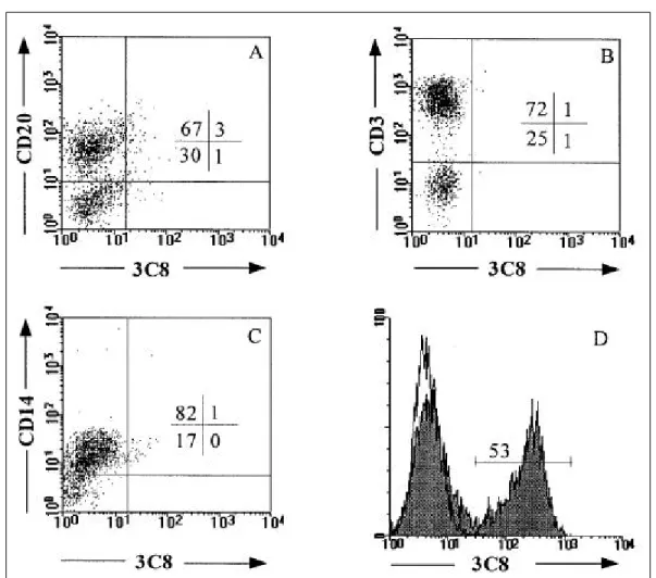
B cells, T cells, and m onocytes were purified from peripheral blood . Con sistent with the cell-based ELISA results, 3C8 did not bind to CD 20+ B cell, CD 3+ T cells, or CD 14+ m onocytes (Fig . 2A ~ 2C) . M oreover, the strong reactivity of 3C8 to HK cells was confirmed by flow cytom etric analy sis (Fig . 2D) . How ever , 3C8 exhibited no reactivity to murine fibroblast line L 929 and little reactivity to hum an skin fibroblasts (data not shown) . These results suggest that the 3C 8 antigen is specifically expressed by HK cells and may be important in the function of FD Cs.
3 . 3C 8 s p e cif ic a lly r e c og n ize s F D C n et w or k s w it h in t on s illa r g er m in a l cen t er s
To provide further evidence upon the relevance of HK cells to FDCs and to characterize the 3C8 antigen , wes tained ton sil section s w ith isotype control, 3C8, and DRC - 1, which was already known to stain the GC ( 10) . A s shown in Fig . 3, 3C 8 stained the same GC of serial ton sil sections as DRC - 1, but 3C8 did not stain T cells, B cells, m acrophages, or dendritic cells outside the GC . It w as clear that 3C8 stain s the dendritic network surrounding GC B cells, but not GC B cells them selves, Fig . 1 . Re a ct iv ity of 3C 8 w it h t o ns illa r mo no n uc le a r c e lls (MNC ), s kin fib ro b la s t s (S K), a nd HK ce lls . C e ll- ba s e d E LIS A w a s pe rfo rme d w it h c u lt ure d c e lls , a s d e s c ribe d in Ma t e ria ls a nd Me t ho d s . Mo us e IgG 1 a nt ibo dy w a s inc lud e d a s a n is oty pe - ma tc he d co nt ro l a nd a nt i-C D4 4 a nt ibo dy a s a pos it iv e co nt ro l. A re p re s e nta t iv e re s u lt of t h re e re pro d uc ible re s u lt s is s how n .
Fig . 2 . Two-color flowcytometric a na lys is reve a ls t he s pe cific re act ivity of 3C8 w ith HK ce lls . Huma n B ce lls (A), T ce lls (B), a nd monocyte s (C) we re purified from pe riphe ra l blood. Double sta ining wa s pe rforme d w ith the indicate d a ntibody a nd 3C8. Histogra ms (D) indicate sta ining of HK ce lls w it h 3C8 a nd the is otype control. The re s ults s how n a re from one of five re producible expe rime nts .
Fig . 3 . Immunoe nzymatic a na lys is of s e ria l tons il s e ctions w ith 3C8. Huma n tons il s e ctions we re sta ine d w ith t he is otype control (A), 3C8 (B), a nd DRC-1 (C). Origina l ma gnificat ion, ×10.
which suggests that 3C8 may stain FDC network of the GC.
DI SCUSSI ON
FDCs play pivotal roles in the survival, proliferation, and differentiation of GC B cells. For instance, the disruption of FDC-B cell clusters results in the apoptosis of B cells, and purified human FDCs enhance cytokine-dependent growth and Ig production in CD40-activated B cells (1,8). The aim of the current study was to develop a monoclonal antibody specific to an FDC line HK and eventually to FDC in order to understand the ontogeny of FDCs and the molecular signals involved in the complex GC reactions. The 3C8 antibody that was developed against HK cells did not react with T cells, B cells, monocytes, (Figure 2) and NK cells (data not shown). It did not bind to murine L929 fibroblasts, but displayed different reactivity to human fibroblasts. Human skin fibroblasts CCD-SK25 obtained from ATCC did not bind 3C8 antibody, whereas another skin fibroblasts SNU-20SK from the Seoul National University Hospital exhibited 3C8 binding reactivity comparable to that of HK cells. In addition, 3C8 antibody intensely stained tonsillar GC but not the extrafollicular areas.
The result that tonsillar GC was intensely stained by 3C8 is consistent with the finding that 3C8 did not react with mononuclear cells. Since the 3C8 staining was directed against the dendritic networks of the GC, the 3C8 antigen appears to be expressed on FDCs. It remains to be confirmed whether 3C8 antibody binds to purified FDCs. Liu et al. recently described the first characterized human FDC-specific molecule, 7D6, that turned out to be the long human CR2/CD2 1 isoform (CD2 1L)(11).
Interestingly, DRC-1 and KiM4, which have been widely used as human FDC-specific antibodies, stained COS7 cells transfected with CD2 1L cDNA, indicating that 7D6, DRC-1, and KiM4 recognized the same target antigen.
On the basis of the following results, 3C8 antigen appears to differ from CD2 1L. DRC-1 is cross-reactive between FDCs and B cells, whereas 3C8 is clearly
negative for B cells. The staining of tonsil sections showed that the area stained with DRC-1 was absolutely restricted to the GC, while 3C8 reacts with minor fibroblast cells outside the GC (data not shown) as well as the maj or dendritic networks in the GC. In addition, the molecules recognized by DRC-1 and 3C8 antibodies have different molecular weights by SDS-PAGE analysis, i.e., 145 kD and 58 kD, respectively (manuscript in preparation). Further characterization of 3C8 is required to identify and to understand the biological function of this antigen.
Unlike the non-reactivity of the 3C8 antibody with CCD-25SK fibroblasts, which were used in the original screening step, 3C8 antibody reacted with SNU-20SK fibroblasts. Since 3C8 did not bind to murine fibroblasts, the antigen detected by 3C8 antibody seems to be specific to humans. However, further study is necessary to confirm that 3C8 antigen is indeed expressed in humans only and to determine the distribution of this antigen in human tissue. Whether the expression of the 3C8 antigen is limited to certain fibroblast cells or not, the 3C8 positivity of HK cells (this line may in fact be tonsillar fibroblasts that have the functional features of FDCs) and a skin fibroblast cell line sheds some clues on the origin of FDCs. It is currently accepted that FDCs originate from the surrounding reticular meshwork but not from bone marrow. Stromal cells such as fibroblasts may function not only as a tissue framework but provide important microenvironments in lymphoid tissue during immune response. If stromal fibroblasts undergo functional transition during immune response, it would be interesting to know what kind of signals trigger the transition. Regarding this aspect, a report by Matsumoto et al. and another by Lindhout et al. are worth mentioning. The former demonstrated that FDC organization and GC formation are controlled by both lymphotoxin-α-expressing bone marrow-derived cells and TNFR-I-expressing non-bone marrow-derived cells ( 12), while the latter reported that some FDC phenotypes are induced from fibroblast-like synoviocytes after stimulating with TNF-α and IL-1β ( 13).
ACKNOWLEDGEMENT
We thank Dr. Jin-Ho Chung for providing human skin fibroblasts.
REFERENCES
1. MacLennan ICM: Germinal centers Annu Rev Immunol 12; 117-139, 1994
2. Nossal GJV, Abbot A, Mitchell J, Lummus Z: Antigen in immunity. XV. Ultrastructural features of antigen capture in primary and secondary lymphoid follicles, J.
Exp. Med 127; 277-290, 1968
3. Humphrey JH, Grennan D, Sundaram V: The origin of follicular dendritic cells in the mouse and mechanism of trapping immune complexes on them. Eur. J. Immunol 14; 859-864, 1984
4. Yoshida K, Kaj i M, Takahashi T, van Den Berg TK, Dij kstra CD: Host origin of follicular dendritic cells induced in the spleen of SCID mice after transfer of allogeneic lymphocytes. Immunol 84; 117-126, 1995 5. Choe J, Li L, Zhang X, Gregory CD, Choi YS: Distinct
role of follicular dendritic cells and T cells in the proliferation, differentiation, and apoptosis of a centroblast cell line, L3055. J. Immunol 164; 56-63, 2000
6. Kim HS, Zhang X, Choi YS: Activation and prolifera- tion of follicular dendritic cell-like cells by activated T lymphocytes. J. Immunol 153; 2951-2961, 1994 7. Kim HS, Zhang X, Klyushnenkova E, Choi YS: Sti-
mulation of germinal center B lymphocyte proliferation by an FDC-like cell line, HK. J. Immunol 155;
1101-1109, 1995
8. Choe J, Kim HS, Zhang X, Armitage RJ, Choi YS:
Cellular and molecular factors that regulate the
differentiation and apoptosis of germinal center B cells.
Anti-Ig down-regulates Fas expression on CD40 ligand-stimulated germinal center B cells and inhibits Fas-mediated apoptosis. J. Immunol 157; 1006-1016,
1996
9. Choe J, Kim HS, Armitage RJ, Choi YS: The functional role of B cell antigen receptor stimulation and IL-4 in the generation of human memory B cells from germinal center B cells. J. Immunol 159; 3757-3766, 1997 10. Naiem M, Gerdes J, Abdulaziz Z, Stein H, Mason DY:
Production of a monoclonal antibody reactive with human dendritic reticulum cells and its use in the immunohistological analysis of lymphoid tissue. J. Clin.
Pathol. 36; 167-175, 1983
11. Liu YJ, Xu J, de Bouteiller O, Parham CL, Grouard G, Dj ossou O, de Saint-Vis B, Lebecque S, Banchereau J, Moore KW: Follicular dendritic cells specifically express the long CR2/CD21 isoform. J. Exp. Med 185;
165-170, 1997
12. Matsumoto M, Fu YX, Molina H, Huang G, Kim J, Thomas DA, Nahm MH, Chaplin DD: Distinct roles of lymphotoxin α and the type I tumor necrosis factor (TNF) receptor in the establishment of follicular dendritic cells from non-bone marrow-derived cells. J.
Exp. Med 186; 1997-2004, 1997
13. Lindhout E, van Eijk M, van Pel M, Lindeman J, Dinant HJ, de Groot C: Fibroblast-like synoviocytes from rheumatoid arthritis patients have intrinsic properties of follicular dendritic cells. J. Immunol 162;
5949-5956, 1999
FOOTNOTES
This work was supported by grant No. 0100086- 1- 1 from the Basic Research Program of the Korea Science
& Engineering Foundation.