Blood Res2017;52:316-39. bloodresearch.or.kr
332 Letters to the Editor
Ankur Jain1, Pankaj Malhotra1, Vikas Suri1, Ritesh Agarwal2, Amanjit Bal3, Subhash Varma1
Departments of 1Internal Medicine, 2Pulmonary Medicine, and 3Histopathology, Nehru Hospital, PGIMER, Chandigarh, India
Correspondence to: Pankaj Malhotra Department of Internal Medicine, Nehru Hospital,
PGIMER, Chandigarh 160012, India E-mail: [email protected]
Received on Jan. 11, 2017; Accepted on Apr. 11, 2017 https://doi.org/10.5045/br.2017.52.4.329
AuthorsÊ Disclosures of Potential Conflicts of Interest No potential conflicts of interest relevant to this article were reported.
REFERENCES
1. Fowler AA, Hamman RF, Good JT, et al. Adult respiratory distress syndrome: risk with common predispositions. Ann Intern Med 1983;98:593-7.
2. Chusid MJ, Dale DC, West BC, Wolff SM. The hypereosinophilic syndrome: analysis of fourteen cases with review of the literature. Medicine (Baltimore) 1975;54:1-27.
3. Roufosse FE, Goldman M, Cogan E. Hypereosinophilic syndromes. Orphanet J Rare Dis 2007;2:37.
4. Gotlib J. World Health Organization-defined eosinophilic dis- orders: 2014 update on diagnosis, risk stratification, and management. Am J Hematol 2014;89:325-37.
5. Gotlib J, Cools J, Malone JM 3rd, Schrier SL, Gilliland DG, Coutré SE. The FIP1L1-PDGFRalpha fusion tyrosine kinase in hyper- eosinophilic syndrome and chronic eosinophilic leukemia: im- plications for diagnosis, classification, and management. Blood 2004;103:2879-91.
6. Dulohery MM, Patel RR, Schneider F, Ryu JH. Lung involvement in hypereosinophilic syndromes. Respir Med 2011;105:114-21.
7. Winn RE, Kollef MH, Meyer JI. Pulmonary involvement in the hypereosinophilic syndrome. Chest 1994;105:656-60.
8. Savage N, George TI, Gotlib J. Myeloid neoplasms associated with eosinophilia and rearrangement of PDGFRA, PDGFRB, and FGFR1: a review. Int J Lab Hematol 2013;35:491-500.
Does the c.-273T>C variant in the upstream region of the HBB gene cause a thalassemia phenotype?
TO THE EDITOR: Beta thalassemia is a hereditary disease that results from mutations in the HBB gene, leading to genetic defects in the production of beta-globin chains [1, 2]. HBB encodes beta-globin, a subunit of hemoglobin. In
adults, hemoglobin is normally made up of four protein subunits, including two subunits of beta-globin and two subunits of alpha-globin, with the latter produced from HBA. Mutations in HBB can result in either beta-plus (B+) thalassemia that is responsible for a less severe form of thalassemia (caused by a decrease in beta-globin production) or beta-zero (B0) thalassemia that is the severe type of the disease (caused by a total lack of beta-globin) [3-8].
The mutations usually include missense or nonsense types, but other types, such as deletions of the beta-globin gene and surrounding regions, also have been identified in tha- lassemia patients. According to the Human Gene Mutation Database (HGMD), currently, 835 disease-causing mutations have been found in HBB, including 404 missense/nonsense, 118 small deletions, 97 gross deletions, 73 regulatory, 53 splicing, 44 small insertions, 21 complex rearrangements, 19 small indels, and six gross insertions (http://www.hgmd.
cf.ac.uk/ac/gene.php?gene=HBB).
In addition, different studies have reported variants with unknown significance in the 5΄ region, near the splice sites, and in the 3΄ area of HBB, including c.-273T>C (upstream of the gene) [9]; until date, there have been no compre- hensive data regarding its role in the phenotype of thalassemia. The goal of this study was to clarify the sig- nificance of this variant using segregation and bioinformatics analysis. Thus, from our large number of samples recruited from cases of minor thalassemia, we collected data regarding their laboratories and genetic studies. All patients provided informed consent before undergoing molecular testing for HBB and HBA mutation analysis. This study was approved by the institutional review board of the Comprehensive Medical Genetics Center, Shiraz University of Medical Sciences (Approval No. 95.113.). Genomic DNA was ex- tracted from the peripheral blood lymphocytes of these sam- ples using DNA Extraction Kits (Yekta Tajhiz, Iran) accord- ing to the manufacturer's instructions, and the DNA concen- tration was measured by NanoDrop (ND1000, USA) and stored at -20oC until use.
Sequences covering all coding and important non-coding regions of HBB and HBA 1 and 2 genes were amplified by PCR. The total volume used for the PCR was 50 μL including 1 μL of each primer (20 pmol/μL), 2 μL DNA template (50–200 ng), 25 μL TEMPase Hot Start 2x Master Mix Blue (Ampicon, A290806), and 21 μL dH2O. The PCR reactions were carried out according to Amplicon TEMPase Hot Start protocol and programs. Ten microliters of the PCR products were visualized on 2% agarose gel containing SYBR Safe. For mutation analysis for HBA 1 and 2 genes, ViennaLab StripAssays was used to locate all important dele- tions that were not detected by standard PCR.
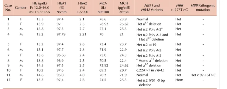
From 200 samples of different types of suspected minor thalassemia, we found 12 cases with c.-273T>C. It is worth noting that one of the cases had this variant in a homozygous pattern and another of the cases with c.-273T>C had a pathogenic mutation. All heterozygous cases for c.-273T>C (cases 1–10) and also that of the homozygous form, including
bloodresearch.or.kr Blood Res 2017;52:316-39.
Letters to the Editor 333
Table 1. Laboratory findings and identified variants in HBB and HBA1. Case
No. Gender
Hb (g/dL) F: 12.0–16.0 M: 13.5–17.5
HbA1(%) 95–98
HbA2(%) 1.5–3.0
MCV(fL) 80–100
(pg/cell)MCH 26–34
HBA1 and
HBA2 Variants HBB
c.-273T>C HBB Pathogenic mutation
1 F 13.3 97.4 2.1 76.6 23.9 Normal Het -
2 F 13.9 97 2.5 78.92 25.62 Het a3.7 deletion Het -
3 M 15.8 97.3 2.7 77.1 25.5 Het α2 Poly A-2a) Het -
4 M 13.2 97.79 2.21 70 21 Het α2 Poly A-2 and
Het a3.7 deletion
Het -
5 F 13.2 97.4 2.6 73.4 23.7 Het α2 cd19 Het -
6 M 15.1 97.7 2.3 71.9 22.9 Het α2 Poly A-2 Het -
7 F 13.8 96.68 2.4 75.0 24.3 Het α2 Poly A-2 Het -
8 M 13.8 96.9 2.5 70.5 22.4 e Homo a3.7 deletion Het -
9 M 14.3 97.5 2.5 75.92 24.62 Het a3.7 deletion Het -
10 F 10.2 97.6 2.4 69.3 20.7 c.22A>T in HBA2 Het -
11 M 14.6 96.0 4.0 70.2 21.9 Normal Het Het c.92+6T>C
12 F 13.3 97.4 2.6 74.5 25.3 Het α2 IVS1 -5 bp
deletion
Hom -
a)α2 Poly A-2: AATAAA>AATGAA
Abbreviations: F, female; Het, Heterozygous; Hom, Homozygous; M, male.
case 12 without any pathogenic mutations in HBB, had HBA2 levels in the normal range (Table 1). However, the case with c.-273T>C (case 11) that also had a pathogenic c.92+6T>C mutation showed an HBA2 level of 4.0%, in- dicating that c.-273T>C has no effect on HBB protein.
In HBA1 and 2, we found the different mutations listed in Table 1, which shows why some cases without any muta- tions in HBB had positive laboratory findings.
We also used bioinformatics software, such as FATHMM, CADD, and PhastCons programs, to predict the effects of this variant in the non-coding region of HBB. The FATHMM program (http://fathmm.biocompute.org.uk/disease.html) uses information concerning sequence homology and its score ranges between -16.13 and 10.64. If a score is lower than -1.5, then the corresponding nonsynonymous SNP (NS) is predicted as DAMAGING. The non-coding score of FATHMM for this variant was 0.042, which predicts that this variant does not have functional effects on the HBB protein. For CADD program, mutations with scores ≥10 are predicted to be the 10% most deleterious substitutions, whereas scores ≥20 signify the 1% most deleterious effects.
Using SeattleSeq Annotation (http://snp.gs.washington.edu/
SeattleSeqAnnotation138/), we found that the CADD score for this variant was 0.146, which predicts that c.-273T>C is a benign variant.
The phastCons program was also used to identify con- served genomic regions. Higher scores indicate a higher probability that the base is in a conserved element. The PhastCons score of the c.-273T>C variant was 0.250, sug- gesting that the region of the variant is less conserved among vertebrates.
In conclusion, our data confirmed that c.-273T>C does not impose any effect on the function of HBB protein and
should be considered as a benign variant.
Hassan Dastsooz1, Mohsen Alipour1, Sanaz Mohammadi1, Fatemeh Dehghanian1, Fatemeh Kamgarpour1, Majid Fardaei1,2
1Comprehensive Medical Genetic Center, 2Department of Medical Genetics, Shiraz University of Medical Sciences, Shiraz, Iran
Correspondence to: Majid Fardaei Department of Medical Genetics, Shiraz University of
Medical Sciences, Shiraz, Fars 7134853185, Iran E-mail: [email protected]
Received on Mar. 12, 2017; Revised on Apr. 4, 2017; Accepted on Jun. 15, 2017 https://doi.org/10.5045/br.2017.52.4.332
Acknowledgments
We wish to thank all the patients for their willingness to take part in this study. This study was supported by the Comprehensive Medical Genetic Center, Shiraz Univer- sity of Medical Sciences, Shiraz, Iran.
AuthorsÊ Disclosures of Potential Conflicts of Interest No potential conflicts of interest relevant to this article were reported.
REFERENCES
1. Rachmilewitz EA, Giardina PJ. How I treat thalassemia. Blood 2011;118:3479-88.
2. Galanello R, Sanna S, Perseu L, et al. Amelioration of Sardinian beta0 thalassemia by genetic modifiers. Blood 2009;114:3935-7.
Blood Res2017;52:316-39. bloodresearch.or.kr
334 Letters to the Editor
3. Taher A, Isma'eel H, Cappellini MD. Thalassemia intermedia:
revisited. Blood Cells Mol Dis 2006;37:12-20.
4. Huehns ER, Dance N, Beaven GH, Heclht F, Motulsky AG.
Human embryonic hemoglobins. Cold Spring Harb Symp Quant Biol 1964;29:327-31.
5. Villegas A, Ropero P, González FA, Anguita E, Espinós D. The thalassemia syndromes: molecular characterization in the Spanish population. Hemoglobin 2001;25:273-83.
6. Nadkarni A, Gorakshakar AC, Lu CY, et al. Molecular patho- genesis and clinical variability of beta-thalassemia syndromes among Indians. Am J Hematol 2001;68:75-80.
7. Rund D, Rachmilewitz E. Beta-thalassemia. N Engl J Med 2005;353:1135-46.
8. Schechter AN. Hemoglobin research and the origins of molecular medicine. Blood 2008;112:3927-38.
9. Nagar R, Sinha S, Raman R. Genotype-phenotype correlation and report of novel mutations in β-globin gene in thalassemia patients. Blood Cells Mol Dis 2015;55:10-4.
Comparison of the acute erythropoietic capacities of erythropoietin and U-74389G in terms of hemoglobin levels
TO THE EDITOR: This study compared the erythropoietic capacities of erythropoietin (EPO) and antioxidant drug U-74389G based on the findings of 2 preliminary studies.
Hemoglobin augmentation was evaluated using a hypoxia reoxygenation (HR) protocol in an animal model. Hemoglo- bin levels were evaluated at the 60th reoxygenation minute (for groups A, C, and E) and at the 120th reoxygenation minute (for groups B, D, and F) in 60 rats. Groups A and B were administered no drugs, groups C and D were ad- ministered EPO, and groups E and F were administered U-74389G. The first preliminary study of EPO did not show a significant increase in hemoglobin levels. However, the second preliminary study of U-74389G showed a significant increase in hemoglobin levels by 2.5±1.3% (P=0.0423).
These 2 studies were co-evaluated because they were con- ducted in the same experimental setting. In non-deficient EPO rats, U-74389G demonstrated an approximately 2-times higher erythropoietic potency than EPO (P=0.0000). This is because the anti-oxidant capacity of U-74389G increased the acute erythropoietic potency.
A previous study claims that U-74389G harbors a remark- able acute erythropoietic capacity [1]. U-74389G is a novel antioxidant factor and has shown tissue protective effects in tissue hypoxia and reoxygenation (HR) experiments.
U-74389G, also known as 21-[4-(2,6-di-1-pyrrolidinyl-4- pyrimidinyl)-1-piperazinyl]-pregna-1,4,9(11)-triene- 3,20-dione maleate salt, prevents both arachidonic acid-in-
duced and iron-dependent lipid peroxidation. It has been shown to protect against HR injury in animal heart, liver, and kidney models. These membrane-associating anti- oxidants are particularly effective in preventing perme- ability changes in brain microvascular endothelial cells monolayers. Lazaroids, or 21-aminosteroids, a novel series of glucocorticoid compounds, scavenge free radicals. U- 74389G is one of the 132 similar lazaroid compounds. It has a molecular weight of 726.90406 g/mol and demonstrates selective action on the vascular endothelium with vitamin E-like properties.
However, the erythropoietic capacity of U-74389G ap- pears more comprehensible when compared with that of a standard known drug. One such well-studied drug, where- in erythropoietic capacity was confirmed (P=0.3984), is EPO. Indeed, EPO has been implicated in over 29,946 known biomedical studies. Among these studies, 30.65% concern tissue HR experiments. However, only a few reports that were found to be related with this study did not completely address the specific matter of antioxidant factors. The aim of this experimental work was to compare the acute eryth- ropoietic capacities of U-74389G and EPO in a non-deficient EPO rat model using an HR protocol. Their effects were assessed on the basis of increase in hemoglobin levels.
The veterinarian licenses for the research were provided under the 3693/12-11-2010 & 14/10-1-2012 decisions. The institute and place of experiment are mentioned in the re- lated references [1, 2]. The experimental protocol, which involved Albino female Wistar rats, adhered to the ethical rules of the relevant organization. For 7 days pre-ex- perimentally, the rats were placed under normal housing and fed ad libitum in the laboratory. Continuous intra-ex- perimental general anesthesia, oxygen supply, electro- cardiography, acidometry, and post-experimental eutha- nasia were provided. Subsequently, 16–18-week-old rats were randomly divided into 6 groups (N=10) according to the HR protocol: hypoxia for 45 minutes followed by reox- ygenation for 60 minutes (group A); hypoxia for 45 minutes followed by reoxygenation for 120 minutes (group B); hypo- xia for 45 minutes followed by immediate intravenous (IV) EPO administration and reoxygenation for 60 minutes (group C); hypoxia for 45 minutes followed by immediate IV EPO administration and reoxygenation for 120 minutes (group D); hypoxia for 45 minutes followed by immediate U-74389G IV administration and reoxygenation for 60 mi- nutes (group E); hypoxia for 45 minutes followed by immedi- ate U-74389G IV administration and reoxygenation for 120 minutes (group F). The dose height selection criteria for EPO and U-74389G were assessed in preliminary studies as 10 mg/kg body mass for both drugs.
Hypoxia was induced by laparotomic clamping of the inferior aorta over the renal arteries with forceps for 45 minutes. Clamp removal restored the inferior aorta patency and reoxygenation. After exclusion of the blood flow, the HR protocol was followed as described above for each ex- perimental group. The drugs were administered at the time