309
This is an Open Access article distributed under the terms of the Creative Commons Attribution Non-Commercial License (http://creativecommons.org/licenses/by-nc/3.0) which permits unrestricted non-commercial use, distribution, and reproduction in any medium, provided the original work is properly cited.
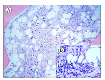
Fig. 1. Serous atrophy in a diffuse and severe hypocellular background (A). Reduced fat cells, a diffuse extracellular gelatinous amorphous material, small typical lymphocytes, and mature plasma cells were also observed (B). Small islets of hematopoietic cells were detected at a level of <5%.
http://dx.doi.org/10.5045/kjh.2012.47.4.309
The Korean Journal of Hematology Volume 47ㆍNumber 4ㆍDecember 2012
Letter to the Editor
Long-term survival of a patient with bone marrow gelatinous degeneration of idiopathic ori- gin
TO THE EDITOR: Gelatinous bone marrow transformation (GMT) is a rare bone marrow (BM) disorder of unknown pathogenesis. It is characterized by fat cell atrophy, focal loss of hematopoietic cells, and deposition of extracellular gelatinous substances (mucopolysaccharides rich in hyalur- onic acid) [1]. The pathogenesis of GMT involves the deposi- tion of hyaluronic acid, which hampers hematopoiesis by altering the BM microenvironment and stroma and dis- ruption of the interactions between BM cells and cell signal- ing molecules [1]. GMT has been reported in association with chronic debilitating diseases, such as anorexia nervosa, malnutrition, and human immunodeficiency virus (HIV) infection, and after cytotoxic treatments [2]; in addition, GMT has been described in patients with myelodysplastic syndrome [3], acute myeloblastic leukemia [3], and idio- pathic myelofibrosis [4]. However, idiopathic GMT has also been reported [5]. Herein, we report a case of idiopathic GMT in a patient who had an unusual clinical course and long-term survival. The patient was a 64-year-old Caribbean woman who had been living in Rome for more than 30 years. She was referred to our clinic in December 2005 because of anemia. Her past medical history was unremark- able. She did not use alcohol, drugs, or tobacco, and her nutritional status was excellent. Physical examination was unremarkable with the exception of pallor; the patient did not have either spleen or liver enlargement. A complete blood count at admission showed normochromic-normo- cytic anemia with reticulocytopenia and normal white blood cell and platelet counts. Total and unconjugated bilirubin and serum haptoglobin levels were normal. Direct and in-
direct Coombs tests were negative. Serological tests for HIV, cytomegalovirus, Epstein-Barr virus, parvovirus B19, and hepatitis B and C viruses were negative. In addition, serum levels of tri-iodothyronine were normal. Moreover, no clin- ical or laboratory features of autoimmune disease were detected. Examination of peripheral blood smears revealed normal RBC; erythrocyte fragments were not detectable.
Radiological examinations, which included a whole-body computed tomography scan, revealed no abnormalities.
Occult blood loss was ruled out by fecal and urine analyses.
A BM aspirate performed at admission resulted in a dry tap. Therefore, a BM trephine biopsy was taken. Histological examination of the BM sample revealed GMT (Fig. 1). The patient did not respond to any potentially disease-modifying treatment, including erythropoietin, prednisone, and cyclosporine. Therefore, she was managed with supportive measures, primarily transfusions. The clinical course was
Korean J Hematol 2012;47:309-10.
310 Letter to the Editor
uncomplicated and her hematological status has remained stable. To date, 80 months after the diagnosis, she is managed only with supportive measures, consisting of 2 units of packed RBC every 3 weeks, along with iron chelation therapy. In addition, her BM features, which have been evaluated annually by BM trephine biopsies, have remained stable. In conclusion, we have described a case of an appa- rently healthy woman who presented with GMT approx- imately 7 years ago, in whom anemia was the only clinical manifestation of hematopoietic impairment. To the best of our knowledge, no such case of GMT has been reported previously as an idiopathic primary condition.
Pasquale Niscola, M.D.1, Massimiliano Palombi, M.D.1, Stefano Fratoni, M.D.2, Malgorzata Monika Trawinska, M.D.1, Laura Scaramucci, M.D.1, Andrea Tendas, M.D.1, Marco Giovannini, M.D.1, Alessio Perrotti, M.D.1, Paolo de Fabritiis, M.D.1
1Haematology, 2Pathology Department, Sant’ Eugenio Hospital, Piazzale dell’ Umanesimo 10, 00144 Rome, Italy
Tel: +390651003241, E-mail: pniscola@gmail.com
1. Böhm J. Gelatinous transformation of the bone marrow: the spec- trum of underlying diseases. Am J Surg Pathol 2000;24:56-65.
2. Aisa Y, Mori T, Nakazato T, Yamazaki R, Ikeda Y, Okamoto S.
Gelatinous degeneration in a recipient of allogeneic bone marrow transplantation. Int J Hematol 2006;84:465-7.
3. Arranz R, Gil-Fernandez JJ, Acevedo A, Tomas JF, Alegre A, Fernandez-Rañada JM. Gelatinous degeneration presenting as a preleukaemic syndrome. J Clin Pathol 1996;49:512-4.
4. Böhm J, Schmitt-Gräff A. Gelatinous bone marrow transformation in a case of idiopathic myelofibrosis: a morphological paradox.
Pathol Res Pract 2000;196:775-9.
5. Niscola P, Maurillo L, Palombi M, et al. Gelatinous degeneration of the bone marrow: two case reports showing different hemato- logical features and clinical outcomes. Acta Haematol 2007;118:
165-6.