ABSTRACT
Objective: To compare the survival outcomes of adjuvant radiotherapy and chemotherapy in women with uterine-confined endometrial cancer with uterine papillary serous carcinoma (UPSC) or clear cell carcinoma (CCC).
Methods: Medical records of 80 women who underwent surgical staging for endometrial cancer were retrospectively reviewed. Stage I UPSC and CCC were pathologically confirmed after surgery. Survival outcomes were compared between the adjuvant radiotherapy and chemotherapy groups.
Results: Fifty-four (67.5%) and 26 (32.5%) women had UPSC and CCC, respectively. Adjuvant therapy was administered to 59/80 (73.8%) women (25 radiotherapy and 34 chemotherapy).
High preoperative serum cancer antigen-125 level (25.1±20.2 vs. 11.5±6.5 IU/mL, p<0.001), open surgery (71.2% vs. 28.6%, p=0.001), myometrial invasion (MI) ≥1/2 (33.9% vs. 0, p=0.002), and lymphovascular space invasion (LVSI; 28.8% vs. 4.8%, p=0.023) were frequent in women who received adjuvant therapy compared to those who did not. However, the histologic type, MI ≥1/2, and LVSI did not differ between women who received adjuvant radiotherapy and those who received chemotherapy. The 5-year progression-free survival (78.9% vs. 80.1%, p>0.999) and overall survival (77.5% vs. 87.8%, p=0.373) rates were similar between the groups. Neither radiotherapy (hazard ratio [HR]=1.810; 95% confidence interval [CI]=0.297–11.027; p=0.520) nor chemotherapy (HR=1.638; 95% CI=0.288–9.321; p=0.578) after surgery was independently associated with disease recurrence.
Conclusion: Our findings showed similar survival outcomes for adjuvant radiotherapy and chemotherapy in stage I UPSC and CCC of the endometrium. Further large study with analysis stratified by MI or LVSI is required.
Original Article
Received: Jun 20, 2018 Revised: Dec 12, 2018 Accepted: Dec 12, 2018 Correspondence to Yong Beom Kim
Department of Obstetrics and Gynecology, Seoul National University Bundang Hospital, 82 Gumi-ro 173beon-gil, Bundang-gu, Seongnam 13620, Korea.
E-mail: [email protected] Copyright © 2019. Asian Society of Gynecologic Oncology, Korean Society of Gynecologic Oncology
This is an Open Access article distributed under the terms of the Creative Commons Attribution Non-Commercial License (https://
creativecommons.org/licenses/by-nc/4.0/) which permits unrestricted non-commercial use, distribution, and reproduction in any medium, provided the original work is properly cited.
ORCID iDs Miseon Kim
https://orcid.org/0000-0002-5118-9275 Byung Su Kwon
https://orcid.org/0000-0002-9586-0200 Ha Kyun Chang
https://orcid.org/0000-0003-3138-1697 Seungmee Lee
https://orcid.org/0000-0003-1405-2656 Suk-Joon Chang
https://orcid.org/0000-0002-0558-0038 Jin Young Choi
https://orcid.org/0000-0003-1522-3111 Sang-Yoon Park
https://orcid.org/0000-0001-8325-6009
Miseon Kim ,1 Byung Su Kwon ,2 Ha Kyun Chang ,3 Seungmee Lee ,4 Suk-Joon Chang ,5 Jin Young Choi ,6 Sang-Yoon Park ,3 Maria Lee ,7 Hee-Sug Ryu ,5 Yong Beom Kim 8
1 Department of Obstetrics and Gynecology, CHA Gangnam Medical Center, CHA University School of Medicine, Seoul, Korea
2Department of Obstetrics and Gynecology, Pusan National University Hospital, Busan, Korea
3Center for Uterine Cancer, Research Institute and Hospital, National Cancer Center, Goyang, Korea
4Department of Obstetrics and Gynecology, Keimyung University School of Medicine, Daegu, Korea
5Department of Obstetrics and Gynecology, Ajou University School of Medicine, Suwon, Korea
6Department of Obstetrics and Gynecology, Chungbuk National University Hospital, Cheongju, Korea
7Department of Obstetrics and Gynecology, Seoul National University College of Medicine, Seoul, Korea
8Department of Obstetrics and Gynecology, Seoul National University Bundang Hospital, Seongnam, Korea
Survival outcomes of adjuvant
radiotherapy and chemotherapy in
women with stage I serous papillary and clear cell carcinoma of the endometrium:
a Korean multicenter study
Maria Lee
https://orcid.org/0000-0002-8017-3176 Hee-Sug Ryu
https://orcid.org/0000-0003-1984-2614 Yong Beom Kim
https://orcid.org/0000-0003-1196-369X Funding
This study was supported by a grant from the Seoul National University College of Medicine Research Fund 2016.
Conflict of Interest
No potential conflict of interest relevant to this article was reported.
Author Contributions
Conceptualization: K.M., K.Y.B.; Data curation:
K.M., K.B.S., C.H.K., L.S., C.S.J., C.J.Y., L.M.;
Formal analysis: K.M.; Funding acquisition:
K.Y.B.; Investigation: K.M., K.B.S., C.H.K., L.S., C.J.Y.; Methodology: K.M., K.Y.B.; Resources:
C.S.J., P.S.Y., L.M., R.H.S., K.Y.B.; Supervision:
K.Y.B.; Visualization: K.M.; Writing - original draft: K.M., K.B.S., C.H.K., L.S., C.J.Y.; Writing - review & editing: K.M., C.S.J., P.S.Y., L.M., R.H.S., K.Y.B.
Keywords: Endometrial Neoplasms; Adenocarcinoma, Papillary; Adenocarcinoma, Clear Cell; Chemotherapy, Adjuvant; Radiotherapy, Adjuvant
INTRODUCTION
Endometrial cancer is the most common malignancies of the female reproductive tract in the United States, with over 65,050 cases estimated to be diagnosed in 2016; its incidence exceeds those of cervical, ovarian and vaginal cancers combined [1]. Although many cases present with early-stage disease, there are 10,470 total annual deaths in the United States from this disease.
Endometrial cancers have been classified into type I and type II cancers based on their risk factors and prognosis. Type I comprises 80% of all endometrial cancers and endometrioid endometrial carcinoma (EEC) accounts for 85% of type I cases. While type I endometrial cancer generally has a favorable prognosis, type II disease, which includes the more aggressive histologies of uterine papillary serous carcinoma (UPSC), and clear cell carcinoma (CCC), portends a poorer prognosis [2-4]. Although UPSC and CCC represent 10% and 3% of endometrial cancer cases, they account for 39% and 8% of disease-related deaths, respectively [5].
Approximately, 10% to 15% of women with endometrial cancer will experience recurrence, even in early-stage disease [6,7]. To reduce the recurrence rate, adjuvant radiotherapy
and/or chemotherapy have been applied after surgery; however, a definite standard of care has not yet been established worldwide. Pelvic external beam radiotherapy (EBRT) has been performed as an adjuvant treatment for women with high-risk endometrial cancer for many decades, although there is a paucity of evidence on the improvement of overall survival (OS) [7-9]. Until now, almost all large-scale studies have reported the prognostic impact of adjuvant therapy for all of intermediate-to-high risk endometrial cancers (International Federation of Gynecology and Obstetrics [FIGO] stage IA grade 3 EEC, FIGO stage IB [grade 1–3] EEC, and all stages with non-endometrioid type). Because UPSC and CCC are very rare to study separately even for all stages, there is a lack of evidence for adjuvant therapy in early-stage disease.
Therefore, we aimed to evaluate the survival outcomes of adjuvant therapy and radiotherapy or chemotherapy alone in uterine-confined endometrial cancer of UPSC and CCC.
MATERIALS AND METHODS
1. Patients
The study cohort was retrospectively recruited from eight institutions. The study protocol was revised and accepted by each institutional ethics committee. We recruited a total of 80 women diagnosed with FIGO stage I endometrial cancer after primary staging surgery between January 2001 and May 2017. All eligible women were pathologically confirmed as having UPSC or CCC after surgery. No women who had undergone prior systemic
chemotherapy or radiotherapy for another malignancy were included in the study population.
The staging surgery included total hysterectomy and bilateral salpingo-oophorectomy;
peritoneal washing cytology and omentectomy were left to the surgeon's discretion. Pelvic and para-aortic lymph node dissections were also omitted at the discretion of the surgeon if there was no evidence of lymph node metastasis in preoperative computed tomography (CT), magnetic resonance imaging (MRI), or positron emission tomography-computed tomography (PET-CT) scans. Lymph nodes with a short diameter ≥1.0 cm in imaging were considered
significantly enlarged. Surgical specimens were reviewed by pathologists specializing in gynecologic pathology who were blinded to the patients' outcomes. We obtained the final pathologic results, which included the histologic type and grade, FIGO stage, lymphovascular space invasion (LVSI), and the presence of malignant cells in peritoneal washing cytology.
In accordance with the clinical policies in each participating institution, adjuvant therapy was administered after surgery to patients whose final pathologic results indicated an intermediate or high risk. Of 80 women, 59 (73.8%) received adjuvant therapy after surgery: 25 radiotherapy and 34 chemotherapy. The radiotherapy included concurrent chemoradiation with a radiosensitizing agent. We excluded five women who received sequential chemoradiation, radiotherapy followed by chemotherapy (n=3) or its reverse (n=2) from the analysis.
2. Statistical analysis
Five-year progression-free survival (PFS) and OS rates were compared using the Kaplan-Meier method. Survival time was counted from the date of surgery until recurrence or death and compared between the two adjuvant therapy method groups by log-rank tests. We classified recurrence as local, regional, or distant. Recurrences limited to the vaginal vault were defined as local metastasis; recurrences beyond the vaginal vault but limited to the pelvic region were defined as regional metastasis; and recurrences beyond the pelvis, the extraperitoneal cavity, and outside the abdominal cavity were defined as distant metastasis.
Student's t-test and χ2 test were used to examine continuous and categorical variables, respectively. For corresponding non-parametric statistics, Mann-Whitney U and Fisher's exact tests were used, respectively. Logistic regression was used to identify the clinicopathological factors associated with disease recurrence. Variables with p≤0.25 were included in the multivariate analysis, and p<0.05 was considered to be significant. IBM SPSS Statistics for Windows, version 24.0 (IBM Corp., Armonk, NY, USA) was used for statistical analyses.
RESULTS
1. Patient characteristics
The median age at diagnosis was 63 years (range, 41–85) and the median follow-up period was 32 months (range, 1–143) in the study population. Comparison of the baseline characteristics between the women who underwent adjuvant therapy after surgery and those who did not are shown in Table 1. The mean serum level of cancer antigen (CA)-125 at diagnosis was higher in women with adjuvant therapy than that in those without (25.1±20.2 vs. 11.5±6.5 IU/mL, p<0.001). Lymph node involvement on preoperative CT or MRI scan were suspected in 4 women in total study population, however, it was not different between the women without adjuvant therapy and with adjuvant therapy (1 [4.8%] vs. 3 [5.1%], p>0.999).
The staging surgery by open approach was frequently performed in women with adjuvant therapy (42 [71.2%] vs. 6 [28.6%], p=0.001). However, peritoneal washing cytology (16 [76.2%]
vs. 52 [88.1%], p=0.188), omentectomy (8 [38.1%] vs. 33 [55.9%], p=0.160), pelvic (18 [85.7%]
vs. 48 [81.4%], p=0.652) and para-aortic lymph node dissection (12 [57.1%] vs. 34 [57.6%], p=0.969) were similarly conducted between women with and without adjuvant therapy.
After surgery, 54 (67.5%) and 26 (32.5%) women were pathologically confirmed as having UPSC and CCC, respectively. The proportions of women with adjuvant therapy did not differ
between the women who had UPSC and CCC (40/54 [74.1%] vs. 19/26 [73.1%], p=0.924).
Instead, myometrial invasion (MI) ≥1/2 (20 [33.9%] vs. 0, p=0.002) and LVSI (17 [28.8%] vs.
1 [4.8%], p=0.023) were more common in women who underwent adjuvant therapy than in those who did not.
2. Clinical course by adjuvant therapy methods: radiotherapy vs.
chemotherapy
The clinical courses after surgery are described in Table 2. A total of 25 and 34 women received radiotherapy and chemotherapy after surgery, respectively. The mean age (63.8±8.8 vs. 62.8±6.5, p=0.6314), body mass index (24.6±4.2 vs. 25.3±2.9 kg/m2, p=0.495), and Table 1. Patient characteristics (n=80)
Parameters No adjuvant therapy (n=21) Adjuvant therapy (n=59) p
Age (yr) 63.3±9.4 63.2±7.5 0.968
Body mass index (kg/m2) 24.9±3.8 25.0±3.5 0.918
Preoperative evaluation
Serum CA-125 level (IU/mL) 11.5±6.5 25.1±20.2 <0.001
Endometrial biopsy (n=74) 0.829
Endometrioid 4 (20.0) 10 (18.5)
Serous papillary 13 (65.0) 31 (57.4)
Clear cell 0 8 (14.8)
Other 3 (15.0) 5 (9.3)
LN involvement on CT or MRI scan >0.999
No 20 (100.0) 56 (94.9)
Yes 1 (4.8) 3 (5.1)
Operation details
Approach 0.001
Open 6 (28.6) 42 (71.2)
Laparoscopic 15 (71.4) 17 (28.8)
WC in pelvic cavity 0.188
No 5 (23.8) 7 (11.9)
Yes 16 (76.2) 52 (88.1)
Omentectomy 0.160
No 13 (61.9) 26 (44.1)
Yes 8 (38.1) 33 (55.9)
Pelvic LN dissection 0.652
No 3 (14.3) 11 (18.6)
Yes 18 (85.7) 48 (81.4)
Retrieved node (No.) 15.3±8.4 17.9±10.4 0.342
Para-aortic LN dissection 0.969
No 9 (42.9) 25 (42.4)
Yes 12 (57.1) 34 (57.6)
Retrieved node (No.) 5.3±5.8 7.7±8.4 0.287
Pathological results
Histology 0.924
Serous papillary 14 (66.7) 40 (67.8)
Clear cell 7 (33.3) 19 (32.2)
MI ≥1/2 0.002
No 21 (100.0) 39 (66.1)
Yes 0 20 (33.9)
LVSI 0.023
No 20 (95.2) 42 (71.2)
Yes 1 (4.8) 17 (28.8)
Malignant cells in WC (n=68) >0.999
No 16 (100.0) 49 (94.2)
Yes 0 3 (5.8)
The values are presented as the median (range) or number (%), unless otherwise indicated.
CA-125, cancer antigen-125; CT, computed tomography; LN, lymph node; LVSI, lymphovascular space invasion; MI, myometrial invasion; MRI, magnetic resonance imaging; WC, washing cytology.
preoperative serum cancer antigen-125 level (27.0±22.6 vs. 23.7±18.4 IU/mL, p=0.543) did not differ significantly between the women who received radiotherapy and those who received chemotherapy. Radiotherapy was mainly performed in women with UPSC, whereas chemotherapy was mainly performed in women with CCC. However, the difference was not statistically significant (p=0.160). In addition, MI ≥1/2 (11 [44.0%] vs. 9 [26.5%], p=0.160), LVSI (5 [20.0%] vs. 12 [35.3%], p=0.200), and malignant cells in washing cytology (1 [4.3%]
vs. 2 [6.9%], p>0.999) were also similar between groups.
Almost all women (23/25 [92.0%]) with radiotherapy underwent EBRT, and no one received vaginal brachytherapy combined with EBRT simultaneously. Concurrent chemoradiation Table 2. Clinical course by adjuvant therapy methods
Parameters Radiotherapy (n=25) Chemotherapy (n=34) p
Age (yr) 63.8±8.8 62.8±6.5 0.631
Body mass index (kg/m2) 24.6±4.2 25.3±2.9 0.495
Serum CA-125 level (IU/mL) 27.0±22.6 23.7±18.4 0.543
Postoperative histology 0.085
Serous papillary 20 (80.0) 20 (58.8)
Clear cell 5 (20.0) 14 (41.2)
Concordance between preoperative
and postoperative histologic results 0.549
No 13 (52.0) 15 (44.1)
Yes 12 (48.0) 19 (55.9)
MI ≥1/2 0.160
No 14 (56.0) 25 (73.5)
Yes 11 (44.0) 9 (26.5)
LVSI 0.200
No 20 (80.0) 22 (64.7)
Yes 5 (20.0) 12 (35.3)
Malignant cells in WC (n=52) >0.999
No 22 (95.7) 27 (93.1)
Yes 1 (4.3) 2 (6.9)
Approach of radiotherapy -
Brachytherapy 2 (8.0) -
EBRT 23 (92.0) -
EBRT with brachytherapy 0 -
Concurrent chemotherapy with
cisplatin -
No 12 (48.0) -
Yes 13 (52.0) -
Platinum based chemotherapy -
No - 1 (2.9)
Yes - 33 (97.1)
Cycles of chemotherapy (cycles) -
<6 - 17 (50.0)
≥6 - 17 (50.0)
Recurrence 0.891
No 21 (84.0) 29 (85.3)
Yes 4 (16.0) 5 (14.7)
Regional 0 3
Distant 4 2
Mean time to recurrence 18.0±11.2 13.8±3.7 0.518
Death 0.386
No 21 (84.0) 32 (94.1)
Yes 4 (16.0) 2 (5.9)
Mean time to death 23.0±12.5 35.6±22.6 0.354
The values are presented as the median (range) or number (%), unless otherwise indicated.
CA-125, cancer antigen-125; EBRT, external beam radiation therapy; LVSI, lymphovascular space invasion; MI, myometrial invasion; WC, washing cytology.
with cisplatin, a radiosensitizing agent, was performed in 13/23 (52.0%) women. Almost all chemotherapy was based on platinum regimens and was applied for at six least cycles in 17/34 (50.0%) women. Recurrence was diagnosed in four (16.0%) and five (14.7%) women with radiotherapy and chemotherapy, respectively (p=0.891). There was no recurrence in two women with brachytherapy. There was no significant difference in recurrence rates between the women who received radiotherapy without and with radiosensitizer (3 [25.0%] vs. 1 [7.7%], p=0.238). Notably, all recurrence in the four women with radiotherapy was distant metastasis. In contrast, three and two women who received chemotherapy had regional and distant metastasis, respectively, at the diagnosis of recurrence. The mean times to recurrence also did not differ significantly (18.0±11.2 vs. 13.8±3.7 months, p=0.518). In addition, disease- related deaths occurred similarly in the two groups (4 [16.0%] vs. 2 [5.9%], p=0.386).
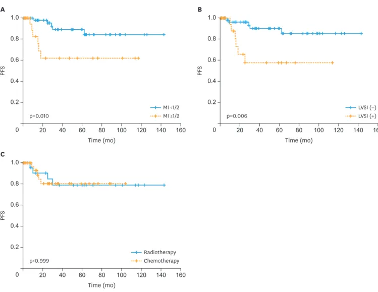
Fig. 1 presents the 5-year PFS rates. The rates were significantly inferior in women with MI
≥1/2 (61.8% vs. 89.0%, p=0.010) and LVSI (57.4% vs. 90.1%, p=0.006). However, there was no significant difference between the women with radiotherapy or chemotherapy after surgery (78.9% vs. 80.1%, p>0.999). In contrast, only MI ≥1/2 was associated with a significantly
B
40
20 60 80 100 120 140 160
Time (mo) 0.2
0
PFS 0.4
0.6 0.8 1.0
LVSI (+) LVSI (−) p=0.006
Time (mo) A
40 20 0.2
0
PFS 0.4
0.6 0.8 1.0
160
80 100 120 140
60
MI ≥1/2 MI <1/2 p=0.010
Time (mo) C
40 20 0.2
0
PFS 0.4
0.6 0.8 1.0
160
80 100 120 140
60
Chemotherapy Radiotherapy p>0.999
Fig. 1. Kaplan-Meier curves of PFS rates by (A) MI (5-year PFS; 61.8% vs. 89.0%, p=0.010), (B) lymphovascular invasion (5-year PFS; 57.4% vs. 90.1%, p=0.006), and (C) adjuvant therapy method (5-year PFS; radiotherapy vs. chemotherapy; 78.9% vs. 80.1%, p>0.999).
LVSI, lymphovascular space invasion; MI, myometrial invasion; PFS, progression-free survival.
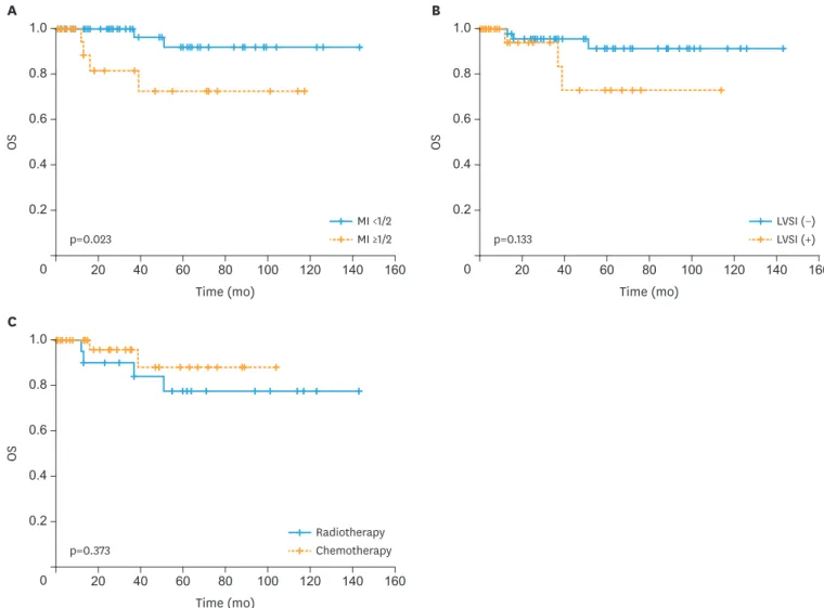
inferior 5-year OS rate (72.4% vs. 91.8%, p=0.023) (Fig. 2). LVSI (91.3% vs. 72.9%, p=0.133) and adjuvant therapy methods (77.5% vs. 87.8%, p=0.373) did not affect the 5-year OS rates.
In addition, survival curves in women with radiotherapy and chemotherapy are presented in stratified groups by LVSI or MI (Supplementary Figs. 1 and 2). There is no difference in PFS and OS according to the adjuvant therapy.
3. Clinicopathologic factors associated with recurrence
Table 3 shows the results of univariate and multivariate analyses of the risk factors for recurrence. In univariate analysis, MI ≥1/2 (hazard ratio [HR]=1.714; 95% confidence interval [CI]=1.254–17.717; p=0.022) and LVSI (HR=5.700; 95% CI=1.492–21.772; p=0.011) were significantly associated with recurrence. However, neither radiotherapy (HR=1.810;
95% CI=0.297–11.027; p=0.520) nor chemotherapy (HR=1.638; 95% CI=0.288–9.321;
p=0.578) was related to recurrence. Multivariate analysis revealed no independent risk factor for predicting recurrence.
B
40
20 60 80 100 120 140 160
Time (mo) 0.2
0
OS
0.4 0.6 0.8 1.0
LVSI (+) LVSI (−) p=0.133
Time (mo) A
40 20 0.2
0
OS
0.4 0.6 0.8 1.0
160
80 100 120 140
60
MI ≥1/2 MI <1/2 p=0.023
Time (mo) C
40 20 0.2
0
OS
0.4 0.6 0.8 1.0
160
80 100 120 140
60
Chemotherapy Radiotherapy p=0.373
Fig. 2. Kaplan-Meier curves of OS rates by (A) MI (5-year OS; 72.4% vs. 91.8%, p=0.023), (B) lymphovascular invasion (5-year OS; 91.3% vs. 72.9%, p=0.133), and (C) adjuvant therapy method (5-year OS; radiotherapy vs. chemotherapy; 77.5% vs. 87.8%, p=0.373).
LVSI, lymphovascular space invasion; MI, myometrial invasion; OS, overall survival.
DISCUSSION
Presently, adjuvant radiotherapy is generally considered standard care after staging surgery for intermediate-to-high risk endometrial cancer. The Post Operative Radiation Therapy in Endometrial Carcinoma (PORTEC)-1 trial showed that adjuvant radiotherapy reduces locoregional recurrence in women with early-stage endometrial cancer, including grade 1 with MI ≥1/2, grade 2 with any invasion, or grade 3 with MI <1/2 [7]. The 5-year locoregional recurrence rate was significantly lower in women with adjuvant radiotherapy than that in women without adjuvant therapy (4% vs. 14%, p<0.001) but there was no difference in OS rates (81% vs. 85%, p=0.031). The Gynecologic Oncology Group (GOG)-99 study also showed that adjuvant radiotherapy decreases the 2-year cumulative recurrence rate in early- stage intermediate risk endometrial cancer (3% vs. 12%, p=0.007) [9]. However, these two fundamental randomized trials mainly included women with endometrial cancer women with endometrioid cell type and did not show stratified results by histologic type. Of 714 women with endometrial cancer in the PORTEC-1 trial, only one and four women were diagnosed with UPSC and CCC, respectively. There was no confirmed UPSC and CCC in the GOG-99 study.
While recurrence in early-stage EEC generally occurs in the vagina or pelvis, the majority of UPSC and CCC patients relapse in multiple sites beyond the pelvis [10]. Furthermore, UPSC and CCC have a worse prognosis resulting from the different histologic patterns compared to that of EEC. Hendrickson et al. [11] showed early that stage 1 UPSC has a poor PFS compared to those of non-papillary pathologic grade 2 or 3 stage I disease (p<0.001).
Similarly, Hamilton et al. [5] reported poorer 5-year PFS rates of 55% and 68% in women with UPSC and CCC, respectively, compared to 77% in women with grade 3 EEC. Regarding its aggressive behavior, frequent p53 gene mutations and HER-2/neu gene amplification have been demonstrated as characteristic molecular genetic profiles of UPSC and CCC [12,13].
Psammoma bodies with a prominent papillary architecture in uterine specimens resemble serous papillary ovarian carcinoma; therefore, it may be related to the clinical features of UPSC [11]. CCC of the endometrium is a rare, but also aggressive variant [14,15]. However, there are conflicting studies on the prognosis of UPSC and CCC [3,16-18]. Fader et al. [19]
suggested that an increased UPSC percentage in uterine specimens is not relevant to high recurrence rates and poor survival outcomes; therefore, adjuvant therapy may not provide any additional benefit to women with UPSC. Creasman et al. [17] also reported 5-year UPSC and CCC survival rates of 72% and 81%, respectively, which were not inferior to the 76% rate for Table 3. Univariate and multivariate analysis for predicting the recurrence in the study population
Characteristics Univariate Multivariate
HR (95% CI) p HR (95% CI) p
Age >59 yr 1.333 (0.323–5.506) 0.691 - -
Serum CA-125 level >35 IU/mL 1.350 (0.318–5.726) 0.684 - -
Clear cell histology 0.417 (0.083–2.085) 0.287 - -
Lymphadenectomy 2.321 (0.272–19.777) 0.441 - -
MI ≥1/2 1.714 (1.254–17.717) 0.022 3.179 (0.770–13.119) 0.110
LVSI 5.700 (1.492–21.772) 0.011 4.073 (0.985–16.842) 0.052
Malignant cells in WC 3.562 (0.289–43.921) 0.322 - -
No adjuvant therapy - -
Radiotherapy 1.810 (0.297–11.027) 0.520
Chemotherapy 1.638 (0.288–9.321) 0.578
CA-125, cancer antigen-125; CI, confidence interval; LVSI, lymphovascular space invasion; MI, myometrial invasion; HR, hazard ratio; WC, washing cytology.
grade 3 EEC. They suggested that radiotherapy after surgery for stage I UPSC and CCC may not significantly improve survival.
Because the pattern and frequency of recurrence in women with UPSC and CCC differ from those of EEC, authors have suggested that adjuvant therapy have to be more systemically applied, even in early-stage disease. Until now, few studies have assessed the prognostic impact of adjuvant chemotherapy in women with early-stage UPSC and CCC. Dietrich et al.
[20] showed that paclitaxel/carboplatin chemotherapy after surgery is effective in reducing recurrence in stage I UPSC. Among 21 women with stage I UPSC treated with a combination of carboplatin (AUC 6 [n=16], AUC 5 [n=5]), and paclitaxel (135 mg/m2 [n=2], 150 mg/m2 [n=1], and 175 mg/m2 [n=18]), only one patient was diagnosed vaginal recurrence at 4 months after adjuvant chemotherapy of three cycles during the follow-up period (mean 41 months).
In women with stage II UPSC, Fader et al. [21] reported recurrence rates of 50% (5/10), 50%
(13/26), and 10.5% (2/19) in women who received no adjuvant therapy, radiotherapy, and chemotherapy ± radiotherapy, respectively. However, these 2 retrospective studies are based on small populations and the survival outcomes were not directly compared between women who received chemotherapy and those who received radiotherapy. Meanwhile, in a national cancer data-based study by Xu et al. [22], neither radiotherapy (HR=1.02; 95% CI=0.70–1.48;
p=0.936) nor chemotherapy (HR=1.15; 95% CI=0.60–2.18; p=0.678) improved the survival outcomes compared to that of observation in 1,672 women with stage I–II CCC. Therefore, they suggested that radiotherapy and chemotherapy should be combined to minimize both locoregional and distant metastasis. Hong et al. [23] also reported a national cancer data- based study in recent. In 5,432 women with stage I UPSC, brachytherapy (HR=0.71; 95%
CI=0.59–0.86; p<0.001) and chemotherapy (HR=0.92; 95% CI=0.80–1.07; p=0.26) were associated with increased survival, however, not in CCC. It is still unclear that additional chemotherapy with radiotherapy improves the survival outcomes in early-stage UPSC or CCC, contrary to their effects in advanced disease [24,25].
This study evaluated the survival outcomes in women diagnosed with stage I UPSC and CCC. The 5-year PFS (78.9% vs. 80.1%, p>0.999) and OS (77.5% vs. 87.8%, p=0.373) did not differ between the groups who received radiotherapy or chemotherapy alone. However, this study has some limitations due to its retrospective nature. Firstly, although we showed the stratified results of sub-analysis by LVSI or deep MI or LVSI (Supplementary Figs. 1 and 2), its statistical power is weak because of the small population size. While it appears that any adjuvant therapy was ineffective in improving survival outcomes in this study population (Table 3), there is a strong possibility of a selection bias. Women who received any adjuvant therapy, radiotherapy or chemotherapy, more often had MI ≥1/2 (33.9% vs. 0%, p=0.002) and LVSI (28.8% vs. 4.8%, p=0.023) and these two factors were significantly associated with recurrence (Table 3). Secondly, the therapeutic protocols of radiotherapy or chemotherapy were not homogenized across the eight institutions. Only a half of the women who received radiotherapy received concurrent cisplatin. Similarly, women who received chemotherapy underwent various treatment cycles.
Our findings showed that the survival outcomes between women who received
radiotherapy or chemotherapy after staging surgery were similar in stage I UPSC and CCC of endometrial cancer. In addition, chemotherapy did not significantly decrease regional or distant metastases, as expected. Significant findings might be revealed in stratified analysis of women with deep MI or LVSI; therefore, further studies with larger populations are required.
SUPPLEMENTARY MATERIALS
Supplementary Fig. 1
Kaplan-Meier curves of PFS rates by adjuvant therapy in stratified groups by LVSI and MI.
Click here to view Supplementary Fig. 2
Kaplan-Meier curves of OS rates by adjuvant therapy in stratified groups by LVSI and MI.
Click here to view
REFERENCES
1. Siegel RL, Miller KD, Jemal A. Cancer statistics, 2016. CA Cancer J Clin 2016;66:7-30.
PUBMED | CROSSREF
2. Felix AS, Scott McMeekin D, Mutch D, Walker JL, Creasman WT, Cohn DE, et al. Associations between etiologic factors and mortality after endometrial cancer diagnosis: the NRG Oncology/Gynecologic Oncology Group 210 trial. Gynecol Oncol 2015;139:70-6.
PUBMED | CROSSREF
3. Fader AN, Java J, Tenney M, Ricci S, Gunderson CC, Temkin SM, et al. Impact of histology and surgical approach on survival among women with early-stage, high-grade uterine cancer: An NRG Oncology/
Gynecologic Oncology Group ancillary analysis. Gynecol Oncol 2016;143:460-5.
PUBMED | CROSSREF
4. McGunigal M, Liu J, Kalir T, Chadha M, Gupta V. Survival differences among uterine papillary serous, clear cell and grade 3 endometrioid adenocarcinoma endometrial cancers: a National Cancer Database analysis. Int J Gynecol Cancer 2017;27:85-92.
PUBMED | CROSSREF
5. Hamilton CA, Cheung MK, Osann K, Chen L, Teng NN, Longacre TA, et al. Uterine papillary serous and clear cell carcinomas predict for poorer survival compared to grade 3 endometrioid corpus cancers. Br J Cancer 2006;94:642-6.
PUBMED | CROSSREF
6. Morrow CP, Bundy BN, Kurman RJ, Creasman WT, Heller P, Homesley HD, et al. Relationship between surgical-pathological risk factors and outcome in clinical stage I and II carcinoma of the endometrium: a Gynecologic Oncology Group study. Gynecol Oncol 1991;40:55-65.
PUBMED | CROSSREF
7. Creutzberg CL, van Putten WL, Koper PC, Lybeert ML, Jobsen JJ, Wárlám-Rodenhuis CC, et al. Surgery and postoperative radiotherapy versus surgery alone for patients with stage-1 endometrial carcinoma:
multicentre randomised trial. PORTEC Study Group. Post Operative Radiation Therapy in Endometrial Carcinoma. Lancet 2000;355:1404-11.
PUBMED | CROSSREF
8. Aalders J, Abeler V, Kolstad P, Onsrud M. Postoperative external irradiation and prognostic parameters in stage I endometrial carcinoma: clinical and histopathologic study of 540 patients. Obstet Gynecol 1980;56:419-27.
PUBMED
9. Keys HM, Roberts JA, Brunetto VL, Zaino RJ, Spirtos NM, Bloss JD, et al. A phase III trial of surgery with or without adjunctive external pelvic radiation therapy in intermediate risk endometrial adenocarcinoma:
a Gynecologic Oncology Group study. Gynecol Oncol 2004;92:744-51.
PUBMED | CROSSREF
10. Boruta DM 2nd, Gehrig PA, Fader AN, Olawaiye AB. Management of women with uterine papillary serous cancer: a Society of Gynecologic Oncology (SGO) review. Gynecol Oncol 2009;115:142-53.
PUBMED | CROSSREF
11. Hendrickson M, Ross J, Eifel P, Martinez A, Kempson R. Uterine papillary serous carcinoma: a highly malignant form of endometrial adenocarcinoma. Am J Surg Pathol 1982;6:93-108.
PUBMED | CROSSREF
12. Santin AD, Bellone S, Gokden M, Palmieri M, Dunn D, Agha J, et al. Overexpression of HER-2/neu in uterine serous papillary cancer. Clin Cancer Res 2002.8:1271-9.
PUBMED
13. Zheng W, Cao P, Zheng M, Kramer EE, Godwin TA. p53 overexpression and bcl-2 persistence in endometrial carcinoma: comparison of papillary serous and endometrioid subtypes. Gynecol Oncol 1996;61:167-74.
PUBMED | CROSSREF
14. Abeler VM, Kjørstad KE. Clear cell carcinoma of the endometrium: a histopathological and clinical study of 97 cases. Gynecol Oncol 1991;40:207-17.
PUBMED | CROSSREF
15. Christopherson WM, Alberhasky RC, Connelly PJ. Carcinoma of the endometrium: I. A clinicopathologic study of clear-cell carcinoma and secretory carcinoma. Cancer 1982;49:1511-23.
PUBMED | CROSSREF
16. Alektiar KM, McKee A, Lin O, Venkatraman E, Zelefsky MJ, McKee B, et al. Is there a difference in outcome between stage I–II endometrial cancer of papillary serous/clear cell and endometrioid FIGO grade 3 cancer? Int J Radiat Oncol Biol Phys 2002;54:79-85.
PUBMED | CROSSREF
17. Creasman WT, Kohler MF, Odicino F, Maisonneuve P, Boyle P. Prognosis of papillary serous, clear cell, and grade 3 stage I carcinoma of the endometrium. Gynecol Oncol 2004;95:593-6.
PUBMED | CROSSREF
18. Halperin R, Zehavi S, Langer R, Hadas E, Bukovsky I, Schneider D. Uterine papillary serous carcinoma (pure and mixed type) compared with moderately and poorly differentiated endometrioid carcinoma. A clinicopathologic study. Eur J Gynaecol Oncol 2002;23:300-4.
PUBMED
19. Fader AN, Starks D, Gehrig PA, Secord AA, Frasure HE, O'Malley DM, et al. An updated clinicopathologic study of early-stage uterine papillary serous carcinoma (UPSC). Gynecol Oncol 2009;115:244-8.
PUBMED | CROSSREF
20. Dietrich CS 3rd, Modesitt SC, DePriest PD, Ueland FR, Wilder J, Reedy MB, et al. The efficacy of adjuvant platinum-based chemotherapy in stage I uterine papillary serous carcinoma (UPSC). Gynecol Oncol 2005;99:557-63.
PUBMED | CROSSREF
21. Fader AN, Nagel C, Axtell AE, Zanotti KM, Kelley JL, Moore KN, et al. Stage II uterine papillary serous carcinoma: carboplatin/paclitaxel chemotherapy improves recurrence and survival outcomes. Gynecol Oncol 2009;112:558-62.
PUBMED | CROSSREF
22. Xu KM, Gill BS, Balasubramani GK, Sukumvanich P, Kelley JL, Beriwal S. Utilization and role of adjuvant radiotherapy and chemotherapy for uterine clear cell carcinoma: A National Cancer Data Base analysis.
Int J Gynecol Cancer 2016;26:472-82.
PUBMED | CROSSREF
23. Hong JC, Foote J, Broadwater G, Gaillard S, Havrilesky LJ, Chino JP. Impact of chemotherapy and radiotherapy on management of early stage clear cell and papillary serous carcinoma of the uterus. Int J Gynecol Cancer 2017;27:720-9.
PUBMED | CROSSREF
24. Einstein MH, Frimer M, Kuo DY, Reimers LL, Mehta K, Mutyala S, et al. Phase II trial of adjuvant pelvic radiation “sandwiched” between combination paclitaxel and carboplatin in women with uterine papillary serous carcinoma. Gynecol Oncol 2012;124:21-5.
PUBMED | CROSSREF
25. Mahdi H, Nutter B, Abdul-Karim F, Amarnath S, Rose PG. The impact of combined radiation and chemotherapy on outcome in uterine papillary serous carcinoma compared to chemotherapy alone. J Gynecol Oncol 2016;27:e19.
PUBMED | CROSSREF