Copyright © 2012 Korean Neurological Association 75
Print ISSN 1738-6586 / On-line ISSN 2005-5013 http://dx.doi.org/10.3988/jcn.2012.8.1.75 CASE REPORT
J Clin Neurol 2012;8:75-78
Introduction
Malignant peripheral nerve sheath tumors (MPNSTs) are ex- ceedingly rare in patients without a history of neurofibroma- tosis - a group of genetic disorders of the nervous system that cause tumors to grow on nerves and other tissues.1 MPNSTs of the spinal accessory nerve may involve the nerve at vari- ous points along its course from the cervical segment of the spinal cord to the trapezius muscle.2-7 The various segments of the nerve are classified as cisternal, foraminal, and intra- sternomastoid.4,6 The extracranial portion of the nerve, spe- cifically its intrasternomastoid segment, is the least common location of the nerve to be involved by a peripheral nerve sheath tumor,4-6 and metastasis to the brain from this location has not been reported. We report here a patient with a well- circumscribed mass in the brain parenchyma, which was a
metastatic lesion of an MPNST of the intrasternomastoid seg- ment spinal accessory nerve - a unique presentation of an ex- ceeding rare tumor.
Case Report
The patient is a 54-year-old white woman previously diag- nosed as having an MPNST of the right spinal accessory nerve. Confirmation of the tumor was made by tissue biopsy of a mass located in the right supraclavicular region approxi- mately 2 years ago. During her initial presentation in July 2009, we performed a wide excision of the mass and a senti- nel lymph node biopsy of the right neck and axillary lymph nodes. Using a marking pen, we drew an elliptical incision around the mass to create a 2-cm margin from its palpable edge. In total, we excised an ellipse of tissue with diameters of 15×10 cm. After resection, the patient received four cycles of radiation therapy to the right supraclavicular region. She had been in good health since the diagnosis, and her outpatient medical regimen consisted only of a proton-pump inhibitor for treatment of gastroesophageal reflux disease. However,
Malignant Nerve Sheath Tumor of the Spinal Accessory Nerve:
A Unique Presentation of a Rare Tumor
Omair A. Sheikh,a Ann Reaves,b Francis A. Kralick,c Ari Brooks,d Rachel E. Musial,e James Gasperinob
aDepartment of Neurology, Section, Critical Care Medicine, Philadelphia, PA, USA
bDepartments of Medicine, cNeurosurgery, dSurgery and ePathology, Drexel University College of Medicine, Philadelphia, PA, USA
Received February 18, 2011 Revised May 3, 2011 Accepted May 3, 2011 Correspondence James Gasperino, MD, PhD Department of Environmental and Occupational Health,
School of Public Health, Drexel University, 245 N. 15th Street,
Mail Stop 107, NCB 12th Floor, Philadelphia, PA 19102, USA Tel +1-215-762-7011 Fax +1-215-762-8728 E-mail James.gasperino@
drexelmed.edu
BackgroundzzMalignant peripheral nerve sheath tumors (MPNSTs), sarcomas originating from tissues of mesenchymal origin, are rare in patients without a history of neurofibromatosis.
Case ReportzzWe report a case of an MPNST of the spinal accessory nerve, unassociated with neurofibromatosis, which metastasized to the brain. The tumor, originating in the intrasternomas- toid segment of the spinal accessory nerve, was removed. Two years later, the patient presented with focal neurological deficits. Radiographic findings revealed a well-defined 2.2×2.2×2.2 cm, homogeneously enhancing mass in the left parieto-occipital region of the brain surrounded by sig- nificant vasogenic edema and mass effect, culminating in a 1-cm midline shift to the right. The mass was surgically removed. The patient had nearly complete recovery of vision, speech, and memory.
ConclusionszzTo our knowledge, this is the first documented case of an MPNST arising from an extracranial segment of the spinal accessory nerve and metastasizing to the brain.
J Clin Neurol 2012;8:75-78 Key Wordszz malignant nerve sheath tumor, spinal accessory nerve, neurofibromatosis.
Open Access
cc This is an Open Access article distributed under the terms of the Cre- ative Commons Attribution Non-Commercial License (http://creative- commons.org/licenses/by-nc/3.0) which permits unrestricted non-com- mercial use, distribution, and reproduction in any medium, provided the ori- ginal work is properly cited.
Malignant Nerve Sheath Tumor
76 J Clin Neurol 2012;8:75-78
nearly 1 month before admission, the patient became symp- tomatic and presented to the emergency department with com- plaints of headache, visual disturbances, loss of short-term memory, and ataxic gait. In addition, she complained of visual loss on rightward gaze and loss of balance with head move- ment.
On presentation to the emergency department, her vital signs remained within normal limits. Physical examination revealed evidence of both long- and short-term memory defi- cits as well as a right visual field cut, and results of laborato- ry studies including an electrocardiogram were all within normal limits. However, radiographic examination of the brain by contrast-enhanced magnetic resonance imaging (MRI) revealed a well-defined 2.2×2.2×2.2 cm, homoge- neously enhancing mass in the left parieto-occipital region surrounded by significant vasogenic edema. There was also evidence of mass effect, culminating in a 1-cm midline shift to the right; these findings were not present on an MRI ob- tained 5 months prior to admission (Fig. 1). The patient was subsequently admitted to the neurological intensive care unit (ICU) for neurological monitoring, and a neurosurgical con- sultation was obtained. As part of an evaluation for distant metastases, a contrast-enhanced CT scan of the chest revealed a new 1.1-cm nodule in the left lower lobe and a stable 0.4-cm nodule in the same location (Fig. 2). The patient’s course in the neurological ICU was remarkable for only one episode of hypertension on the second day. Routine laboratory stud- ies were obtained and were within normal limits.
On day 7 in the neurological ICU, we performed a cranioto- my with complete removal of the mass. A biopsy of the mass was obtained in the operating room, and a pathologist con- firmed the diagnosis of a metastatic nerve sheath tumor (Fig.
3). Histopathological findings revealed malignant tissue with poorly defined cellular and cystic components. The malignant cells expressed vimentin and S-100 but not glial fibrillary acidic protein, neurofilaments, melanoma marker, actin, smooth muscle antibody, pan-cytokeratin, or Melan-A. Con- firmation of the brain tumor as a metastatic lesion of the orig- inal tumor of the spinal accessory nerve was based both on findings of immunohistochemical tests and the nearly identi- cal morphological characteristics shared by the 2 tumors.
On postoperative day 1, the patient reported marked im- provement in vision, speech, and memory, although not to baseline level. It is noteworthy that the brain lesion was consid- ered to be a new tumor, because the results of an MRI scan of the brain obtained 5 months before this admission were within normal limits (Fig. 1B). The patient’s postoperative course was uneventful and included a 2-week course of radiation therapy. On postoperative day 6, she was transferred to the on- cology floor for further treatment.
Discussion
To our knowledge, this is the first documented case of an MPNST arising from an extracranial segment of the spinal accessory nerve and metastasizing to the brain.1-4
Kurokowa et al.4 described the first case of a spinal accesso- ry schwannoma arising in the fourth ventricle in a 50-year-old man presenting with neck pain after trauma. Physical examina- tion findings from the patient described in their report were not consistent with cranial nerve involvement; and they did not re- port upper or lower extremity weakness or gait disturbance. The
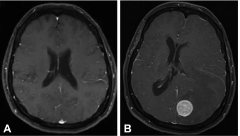
Fig. 1. A: Initial MRI performed in 2009 as part of staging for pe- ripheral nerve sheath tumor. T1 weighted axial image post gadolin- ium injection was negative for abnormal intracranial enhancement.
B: Repeat MRI performed August 2010 on admission to neurologi- cal intensive care unit. T1 weighted axial image post gadolinium in- jection revealed 2.2×2.2×2.2 cm, well-circumscribed, homoge- neously enhancing lesion identified in the left occipital lobe with extensive vasogenic edema. A midline shift to the right of approxi- mately 1 cm with subfalcine herniation is evident. Mass effect is identified on the occipital horn of the lateral ventricle.
B A
Fig. 2. Contrast-enhanced CT scan of the chest at the time of ad- mission to the neurological intensive care unit. Scan revealed a new 1.1-cm left lower lobe solid nodular density adjacent to the lateral basilar segment artery and bronchus. A stable 0.4-cm nodule was seen peripheral to the left lower lobe.
Sheikh OA et al.
www.thejcn.com 77 authors speculate that the accessory nerve tumor initially in-
volved the intracisternal segment of the nerve and subsequent- ly grew into the fourth ventricle. In their review of the literature, they identified only 30 cases of accessory schwannomas unas- sociated with neurofibromatosis published since 1975;4 how- ever, only two of these cases presented with a schwannoma of the nerve in the region of the neck (i.e., intrasternomastoid por- tion), and neither of these cases presented with metastasis to the brain.6,7 Moreover, in both cases, no focal neurological def- icits were reported on physical examination.6,7
MPNSTs are sarcomas originating from tissues of mesenchy- mal origin, i.e., peripheral nerves or cells of the nerve sheath such as Schwann cells or perineural cells.8 Hence, the terms malignant schwannoma, neurogenic sarcoma, or neurofibrosar- coma are used interchangeably.8-10 MPNST tumors commonly present a diagnostic challenge to pathologists because they arise from different types of cells (Schwann cells, fibroblasts, peri- neural cells) and may therefore produce a variable histological appearance.10 According to Geller and Gebhardt, at least one of the following has to be present for the diagnosis of MPNST:
“1) a tumor arising from peripheral nerves; 2) a tumor arising from existing benign or other MPNST; or 3) a tumor that dis- plays histological features of Schwann cells or perineural dif- ferentiation as revealed by immunohistochemical analysis or electron microscopic examination.”10 MPNSTs comprise almost 10% to 12% of all soft tissue sarcomas, which are primarily associated with neurofibromatosis type 1 with a gene muta- tion of the same name; however, they can arise sporadically without neurofibromatosis type 1, in almost 50% of patients, as in the case of our patient. A strong relationship exists be- tween a history of radiation therapy and MPNST.11,12 This tu- mor has a high percentage of recurrence at the primary or new site, and metastasis to the lungs, bones, or intra-abdominal or intrathoracic organs is not uncommon. However, metastasis to the brain is highly unusual; only a handful of cases have been reported to date, making this case of an MPNST of the spinal accessory nerve a novel finding.
MPNST usually occurs in patients between the ages of 20 to 50, but in an extreme case, it has been reported in an 11- month-old patient.9,10 These tumors present with pain, palpa- ble mass, hemiparesis, or paresthesias, usually in a dermato- mal pattern. Not surprisingly, an MPNST affects large peri- pheral nerves such as the sciatic nerve or the brachial or sacral plexus. Large nodular lesions in large peripheral nerves or plex- us have greater potential for malignant transformation.10 Diag- nosis and staging are usually done by surgical resection and bi- opsy. MRI and CT scans show the extent of the lesion and the distant metastasis and are therefore valuable in staging the le- sion. Large tumors (>5 cm), invasion of fat planes, heterogene- ity, ill-defined margins, and edema surrounding the lesion sug- gest an MPNST.13,14
According to Geller and Gebhardt, staging of soft tissue sar- comas is dependent on a number of factors including histolog- ical grade, tumor size, tumor depth, and whether or not metas- tases have been identified.10 In patients without detectable metastases, histological grade, tumor size, and tumor depth ap- pear to predict subsequent metastasis.10 In addition, tumor stag- ing is also based on the findings of imaging studies, which are frequently needed to define location and extent of disease.10,15
It is postulated that metastasis to the brain, though rare, hap- pens by the following three routes:7 1) direct invasion; 2) he- matogenous spread; 3) dissemination through the cerebrospi- nal fluid.16 It is not known whether the 9-mm pulmonary nodule identified on CT scans of the chest in the present case was a metastatic lesion, but by inference it raises the possibility of metastasis to the brain by a hematogenous route.16
It remains unclear why the brain is an infrequent site of me- tastasis. Although negative surgical margins and location of the tumor are considered important prognostic factors, patients frequently have metastases from negative surgical margins at the primary lesion site, as in the case of our patient. Radiation is still the mainstay of therapy because of high local recurrence and pulmonary involvement, and the role of chemotherapy in the treatment of this tumor still needs to be firmly established.
B A
Fig. 3. A: Photomicrograph of an S-100 immunohistochemically stained slide (magnification ×400) of tissue from the original tumor, which con- firms the tumor as an MPNST. B: Photomicrograph of an H & E slide (magnification ×400) of tissue from the cerebral mass. It shows a cellular neoplasm exhibiting storiform architecture with moderate nuclear atypia. Other areas show increased mitotic activity and necrosis. The diagno- sis of MPNST was based on expression of similar immunohistochemical staining and nearly identical morphological characteristics shared be- tween the original tumor and the metastatic tumor.
Malignant Nerve Sheath Tumor
78 J Clin Neurol 2012;8:75-78
The overall prognosis for a patient with this tumor is poor be- cause most of the patients succumb to pulmonary complica- tions; the 5-year survival rate ranges from 16% to 52%.16 Conflicts of Interest
The authors have no financial conflicts of interest.
REFERENCES
1. Fenzi F, Moretto G, Zamboni G, Passarin MG, Rizzuto N. Brain metas- tases from post-radiation malignant peripheral nerve sheath tumour.
Ital J Neurol Sci 1995;16:495-498.
2. Tilgner J, Müller K, Ghanem N, Lutterbach J, Vesper J. Brain metasta- ses as primary manifestation of a melanocytic malignant peripheral nerve sheath tumor in a 60-year-old man. BMC Neurol 2007;7:2.
3. McClatchey AI. Neurofibromatosis. Annu Rev Pathol 2007;2:191-216.
4. Kurokawa R, Tabuse M, Yoshida K, Kawase T. Spinal accessory schwannoma mimicking a tumor of the fourth ventricle: case report.
Neurosurgery 2004;54:510-514; discussion 514.
5. Agrawal A, Rao KS, Makannavar JH, Shetty L, Raveendra VM. Intra- sternomastoid spinal accessory nerve schwannoma: clinical and radio- logical correlation. Neurol India 2005;53:347-348.
6. McShane D, Noyek AM, Chapnik JS, Steinhardt MI, Cooter N.
Schwannoma of the intrasternomastoid portion of the spinal accessory nerve: sophisticated preoperative CT diagnosis and appropriate surgi- cal management. J Otolaryngol 1986;15:282-285.
7. Noyek AM, Chapnik JS, Wortzman G, Kandel R. Schwannoma of the intrasternomastoid portion of the spinal accessory nerve: sophisticated pre-operative MRI diagnosis and appropriate surgical management. J
Otolaryngol 1992;21:286-289.
8. D’Agostino AN, Soule EH, Miller RH. Sarcomas of the peripheral nerves and somatic soft tissues associated with multiple neurofibroma- tosis (von recklinghausen’s disease). Cancer 1963;16:1015-1027.
9. Park SK, Yi HJ, Paik SS, Kim YJ, Ko Y, Oh SJ. Metastasizing malig- nant peripheral nerve sheath tumor initially presenting as intracerebral hemorrhage. Case report and review of the literature. Surg Neurol 2007;
68:79-84; discussion 84.
10. Geller DS, Gebhardt M. Malignant peripheral nerve sheath tumors.
Electronic Sarcoma Update Newsletter. Ossining, New York: Liddy Shriver Sarcoma Initiative, 2006;3. (Accessed 17 Feb 2011 at http://sar- comahelp.org/learning_center/mpnst.html)
11. Adamson DC, Cummings TJ, Friedman AH. Malignant peripheral nerve sheath tumor of the spine after radiation therapy for Hodgkin’s lymphoma. Clin Neuropathol 2004;23:245-255.
12. Amin A, Saifuddin A, Flanagan A, Patterson D, Lehovsky J. Radiother- apy-induced malignant peripheral nerve sheath tumor of the cauda equina. Spine (Phila Pa 1976) 2004;29:E506-E509.
13. Friedrich RE, Kluwe L, Fünsterer C, Mautner VF. Malignant peripher- al nerve sheath tumors (MPNST) in neurofibromatosis type 1 (NF1):
diagnostic findings on magnetic resonance images and mutation analy- sis of the NF1 gene. Anticancer Res 2005;25:1699-1702.
14. Poyhonen M, Niemela S, Herva R. Risk of malignancy and death in neurofibromatosis. Arch Pathol Lab Med 1997;121:139-143.
15. Stojadinovic A, Yeh A, Brennan MF. Completely resected recurrent soft tissue sarcoma: primary anatomic site governs outcomes. J Am Coll Surg 2002;194:436-447.
16. Gupta G, Mammis A, Maniker A. Malignant peripheral nerve sheath tumors. Neurosurg Clin N Am 2008;19:533-543, v.