297 책임저자:김 신, 690-756, 제주도 제주시 제주대학로 66
제주대학교 에너지공학과
Tel: 064-754-3647, Fax: 064-757-9276 E-mail: sinkim@jejunu.ac.kr
접수일:2009년 10월 7일, 게재승인일:2009년 10월 19일
Correspondence to:Sin Kim
Department of Nuclear and Energy Electronic Engineering, Jeju National University, Jeju Daehakro 66, Jeju 690-756, Korea
Tel: +82-64-754-3647, Fax: +82-64-757-9276 E-mail: sinkim@jejunu.ac.kr
유방암 검출을 위한 전기 임피던스 방법에 관한 연구
1제주대학교 에너지공학과, 2제주대학교 의학전문대학원, 3제주대학교 전자공학과
이정성1ㆍ김우건2ㆍ강경아2ㆍ현진원2ㆍ장원영2ㆍ이창현2ㆍ김경연3ㆍ김 신1
A Study on Electrical Impedance Technique for Breast Cancer Detection
Jeong Seong Lee1, Woo Kun Kim2, Kyoung Ah Kang2, Jin Won Hyun2, Weon Young Chang2, Chang Hyun Lee2, Kyung Youn Kim3 and Sin Kim1
1Department of Nuclear and Energy Engineering, 2School of Medicine,
3Department of Electronic Engineering, Jeju National University, Jeju 690-756, Korea
There have been many studies on the impedance characteristics of body tissues for decades. Since the breast tissue is relatively uniform compared to other tissues and the electrical characteristics of normal and tumor tissues are obviously different, for the breast cancer detection the electrical impedance method seems to be promising. The purpose of this research is to investigate the feasibility of the detection of a cancerous tissue in a normal tissue with a numerical method based on the finite element method and to design the electrode configuration. The effect of the skin tissue having high permittivity on the measurement of the electrical signals have also been studied. The results of this study can be used as the fundamental data for the development of the electrical impedance method for the beast cancer detection. (Cancer Prev Res 14, 297-302, 2009)
Key Words: Breast cancer detection, Electric impedance, Permittivity, Conductivity
서 론
인체 조직의 유전율 특징에 대한 연구는 많은 연구자들 에 의해 세계적으로 광 범위하게 진행되어 왔다.1∼5) 생체 조직의 전기적 특성은 세포막의 변화와 조직의 화학조성 에 의해 결정된다. 주파수에 따른 생체조직의 전기적 특성 은 낮은 주파수영역에서 세포막 내 이온 확산과 관련이 있 는 α 분산, 수백 kHz 영역에 해당하는 유기 고분자와 세포 막의 분극(Polarization) 때문에 일어나는 β 분산, GHz 영역 에서 체액의 분자 분극 때문에 일어나는 γ 분산이라고 알 려진 세 가지 영역으로 나눌 수 있다. 이미 여러 연구자들 에 의해 비 정상 조직에 대한 유전율과 도전율에 대한 측
정 결과가 보고된 바 있으며6,7) 정상조직과 암 조직은 전기 적 특성이 분명히 다르다고 보고되었다.8∼12) 특히 유방 조 직은 다른 신체부위와 비교할 때 균질한 특성을 가지고 있 고 정상 조직과 종양 조직 사이의 물질 특성치 차가 분명 하므로 전기적 방법이 암조직 검출 기술에 적합하다고 할 수 있다.13∼17) 본 연구에서는 정상 조직 내에 존재하는 종 양 조직을 전기적인 방법으로 검출할 수 있는 지를 평가하 고 검출에 적합한 전극 배열을 설계하고자 한다. 조직 내에 서의 전기장을 해석하기 위하여 유한요소법 수치해석 프 로그램인 COMSOL을 이용하였다. 실제의 경우에는 조직이 3차원 구조를 가지나 본 연구에서는 전기적 암검출 방법의 타당성을 살펴보는 것을 주요 목적으로 하고 있으므로 문 제를 단순화하기 위하여 2차원 구조를 가정하였다. 본 연
298 Cancer Prevention Research Vol. 14, No. 4, 2009
Fig. 1. Schematic of 4-electrode system. Fig. 2. Various cancer tissues for numerical simulation.
Table 1. Electrical properties tissues Electrical conductivity [S/m]
Relative permittivity [−]
Normal tissue Tumor tissue Skin tissue
0.1 0.8 0.012
10 50 990 구의 결과는 전기 임피던스법을 이용한 유방암 검출 기술
개발을 위한 기초자료로 활용될 수 있을 것이다.
재료 및 방법
정확한 임피던스의 측정을 위해서는 직렬구조의 임피 던스 배열에서 나타나는 전극의 분극현상을 줄이는 것 은 필수적이다. 이러한 분극 현상을 줄이기 위해서는 4- 전극법이 사용되어야 한다.18∼20) 4-전극법에서는 Fig. 1과 같이 전류를 주입하기 위한 외곽의 전극 2개와 전압을 측정하기 위한 전극 2개가 사용된다.
본 연구에서는 임피던스의 분석에 있어서 주요한 출 력값인 전압값만 고려하였다. 다양한 전극 구조와 길이 그리고 주파수에 따른 전압값을 유한요소법 수치해석 프로그램인 COMSOL 이용하여 다음과 같이 계산하였다.
COMSOL 모델링 순서는 다음과 같다.
1. Fig. 1과 같이 정상조직을 모사하기 위한 1 cm2 크기 의 정사각형을 생성하고 전극 4개를 생성하였다.
2. 소영역 설정에서 Table 1의 전기적 물성치를 입력하 였다.
3. 경계조건 설정에서 외부 경계는 절연을 선택하고, 내부 경계는 연속성 조건을 부여한다. 그리고 전류주입
을 위해 외곽에 위치한 두 전극에 1 V의 전위차 설정하 였다.
4. 메쉬 생성 후 주파수를 매개변수로 하여 1 Hz∼100 MHz 범위에서 계산을 수행한다.
5. 후처리에서, 전압을 측정하기 위한 내부의 두 전극 사이의 전압을 계산하였다.
6. 암조직이 정상조직 내부에 있는 경우를 고려하기 위하여 Fig. 2에서처럼 다양한 크기의 원을 생성하고 Table 1의 종양조직 물성치 입력하였다.
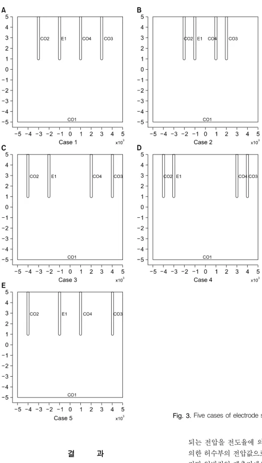
7. 전극구조와 길이에 따른 분석을 위해 Fig. 2와 Fig.
3에 보이는 것과 같이 전극의 구조와 길이를 변경하였다.
8. 전압을 측정하기 위한 전극 사이의 전압을 계산하 여 비교하였다.
전극의 구조에 따른 비교를 위하여 1 cm2 크기의 정상 조 직 내부에 종양조직이 없는 경우에서부터 반경을 0.5 mm씩 증가시켜 종양조직 반경이 3 mm일 때까지 계산을 하였다.
Fig. 3에 5가지 경우의 전극구조를 나타내었다. 그리고 Table 1에 계산에 사용된 조직의 전기적 물성치를 나타내었다.
Fig. 3. Five cases of electrode structures for numerical simulation.
결 과
수치계산에서는 교류전류를 주입시켰을 때 전극에 인가
되는 전압을 전도율에 의한 실수부의 전압값과 유전율에 의한 허수부의 전압값으로 개별적으로 계산할 수 있다. 하 지만 일반적인 계측기에서는 전압의 크기만을 고려하므로 계산결과는 전압값의 크기만을 고려하여 나타내었다.
300 Cancer Prevention Research Vol. 14, No. 4, 2009
Fig. 4. Potential difference with respect to electrode length (tumor radius: 2 mm).
Fig. 5. Potential difference with respect to electrode configurations.
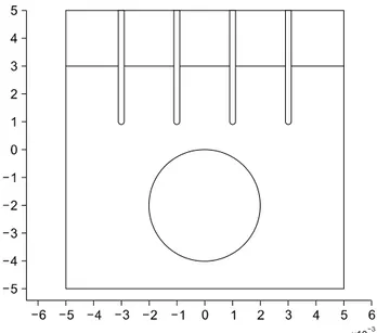
Fig. 6. Schematic of 4-electrode system for tissue with skin (top layer).
우선 전극의 길이에 따른 전압 차를 비교하였다. Fig.
4에 정상조직 내부에 중심이 표면으로부터 7 mm 깊이에 2 mm 반경의 종양조직이 있을 경우 전극의 길이에 따른 전압값 주파수 응답을 나타내었다. 전극의 길이가 0 mm 에서부터 5 mm까지 변화시켜가면서 계산을 하였을 때, 주파수에 따른 전압값의 변화는 없지만 전극 길이에 따 른 측정되는 전압값이 차이는 분명하였다. 전극의 길이 가 3 mm가 될 때까지는 길이가 증가할수록 전압 차가 증가하다가 이후에는 전극의 길이에 따라 전압 차는 감 소하였다. 이는 종양 조직의 전기전도도가 정상 조직에 비해 크므로 종양에 지나치게 가까워지면 저항이 감소
하여 오히려 민감도가 줄어들게 된다는 것을 의미한다.
Fig. 5는 Fig. 3에 나타낸 5가지 경우의 전극 구조에 대 한 계산 결과이다. 주파수에 따른 전압값 차이가 없으므 로 종양 조직의 크기에 따른 전압값 계산결과를 나타내 었다. 결과는 정상 조직보다 높은 전도도를 갖는 종양 조직이 크기가 커질수록 전극에 인가되는 전압 차는 작 은 것으로 나타났다.
피부 조직은 다른 조직에 비해 낮은 전도율과 높은 유 전율를 가지므로 전기 임피던스 측정에 피부조직이 미 치는 영향을 고려하여야 한다. Fig. 6에 2 mm 두께의 피 부조직을 포함한 계산영역의 단면을 나타내었다.
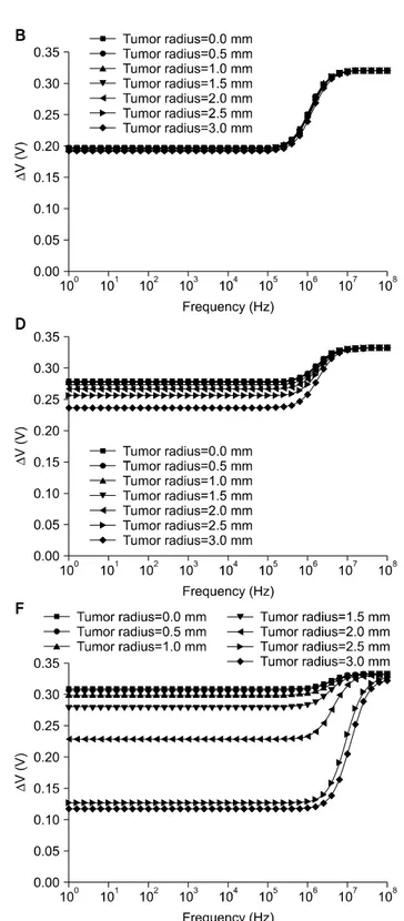
Fig. 7에서 피부를 고려한 경우의 계산결과는 피부조직 의 높은 유전율로 인해 10 kHz 이상 주파수 영역에서 전 압값이 크게 상승되는 것을 확인할 수 있다. 특히 피부조 직의 저항이 증가하여 계산영역 하부에 있는 종양조직에 전류의 영향이 미치지 못하므로 고주파수 영역에서는 종 양조직의 크기에 따른 전압값 차이가 전압값 차이가 없어 지게 된다. 또한 피부 조직을 고려한 경우에는 전극 길이 가 피부조직이 두께보다 작으면 종양조직의 유무에 상관 없이 전압값이 일정하게 계산된다(Fig. 7A, B). 따라서 전 극의 길이는 피부조직 두께보다 길게 설계하여야 한다.
고 찰
유방 조직은 다른 조직에 비해 균질하고 종양 조직의 전 기적 물성이 정상 조직과 상당한 차이를 보이므로 전기 임
Fig. 7. Potential difference with respect to electrode length (0∼5 mm) and tumor size (0∼3 mm radius) for tissue with skin (thickness 2 mm). (A) Electrode length: 0 mm, (B) electrode length: 1 mm, (C) electrode length: 2 mm, (D) electrode length:
3 mm, (E) electrode length: 4 mm, (F) electrode length: 5 mm.
피던스 방법이 유방암 검출에 유용한 방법이 될 수 있을 것이다. 본 연구에서는 이를 위한 기초 연구로 관심 영역에 대한 전기장을 해석하여 전극의 배열 및 크기, 종양 조직의
크기에 따른 전기 신호를 수치해석적으로 분석하여 전기 임피던스 방법의 타당성을 평가하고 적절한 전극의 설계 를 시도하였다. 우선 실제 조직은 3차원 구조를 가지나 본
302 Cancer Prevention Research Vol. 14, No. 4, 2009
연구에서는 방법론의 타당성을 평가하는데 주목적으로 두 고 있으므로 계산의 단순화를 위해 2차원 영역으로 가정하 였다. 따라서 추후에는 보다 현실적인 모델링을 위해 3차 원 해석을 수행하여야 할 것이다. 3차원 해석은 2차원 해석 에 비해 많은 계산 시간과 계산 능력을 필요로 하지만 본 연구에서 시도한 방법론을 동일하게 적용할 수 있다. 또한 본 연구에서는 종양 조직의 위치를 중심으로 전기장을 해 석하였으나 실제의 경우에는 종양 조직의 위치를 정확히 알고 있지 못하므로 전극과 종양 조직의 상대적인 위치를 변화시켜가면서 전기신호를 해석하여 종양 위치를 찾는 방법에 대한 연구가 필요할 것으로 사료된다.
결 론
본 연구에서는 유방암 검출 수단으로서 전기 임피 던스 방법을 도입하고 검출 가능성을 평가하고 적절 한 전극 설계를 시도하였다. 0∼3 mm의 여러 종양조 직 크기와 5가지 경우의 전극 구조와 길이 그리고 주 파수에 따른 전압값을 유한요소 수치해석 소프트웨어 인 COMSOL을 이용하여 계산하였다. 4-전극법을 이용 한 전극구조 중 균등한 간격으로 4개의 전극이 배열된 경우에 종양조직의 크기에 따라 전압값이 차이가 컸 으며, 실험을 수행하고자 하는 주파수영역에서 주파수 에 따른 전압값에는 큰 차이가 없었다. 피부조직을 고 려한 계산에서는 피부조직의 높은 유전율로 인하여 10 kHz 이상의 주파수 영역에서 종양조직에 따른 전 압값을 구분할 수 없고, 낮은 주파수영역에서도 탐침 되는 전극의 길이가 피부의 두께보다 길어야만 종양 조직의 크기에 따른 전압값 차이를 확인할 수 있었다.
본 연구의 결과는 유방암 검출을 위한 전기 임피던스 기법 개발에 기초자료로 활용될 수 있을 것이다.
감사의 글
본 연구는 제주대학교병원 연구비 지원에 의해 연구 를 수행하였습니다.
참 고 문 헌
1) Surowiec A, Stuchly SS, Eidus L, Swarup A. In vitro dielectric properties of human tissues at radiofrequencies. Phys Med Biol 32, 615-621, 1987.
2) Lu Y, Cui H, Yu J, Mashimo S. Dielectric properties of hu- man fetal organ tissues at radio frequencies. Bioelectromagnetics 17, 425-426, 1996.
3) Gabriel C, Gabriel S, Corthout E. The dielectric properties of
biological tissues: I. Literature survey. Phys Med Biol 41, 2231-2249, 1996.
4) Gabriel S, Lau RW, Gabriel C. The dielectric properties of biological tissues: II. Measurements on the frequency range 10 Hz to 20 GHz. Phys Med Biol 41, 2251-2269, 1996.
5) Gabriel S, Lau RW, Gabriel C. The dielectric properties of biological tissues: III. Parametric models for the dielectric spectrum of tissues. Phys Med Biol 41, 2271-2293, 1996.
6) Joines WT, Shrivastav S, Jirtle RL. A comparison using tissue electrical properties and temperature rise to determine relative absorption of microwave power in malignant tissue. Med Phys 16, 840-844, 1989.
7) Joines WT, Zhang Y, Li C, Jirtle RL. The measured electrical properties of normal and malignant human tissues from 50 to 900 MHz. Med Phys 21, 547-550, 1994.
8) Campbell AM, Land DV. Dielectric properties of female breast tissue measured in vitro at 3.2 GHz. Phys Med Biol 37, 193- 210, 1992.
9) Heinitz J, Minet O. Dielectric properties of female breast tumors. Ninth Int Conf on Electrical Bio-Impedance 356-359, 1995.
10) Steleter J, Wtorek J, Nowakowski A, Kopacz A, Jastrzembski T. Complex permittivity of breast tumor tissue. Tenth Int Conf on Electrical Bio-Impedance 59-62, 1998.
11) Jossinet J. Variability of impedivity in normal and pathological breast tissue. Med Biol Eng Comput 34, 346-350, 1996.
12) Jossinet J. The impedivity of freshly excised human breast tissue. Physiol Meas 19, 61-75, 1998.
13) Malich A, Fritsch T, Anderson R, Boehm T, Freesmeyer MG, Fleck M, Kaiser WA. Electrical impedance scanning for classifying suspicious breast lesions: first results. Eur Radiol 10, 1555-1561, 2000.
14) Wersebe A, Siegmann K, Krainick U, 1Fersis N, Vogel U, Claussen CD, Müller-Schimpfle M. Diagnostic potential of targeted electrical impedance scanning in classifying suspicious breast lesions. Invest Radiol 37, 65-72, 2002.
15) Zou Y, Guo Z. A review of electrical impedance techniques for breast cancer detection. Med Eng Phys 25, 79-90, 2003.
16) Stoneman MR, Kosempa M, Gregory WD, Gregory CW, Marx JJ, Mikkelson W, Tjoe J, Raicu V. Correction of electrode polarization contributions to the dielectric properties of normal and cancerous breast tissues at audio/radiofrequen- cies. Phys Med Biol 52, 6589-6604, 2007.
17) Wang K, Wang T, Fu F, Ji ZY, Liu RG, Liao QM, Dong XZ. Electrical impedance scanning in breast tumor imaging:
correlation with the growth pattern of lesion. Chin Med J (Engl) 122, 1501-1506, 2009.
18) Schwan HP. Alternating current electrode polarization.
Biophysik 3, 181-201, 1966.
19) Schwan HP, Onaral B. Linear and nonlinear properties of platinum electrode polarisation. Part 1: frequency dependence at very low frequencies. Med Biol Eng Comput 20, 299-306, 1982.
20) Schwan HP. Linear and nonlinear electrode polarization and biological materials. Ann Biomed Eng 20, 269-288, 1992.
![Table 1. Electrical properties tissues Electrical conductivity [S/m] Relative permittivity [−] Normal tissue Tumor tissue Skin tissue 0.10.8 0.012 10 50 990구의 결과는 전기 임피던스법을 이용한 유방암 검출 기술 개발을 위한 기초자료로 활용될 수 있을 것이다.재료 및 방법 정확한 임피던스의 측](https://thumb-ap.123doks.com/thumbv2/123dokinfo/5454698.239927/2.892.435.811.639.1117/electrical-properties-electrical-conductivity-permittivity-임피던스법을-기초자료로-임피던스의.webp)