Journal of Korean Medicine Rehabilitation
제29권 제2호, 2019년 4월
원 저
91 침술 치료가 모성분리 새끼 쥐의 우울증과 해마의 치상회 세포증식에 미치는 영향 박정식⋅임형호
101 보골탕이 Monosodium Iodoacetate 유도 골관절염과 Interleukin-1β 유도 연골세포에 미치는 보호 효과 성진욱⋅이해웅⋅강경화⋅김경민⋅조성우
115 강황(薑黃) 약침이 Monosodium Iodoacetate 유도 골관절염 흰쥐에 미치는 영향 이종훈⋅우창훈
135 녹제초 추출물이 파골세포 분화 및 골 흡수에 미치는 영향 박정식⋅임형호
종 설
149 경추증성 척수증의 한의학적 치료에 대한 국내외 연구 동향 변다영⋅이기언⋅노지애⋅김효준⋅허석원⋅한시훈
159 마미증후군의 한의학적 치료에 대한 연구 분석 박종한⋅정수현
171 황련해독탕약침 치료에 대한 임상 논문 고찰: 국내 학술지의 증례보고와 대조군 연구 중심으로 이수환⋅김순중
원 저
189 뇌졸중 환자의 교각 운동 시 슬링 줄의 종류에 따른 장력 비교 장권욱⋅장종성
증 례
195 근육 수축/이완 강화 기법과 Instrument Assisted Soft Tissue Mobilization 및 침 치료를 병행하여 치료한 주관절 외상과염 환자 치험 12예
정승현⋅김창곤⋅윤영웅⋅류원형⋅전용현⋅최영준⋅정재현
Journal of Korean Medicine Rehabilitation
Vol. 29 No. 2, April 2019
Original Articles
91 Effect of Acupuncture on Depression and Cell Proliferation in Hippocampal Gyrus Dentatus of Maternal-separated Rat Pups
Jung-Sik Park, Hyung-Ho Lim
101 Protective Effects of Bogol-tang on Monosodium Iodoacetate-induced Osteoarthritis and Interleukin-1β -treated Primary Chondrocytes
Jin Wook Sung, Hai Woong Lee, Kyung Hwa Kang, Kyoung Min Kim, Sung Woo Cho
115 Effects of Curcumae Longae Rhizoma Pharmacopuncture on Monosodium Iodoacetate-induced Osteoarthritis Rats Jong-Hoon Lee, Chang-Hoon Woo
135 Effects of Pyrola japonica Extracts on Osteoclast Differentiation and Bone Resorption Jung-Sik Park, Hyung-Ho Lim
Review Article
149 A Review of the Korean Traditional Medicine for Cervical Spondylotic Myelopathy Da-young Byun, Gi-eon Lee, Ji-ae Roh, Hyo-jun Kim, Suk-won Huh, Si-hoon Han
159 Review of the Studies on the Treatment of Cauda Equina Syndrome Using Korean Medicine Jong-Han Park, Su-Hyeon Jeong
171 An Intensive Review On Clinical Thesis about Hwangryunhaedok-tang Pharmacopuncture Treatment: Focused on Case Reports and Controlled Studies in Korean Academic Journals
Soo-Hwan Lee, Soon-Joong Kim
Original Articles
189 Comparison of Tension According to the Type of Sling Cord during the Bridging Exercise with Sling in Stroke Patients
Gwonuk Jang, Jongsung Chang
Case Reports
195 The Case Report on Twelve Patients of Lateral Epicondylitis Treated with Muscle Contraction/Relaxation Strengthen Technique, Instrument Assisted Soft Tissue Mobilization Treatment and Acupuncture
Seung-Hyun Jeong, Chang-gon Kim, Yeong-Ung Yun, Won-Hyung Ryu, Yong-hyun Jeon, Young-jun Choi, Jae-hyun Chung
Original Article
침술 치료가 모성분리 새끼 쥐의 우울증과 해마의 치상회 세포증식에 미치는 영향
박정식⋅임형호
가천대학교 한의과대학
Effect of Acupuncture on Depression and Cell Proliferation in Hippocampal Gyrus Dentatus of Maternal-separated Rat Pups
Jung-Sik Park, K.M.D., Hyung-Ho Lim, K.M.D.
College of Korean Medicine, Gachon University
이 논문은 2019년도 가천대학교 교내 지원에 의한 결과임.
RECEIVED March, 29, 2019 REVISED April, 3, 2019 ACCEPTED April, 5, 2019 CORRESPONDING TO
Hyung-Ho Lim, College of Korean Medicine, Gachon University, 1342 Seongnam-daero, Sujeong-gu, Seongnam 13120, Korea TEL +82-31-750-8599 FAX +82-31-750-5416 E-mail omdlimhh@gachon.ac.kr Copyright © 2019 The Society of Korean Medicine Rehabilitation
Objectives The loss of maternal care during early postnatal period may increase develop- ment of mood-related disorders, such as depression, anxiety, and personality disorders. In this study, the effect of acupuncture on depression in relation with cell proliferation in the hippocampal gyrus dentatus was investigated using maternal-separated rat pups.
Methods On the postnatal 14th day, rat pups from six dams were grouped into following groups: maternal care group, maternal separation group, maternal separation and non-acupoint-acupunctured group, maternal separation and Zusanli-acupunctured group, and maternal separation and fluoxetine-treated group. Acupuncture was performed from postnatal 28th day to postnatal 37th day. The rat pups that belong in the maternal separa- tion and fluoxetine-treated group were injected subcutaneously with 5 mg/kg fluoxetine hy- drochloride once a day for the same period of time. To evaluate activity of the rat pups, open field test was performed. Immunohistochemistry for serotonin (5-hydroxytryptamine, 5-HT) and tryptophan hydroxylase (TPH) in the dorsal raphe and for 5-bromo-2'-deoxyur- idine (BrdU) in the hippocampal gyrus dentatus was conducted.
Results The present results reveal that the activity was decreased by maternal separation.
In contrast, acupuncture at Zusanli overcame maternal separation-induced hypoactivity.
Maternal separation suppressed TPH expression and 5-HT synthesis in the dorsal raphe and decreased cell proliferation in the hippocampal gyrus dentatus of rat pups. In contrast, acupuncture at Zusanli alleviated maternal separation-induced decrease of 5-HT synthesi- sand TPH expression.
Conclusions The present results demonstrate that acupuncture at Zusanli ameliorated depressive state through increasing cell proliferation and enhancing 5-HT synthesis. (J Korean Med Rehabil 2019;29(2):91-99)
Key words Maternal separation, Acupuncture, Depression, Cell proliferation, Serotonin, Zusanli
Introduction»»»
Children that are subjected to neglect and physical
or emotional abuse may cause changes in the central nervous system and are associated with the develop- ment of psychological alternations and neuropsychiatric
disorders1). Increased rate of depression, post traumatic stress disorder, and attention-deficit hyperactivity dis- order in adulthood are caused by maltreatment in the childhood2). Abnormal mother-infant interaction is a key factor, in both the mother and their offspring, in- creasing weakness to psychological stress3). Maternal separation and social isolation in young animals induce alteration of neurotransmitters in their brains. In partic- ular, the dopaminergic and serotonergic systems are suppressed by maternal separation and social isolation.
Therefore, maternal separation has been applied as the animal model of early life stress and depression4).
Depression is associated with dysfunction of the se- rotonergic neurotransmitter system, such as serotonin (5-hydroxytryptamine, 5-HT), which are involved in the regulation of mood5). 5-HT plays an important role in depressive disorders. Tryptophan hydroxylase (TPH), is an enzyme that limites rate in the biosynthesis of 5-HT6).
Depression is also a common medical condition asso- ciated with neurochemical changes in the hippocampus7). Hippocampus is one of the important brain regions im- plicated in the symptoms of depression8). Reduction in the hippocampal volume is observed in the depressive patients9). Hippocampal neurogenesis is decreased by stress and increased by antidepressants10). Serotonin, N-methyl-D-aspartate receptor antagonists, and physical exercise11) facilitate new formation in the hippocampal denate gyrus. Clinically, depression is treated with anti- depressants, however, not all patients respond to the medication, and sometimes antidepressants cause un- wanted side effects12).
Acupuncture has long been practiced for treating vari- ous disease conditions. Acupuncture has therapeutic ef- fects on depression, anxiety, and substance abuse4). Some acupoints are closely related to brain function. In particular, Zusanli acupoint (ST36) is major acupoint that improves memory function and facilitates cell pro- liferation in the hippocampal gyrus dentatus13). Acupuncture enhanced hippocampal neuronal cell proliferation fol- lowing stress4). In the ischemic model and in the strep-
tozotocin-induced diabetic model, acupuncture at Zusanli acupoint also rasied cell proliferation in the hippocampal gyrus dentatus14).
Although many studies have attempted to explain the effects of acupuncture, the effect of acupuncture on ma- ternal separation-induced depression is not proved. In this study, the effect of acupuncture on depression in relation with cell proliferation in the hippocampal gyrus dentatus was investigated using maternal-separated rat pups.
Materials and Methods»»»
1. Treatements and animals
Female and male Sprague–Dawley rat pups were chosen to be used in this experiment. The experimental procedures were performed according to the animal care guidelines of the Korean Academy of Medical Sciences.
and the National Institutes of Health. The rat pups were maintained under controlled temperature environment of 20±2°C and the lighting (07:00–19:00 hours) conditions.
Water and food were made accessible ad libitum. The delivery day was designated postnatal 0 day. On the postnatal 14th day, the rat pups from six dams were grouped into one of the five groups: the maternal care group, the maternal separation group, the maternal sep- aration and non-acupoint-acupunctured group, the ma- ternal separation and Zusanli-acupunctured group, and the maternal separation and fluoxetine-treated group (n=8 in each group). The rat pups that belong in the maternal separation groups were kept individually while the rat pups that belong in the maternal care group were kept with their respective mothers under standard conditions. The maternal separation procedures began on the postnatal 14th day. 50 mg/kg 5-bromo-2’-deox- yuridine (BrdU) (Sigma Chemical Co., St. Louis, MO, USA) were injected subcutaneously into all the rat pups, once a day at 1 hour prior to starting acupuncture from post-
natal 28th day to postnatal 37th day.
0.3 mm diameter of acupuncture needles were used for acupunture stimulation. For the rat pups that be- long in the maternal separation and non-acupoint-acu- punctured group, were lightly immobilized using hands to minimize stress and the acupuncture needles were inserted 3 mm in depth at both side of hip. The nee- dles were twisted at the speed of twice a second for 30 sec and removed immediately afterwards. For the rat pups that belong in the maternal separation and Zusanli- acupunctured group, the same manipulation was ap- plied to Zusanli acupoint, near the knee joint of hind limb 2 mm lateral to the anterior tubercle of the tibia.
The rat pups that belong in the maternal care group and in the maternal separation group were also lightly immobilized with the same method for 30 sec, and then returned to their cages. Acupuncture was performed from postnatal 28th day to postnatal 37th day. The rat pups that belong in the maternal separation and fluox- etine-treated group were injected subcutaneously with 5 mg/kg fluoxetine hydrochloride (Tocris, Bristol, UK), once a day for the same period of time.
2. Open field test
To evaluate activity, open field test was performed.
The animals were randomly selected to an order of testing and placed in a white square open field arena (100×100 cm) that is made of wood. As previously re- ported15), it was placed under strong illumination (200 lux), enclosed with 40.0 cm high walls. The arena was split into 25 squares of 20×20 cm, defined as 16 pe- ripheral and 9 central squares. The animal was placed in the central part of the arena and w as allow ed to freely explore the environment for 1 minute. After that time, the numbers of squares that the rat crossed were recorded for 5 minutes.
3. Tissue preparation
To begin the sacrificial process, Zoletil 50® (10 mg/kg i.p.; Vibac Laboratories, Carros, France) was used to an- esthetized animals fully. After observing a complete lack of response, 50 mM phosphate-buffered saline (PBS) was used to perfuse the rats pups transcardially, and then, it was fixed with freshly prepared 500 mM phosphate buf- fer (PB, pH 7.4) containing 4% paraformaldehyde. Rat pups’ brains were removed and placed in the same fix- ative overnight. after that, it was transferred into a 30%
sucrose solution for cryoprotection. A freezing micro- tome (Leica, Nussloch, Germany) was used to obtain se- rial coronal section of 40 μm thickness.
4. Immunofluorescence for 5-HT synthesis and TPH expression
Immunofluorescence was carried out to evaluate the TPH-positive cells and the 5-HT-positive in the dorsal raphe as previously described method3). An average of 10 sections within the dorsal raphe region spanning from bregma -7.30 mm to -8.00 mm was obtained from each brain. To start the procedure, the sections w ere incubated in PBS for 10 minutes and it was washed 3 times in the same buffer. after that, Free-floating sec- tions were incubated in 3% hydrogen peroxide for 30 minutes. Next, blocking solution (1% bovine serum al- bumin and 10% goat serum for 5-HT or horse serum for TPH in 0.05 M PBS) w as used to incuate the sec- tions for 90 minutes at room temperature. And then, mouse monoclonal anti-TPH antibody (1:500; Oncogene Research Products, Cambridge, UK) and rabbit poly- clonal anti-5-HT antibody (1:5,000; Immuno Star, Hudson, WI, USA) were used to incubate the sections overnight. The sections were next incubated for 90 mi- nutes with FITC anti-mouse secondary antibody (Jackson ImmunoResearch Laboratories, West Grove, PA, USA) and CY3 anti-rabbit secondary antibody (Vector Laboratories, Burlingame, CA, USA). Gelatin-coated glass slides were
used to mount the sections, and fluorescent mounting medium (DakoCytomation, Carpinteria, CA, USA) was used to mount the coverslips. Confocal laser-scanning microscopy with LSM 510 META (Carl Zeiss, Oberkochen, Germany) was used to capture the slides of the fluo- rescent images.
5. Immunohistochemistry for BrdU
BrdU immunohistochemistry was performed accord- ing to a previously described method14) to detect new - ly generated cells in the gyrus dentatus. First, the brain sections were permeabilized by incubation in 0.5%
Trioton X-100 in PBS for 20 minutes, then pretreated in 50% formamide-2 × standard saline citrate (SSC) at 65°C for 2 hours, denaturated in 2 N Hydrogen chlor- ide at 37°C for 30 minutes. after that, it was washed two times in 100 mM sodium borate (pH 8.5). Thereafter, incubation of the sections was processed overnight at 4°C with mouse monoclonal anti-BrdUantibody (1:600;
Roche, Mannheim, Germany). Then, the sections w ere rinsed 3 times with PBS and incubated for 90 minutes with the biotionylated mouse secondary antibody (1:200;
Vector Laboratories). Then, incubation of the sections were processed with avidin-peroxidase complex (1:100;
Vector Laboratories). For visualization, incubation of the sections were processed in 50 mM Tris-HC1 (pH 7.6) containing 0.02% diaminobenzidine (DAB), 40 mg/mL NiCl2 and 0.03% H2O2 for 5 minutes. After BrdU-specif- ic staining, a mouse monoclonal anti-neuronal nucleic antibody (1:300; Chemicon International, Temecula, CA, USA) was used to perform counter-staining on the same section. The sections were rinsed 3 times with PBS and incubate for 1 hr with a biotinylated anti-mouse secon- dary antibody. For staining, incubation of the section was processed in a reaction mixture containing 0.02%
DAB and 0.03% H2O2 for 5 minutes. Finally, the sec- tions were mounted onto gelatin-coated slides. The slides were air dried overnight at room, and coverslips were mounted with Permount® (Olympus, Tokyo, Japan).
Under a light microscope, the numbers of BrdU-positive cells in the gyrus dentatus was counted hemilaterally, and they were expressed as the number of cells per square mm in the gyrus dentatus. The area of the gy- rus dentatus was measured with the Image-Pro® Plus image analysis system (Media Cyberbetics Inc., Silver Spring, MD, USA).
6. Data analysis
SPSS by the one-way analysis of variance (ANOVA) was used to evaluate difference among the groups, fol- lowed by Duncan’s post-hoc test. The mean±standard error of the mean was used to express all the values.
Statistically significant differences were established at p<0.05.
Results»»»
1. Effects of acupuncture on activity in the open fields test
Figure 1 show s the activity score of the open field.
The activity score was 71.42±11.05 in the maternal care group, 32.42±5.71 in the maternal separation group, 32.57±7.90 in the maternal separation and non-acu- point-acupunctured group, 70.75±10.60 in the maternal separation and Zusanli-acupunctured group and 57.75±
4.92 in the maternal separation and fluoxetine-treated group.
The results present that the activity was decreased by maternal separation. In contrast, acupuncture at Zusanli overcame maternal separation-induced hypoactivity.
Acupuncture at the non-acupoint exerted no significant effect on activity in the maternal separated rat pups.
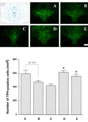
Fig. 2. Effect of acupuncture on the number of 5-hydroxytryptamine (5-HT)-positive cells in the dorsal raphe. Upper: Photomicrographs showing immunofluorescence phycoerythrin (PE) of 5-HT-positive cells in the dorsal raphe. A: maternal care group, B: maternal separation group, C: maternal separation and non-acupoint- acupunctured group, D: maternal separation and Zusanli- acupunctured group, E: maternal separation and fluoxetine- treated group. The scale bar represents 100 µm. Lower: Number of 5-HT-positive cells in the dorsal raphe in each group.
*p<0.05 compared to the maternal separation group. Values are represented as the mean±standard error of the mean.
Fig. 1. Effects of acupuncture on hyperactivity in the open field test. A: maternal care group, B: maternal separation group, C:
maternal separation and non-acupoint-acupunctured group, D:
maternal separation and Zusanli-acupunctured group, E: maternal separation and fluoxetine-treated group. *p<0.05 compared to the maternal separation group. Values are represented as the mean±
standard error of the mean.
2. Effects of acupuncture on the number of 5-HT-positive cells in the dorsal raphe
Photomicrographs of 5-HT-positive cells in the dorsal raphe are illustrated in Figure 2. The number of 5-HT- positive cells in the dorsal raphe was 582.25±34.40/mm2 in the maternal care group, 349.09±13.70/section in the maternal separation group, 394.09±13.70/mm2 in the maternal separation and non-acupoint-acupunctured group, 632.37± 22.62/mm2 in the maternal separation and Zusanli-acupunctured group and 610.92±29.10/mm2 in the maternal separation and fluoxetine-treated group.
The results present that 5-HT synthesis in the dorsal raphe was decreased by maternal separation. In con- trast, acupuncture at Zusanli alleviated maternal separa- tion-induced decrease of 5-HT synthesis. Acupuncture at the non-acupoint exerted no significant effect on 5-HT synthesis in the maternal separated rat pups.
3. Effects of acupuncture on the number of TPH-positive cells in the dorsal raphe
Photomicrographs of TPH-positive cells in the dorsal raphe are illustrated in Figure 3. The number of TPH-
ositive cells in the dorsal raphe was 592.98±50.12/mm2 in the maternal care group, 471.05±16.96/mm2 in the maternal separation group, 420.09±22.97/mm2 in the maternal separation and non-acupoint-acupunctured group, 617.10±25.71/mm2 in the maternal separation and Zusanli- acupunctured group and 558.24±28.40/mm2 in the ma- ternal separation and fluoxetine-treated group.
The results present that TPH expression in the dor- sal raphe was decreased by maternal separation. In con- trast, acupuncture at Zusanli alleviated maternal separa- tion-induced decrease of TPH expression in the dorsal raphe. Acupuncture at the non-acupoint exerted no sig- nificant effect on TPH expression in the maternal sepa- rated rat pups.
Fig. 4. Effects of acupuncture on cell proliferation in the hippocampal gyrus dentatus. Upper: Photomicrographs showing immunostaining of BrdU-positive cells in the dorsal raphe. A:
maternal care group, B: maternal separation group, C: maternal separation and non-acupoint-acupunctured group, D: maternal separation and Zusanli-acupunctured group, E: maternal separation and fluoxetine-treated group. The scale bar represents 50 µm.
Lower: Number of BrdU-positive cells in the dorsal raphe in each group. *p<0.05 compared to the maternal separation group.
Values are represented as the mean±standard error of the mean.
Fig. 3. Effects of acupuncture on the number of tryptophan hydroxylase (TPH)-positive cells in the dorsal raphe. Upper:
Photomicrographs showing immunofluorescence fluorescein isothiocyanate (FITC) of TPH-positive cells in the dorsal raphe.
A: maternal care group, B: maternal separation group, C: maternal separation and non-acupoint-acupunctured group, D: maternal separation and Zusanli-acupunctured group, E: maternal separation and fluoxetine-treated group. The scale bar represents 100 µm.
Lower: Number of TPH-positive cells in the dorsal raphe in each group. *p<0.05 compared to the maternal separation group. Values are represented as the mean±standard error of the mean.
4. Effects of acupuncture on the number of BrdU-positive cells in the hippocampal gyrus dentatus
Photomicrographs of BrdU-positive cells in the hippo- campal gyrus dentatus are illustrated in Figure 4. The number of BrdU-positive cells in the hippocampal gyrus dentatus was 142.07±13.87/mm2 in the maternal care group, 72.26±7.34/mm2 in the maternal separation group, 60.53±5.69/mm2 in the maternal separation and non- acupoint-acupunctured group, 136.79±5.81/mm2 in the maternal separation and Zusanli-acupunctured group and 101.15±4.56/mm2 in the maternal separation and fluoxetine-treated group.
The results present that cell proliferation in the hip- pocampal gyrus dentatus w as decreased by maternal separation. In contrast, acupuncture at Zusanli alleviated maternal separation-induced decrease of cell proliferation.
Acupuncture at the non-acupoint exerted no significant effect on cell proliferation in the maternal separated rat pups.
Discussion»»»
The lack of a mother-infant relationship is known to exert an influence on neonatal development and to in- crease the vulnerability of the offspring to certain neu- ropsychiatric disorders. Maternal deprivation causes cell
death in the infant rat brain16). In this study, the rat pups were separated from their mothers from postnatal 14th day. In order to assess the influence of maternal separation on the activity of rat pups, open field test was performed. In this study, maternal separated rat pups show ed a decreased activity on the open field test.
However, acupuncture at Zusanli for 10 days overcame maternal separation-induced hypoactivity. Maternal sep- aration caused decrease of locomotion activity17). Depression is primarily characterized by a lowering of mood and an inhibition on both mental and physical activity. Many studies suggested that acupuncture improves depres- sion-like behavior4,12). In this study, anti-depression ef- fect of acupuncture w as verified by the findings that acupuncture at Zusanli significantly increased locomo- tor activity in the maternal separation rat pups.
Maternal separation during infancy may influence se- rotonergic neuronal growth and development18). Serotonergic system in the brain is influenced by various stressful stimuli19) and reduced activity in the brain serotonergic system is implicated in the pathophysiology of depres- sion20). In order to confirm whether maternal separa- tion induced depression in this study, we measured 5-HT and TPH expression in the dorsal raphe. In this study, 5-HT level in the dorsal raphe of rat pups w as suppressed by maternal separation, but acupuncture at Zusanli increased the synthesis of 5-HT. This tendency w as supported by Yang et al.21) show ing that 5-HT synthesis in the dorsal raphe was lower in the depres- sion model than that in the normal rats. Reduction in TPH expression leads to a rapid decrease in 5-HT re- lease, indicating that changes TPH activity can influ- ence 5-HT synaptic activity 19). In this study, TPH ex- pression in the dorsal raphe of rat pups was decreased by maternal separation, and acupuncture at Zusanli also increased TPH expression. Acupuncture is known to activate descending serotonergic systems originating in the brainstem22) and acupuncture at Zusanli was effec- tive in the treatment of stress-related physical and men- tal disorders23). The present results suggest that acu-
puncture at Zusanli restored serotonin content in the maternal-separated rat pups.
The hippocampal gyrus dentatus is the brain struc- ture in which cell proliferation and neurogenesis oc- curs24). Stressful experiences, such as maternal separa- tion, are known to suppress neurogenesis in the hip- pocampal gyrus dentatus25). Lee et al.26) showed that a decrease in neurogenesis was related to the patho- genesis of depression. Thus, a decrease in neurogenesis can be considered as a hallmark for depression. In this study, the number of BrdU-positive cells in the hippo- campal gyrus dentatus of rat pups w as decreased by maternal separation. However, acupuncture at Zusanli significantly increased cell proliferation in the hippo- campal gyrus dentatus of maternal-separated rat pups.
Maternal separation suppressed hippocampal cell pro- liferation and impaired cognitive performance27). Park et al.4) reported that acupuncture enhances cell pro- liferation in the maternal-separated rats.
5-HT is also known to modulate neurogenesis in rat brains28). Inhibition of 5-HT synthesis significantly de- creased the number of new ly generated cells in the subventricular zone and the subgranular zone29). Therefore, the acupuncture-induced increase in 5-HT synthesis may contribute to increased cell proliferation, in this study.
The present results demonstrate that maternal separa- tion induced depressive state through decreasing cell proliferation in the hippocampal gyrus dentatus and suppressing 5-HT synthesis in the dorsal raphe of rat pups. However, since this study only observed the ef- fects of acupunture at Zusanli on cell proliferation in the hippocampal gyrus dentatus associated with mech- anism of depression, continuous research will be needed to further investigate the effects of other type of depres- sion and the developmental mechanism of depression.
Conclusion»»»
We investigated the effect of acupuncture on depres-
sion and cell proliferation in hippocampal gyrus denta- tus of maternal-separated rat pups.
Acupuncture at Zusanli acupoint shows that 1. Acupuncture at Zusanli overcame maternal sepa-
ration-induced hypoactivity.
2. Acupuncture at Zusanli alleviated maternal separa- tion-induced decrease of 5-HT synthesis.
3. Acupuncture at Zusanli alleviated maternal separa- tion-induced decrease of TPH expression in the dorsal raphe.
4. Acupuncture at Zusanli alleviated maternal separa- tion-induced decrease of cell proliferation.
Acupuncture at Zusanli ameliorated depressive state through increasing cell proliferation and enhancing 5-HT synthesis. The present results show that acupuncture might be used as the therapeutic strategy for depression patients.
References»»»
1. Aisa B, Elizalde N, Tordera R, Lasheras B, Del Río J, Ramírez MJ. Effects of neonatal stress on markers of synaptic plasticity in the hippocampus: implications for spatial memory. Hippocampus. 2009;19(12):1222-31.
2. Marais L, van Rensburg SJ, van Zyl JM, Stein DJ, Daniels WM. Maternal separation of rat pups increases the risk of developing depressive-like behavior after subsequent chronic stress by altering corticosterone and neurotrophin levels in the hippocampus. Neurosci Res.
2008;61(1):106-12.
3. Sung YH, Shin MS, Cho S, Baik HH, Jin BK, Chang HK, Lee EK, Kim CJ. Depression-like state in maternal rats induced by repeated separation of pups is accom- panied by a decrease of cell proliferation and an in- crease of apoptosis in the hippocampus. Neurosci Lett.
2010;470(1):86-90.
4. Park HJ, Lim S, Lee HS, Lee HJ, Yoo YM, Lee HJ, Kim SA, Yin CS, Seo JC, Chung JH. Acupuncture enhances cell proliferation in dentate gyrus of maternally-sepa- rated rats. Neurosci Lett. 2002;319(3):153-6.
5. Christiansen L, Tan Q, Iachina M, Bathum L, Kruse TA, McGue M, Christensen K. Candidate gene polymorphisms in the serotonergic pathway: influence on depression symptomatology in an elderly population. Biol Psychiatry.
2007;61(2):223-30.
6. Neumeister A. Tryptophan depletion, serotonin, and de- pression: where do we stand? Psychopharmacol Bull.
2003;37(4):99-115.
7. Fairhall SL, Sharma S, Magnusson J, Murphy B. Memory related dysregulation of hippocampal function in major depressive disorder. Biol Psychol. 2010;85(3):499-503.
8. Warner-Schmidt JL, Duman RS. Hippocampal neuro- genesis: opposing effects of stress and antidepressant treatment. Hippocampus. 2006;16(3):239-49.
9. Campbell S, Marriott M, Nahmias C, MacQueen GM.
Lower hippocampal volume in patients suffering from depression: a meta-analysis. Am J Psychiatry. 2004;161(4):
598-607.
10. Duman RS, Monteggia LM. A neurotrophic model for stress-related mood disorders. Biol Psychiatry. 2006;59(12):
1116-27.
11. Baek SS, Jun TW, Kim KJ, Shin MS, Kang SY, Kim CJ.
Effects of postnatal treadmill exercise on apoptotic neu- ronal cell death and cell proliferation of maternal-sepa- rated rat pups. Brain Dev. 2012;34(1):45-56.
12. Dos Santos JG Jr, Kawano F, Nishida MM, Yamamura Y, Mello LE, Tabosa A. Antidepressive-like effects of electroacupuncture in rats. Physiol Behav. 2008;93(1-2):
155-9.
13. Hwang IK, Chung JY, Yoo DY, Yi SS, Youn HY, Seong JK, Yoon YS. Comparing the effects of acupuncture and electroacupuncture at Zusanli and Baihui on cell pro- liferation and neuroblast differentiation in the rat hippocampus. J Vet Med Sci. 2010;72(3):279-84.
14. Kim EH, Kim YJ, Lee HJ, Huh Y, Chung JH, Seo JC, Kang JE, Lee HJ, Yim SV, Kim CJ. Acupuncture in- creases cell proliferation in dentate gyrus after transient global ischemia in gerbils. Neurosci Lett. 2001;297(1):21-4.
15. Durand M, Berton O, Aguerre S, Edno L, Combourieu I, Mormède P, Chaouloff F. Effects of repeated fluoxetine on anxiety-related behaviours, central serotonergic sys- tems, and the corticotropic axis axis in SHR and WKY rats. Neuropharmacology. 1999;38(6):893-907.
16. Zhang LX, Levine S, Dent G, Zhan Y, Xing G, Okimoto D, Kathleen Gordon M, Post RM, Smith MA. Maternal deprivation increases cell death in the infant rat brain.
Brain Res Dev Brain Res. 2002;133(1):1-11.
17. Lim S, Ryu YH, Kim ST, Hong MS, Park HJ. Acupuncture increases neuropeptide Y expression in hippocampus of maternally-separated rats. Neurosci Lett. 2003;343(1):49-52.
18. Braun K, Lange E, Metzger M, Poeggel G. Maternal separation followed by early social deprivation affects the development of monoaminergic fiber systems in the medial prefrontal cortex of Octodon degus. Neuroscience.
2000;95(1):309-18.
19. Lee SH, Chung SH, Lee JS, Kim SS, Shin HD, Lim BV, Jang MH, Kim H, Kim EH, Kim CJ. Effects of acu- puncture on the 5-hydroxytryptamine synthesis and
tryptophan hydroxylase expression in the dorsal raphe of exercised rats. Neurosci Lett. 2002;332(1):17-20.
20. Arborelius L, Hawks BW, Owens MJ, Plotsky PM, Nemeroff CB. Increased responsiveness of presumed 5-HT cells to citalopram in adult rats subjected to prolonged maternal separation relative to brief separation. Psychopharmacology (Berl). 2004;176(3-4):248-55.
21. Yang LM, Hu B, Xia YH, Zhang BL, Zhao H. Lateral ha- benula lesions improve the behavioral response in de- pressed rats via increasing the serotonin level in dorsal raphe nucleus. Behav Brain Res. 2008;188(1):84-90.
22. Takagi J, Yonehara N. Serotonin receptor subtypes in- volved in modulation of electrical acupuncture. Jpn J Pharmacol. 1998;78(4):511-4.
23. Chen A. An introduction to sequential electric acu- puncture (SEA) in the treatment of stress related phys- ical and mental disorders. Acupunct Electrother Res.
1992;17(4):273-83.
24. Eriksson PS, Perfilieva E, Björk-Eriksson T, Alborn AM, Nordborg C, Peterson DA, Gage FH. Neurogenesis in the adult human hippocampus. Nat Med. 1998;4(11):1313-7.
25. Fabricius K, Wörtwein G, Pakkenberg B. The impact of maternal separation on adult mouse behaviour and on the total neuron number in the mouse hippocampus.
Brain Struct Funct. 2008;212(5):403-6.
26. Lee HJ, Kim JW, Yim SV, Kim MJ, Kim SA, Kim YJ, Kim CJ, Chung JH. Fluoxetine enhances cell proliferation and prevents apoptosis in dentate gyrus of maternally separated rats. Mol Psychiatry. 2001;610(6):725-8.
27. Hulshof HJ, Novati A, Sgoifo A, Luiten PG, den Boer JA, Meerlo P. Maternal separation decreases adult hip- pocampal cell proliferation and impairs cognitive per- formance but has little effect on stress sensitivity and anxiety in adult Wistar rats. Behav Brain Res. 2011;
216(2):552-60.
28. Banasr M, Hery M, Brezun JM, Daszuta A. Serotonin me- diates oestrogen stimulation of cell proliferation in the adult dentate gyrus. Eur J Neurosci. 2001;14(9):1417-24.
29. Brezun JM, Daszuta A. Depletion in serotonin decreases neurogenesis in the dentate gyrus and the subventricular zone of adult rats. Neuroscience. 1999;89(4):999-1002.
Original Article
보골탕이 Monosodium Iodoacetate 유도 골관절염과 Interleukin-1β 유도 연골세포에 미치는 보호 효과
성진욱*⋅이해웅
†⋅강경화
‡⋅김경민
§⋅조성우*
동의대학교 한의과대학 한방재활의학과교실*, 예방의학교실†, 생리학교실‡, 한방내과학교실§,
Protective Effects of Bogol-tang on Monosodium Iodoacetate-induced Osteoarthritis and Interleukin-1β-treated Primary Chondrocytes
Jin Wook Sung, K.M.D.*, Hai Woong Lee, K.M.D.†, Kyung Hwa Kang, K.M.D.‡, Kyoung Min Kim, K.M.D.§, Sung Woo Cho, K.M.D.*
Departments of Korean Rehabilitation Medicine*, Preventive Medicine†, Physiology‡, and Oriental Internal Medicine§, College of Korean Medicine, Dong-eui University
RECEIVED March, 14, 2019 REVISED April, 15, 2019 ACCEPTED April, 17, 2019 CORRESPONDING TO Sung Woo Cho, Department of Korean Rehabilitation Medicine, College of Korean Medicine, Dong-eui University, 52-57 Yangjeong-ro, Busanjin-gu, Busan 47227, Korea TEL (051) 850-8671 FAX (051) 867-5162 E-mail luxy@daum.net CO-CORRESPONDING TO Kyung-Hwa Kang, Department of Physiology, College of Korean Medicine, Dong-eui University, 52-57 Yangjeong-ro, Busanjin-gu, Busan 47227, Korea
TEL (051) 850-7423 FAX (051) 853-4036 E-mail ghkang@deu.ac.kr Copyright © 2019 The Society of Korean Medicine Rehabilitation
Objectives Bogol-tang has clinically been used to protect joint cartilage and to treat osteoarthritis. Our objective was to study the protective effect of Bogol-tang extract (BGT) in functional impairment, behavioral disorders, cartilage loss and pathological changes in a monoiodoacetate (MIA)-induced murine osteoarthritis (OA) model and interleukin (IL)-1β -treated primary rat chondrocytes.
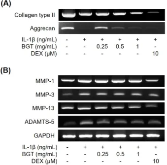
Methods Mouse knee joints were injected with MIA, a chemical that inhibits glycolysis and causes joint inflammation and matrix loss. MIA-OA induced mice orally administered BGT or acetaminophen (AAP) for 18 days by daily. Primary rat chondrocytes were pretreated with BGT or dexamethasone (DEX) and followed by co-incubation with IL-1β (10 ng/mL).
Results In MIA-OA mice model, BGT led to delayed response on hot plate analysis, and suppressed the cartilage loss and damages in joint tissues. BGT suppressed the elevated levels of inflammatory mediators, nitrite and PGE2, the gene expression of matrix degrading enzymes, and extracellular-signal-regulated kinases 1/2 and c-JunN-terminal kinase phos- phorylation in IL-1β-treated primary rat chondrocytes.
Conclusions Our results suggest that BGT improve the knee joint function and delay the cartilage damages by anti-nociceptive, anti-inflammatory and ant-catabolic effects, which indicate BGT could be a potential candidate for osteoarthritis treatment. (J Korean Med Rehabil 2019;29(2):101-113)
Key words Bogol-tang, Osteoarthritis, Monosodium iodoacetate, Nociception
Introduction»»»
Osteoarthritis (OA) is the most common form of joint disorders in aging populations over 65 years of age1).
The development of OA is promoted by the risk fac- tors such as previous knee injury, female gender, aging, obesity2). Due to the progressive destruction of joint cartilage and inflammatory response, patients with OA
experience pain, stiffness, and functional impairment in their damaged joints3,4). It results in a substantial eco- nomic burden and impaired quality of life5).
Although analgesics and non-steroid anti-inflammatory drugs (NSAIDs) are commonly used for symptomatic treatment for OA6), no drug has been approved by other agencies around the world, Food and Drug Administration (FDA) or OA disease modification.
Korean traditional therapies including medications, acupuncture, moxibustion and pharmacopuncture have been applied for degenerative osteoarthritis7-12). Recently, scientific efficacy studies have been active on the treat- ment of Korean medicine for osteoarthritis13).
Animal models and primary rat chondrocytes that mimic degenerative arthritis pathology were used in the experiment. Intra-articular injection of monosodium iodoacetate (MIA) into joints inhibits glyceraldehye-3- phosphate dehydrogenase activity in chondrocytes, caus- ing the disruption of glycolysis and eventual cell death.
The loss of chondrocytes results in pathological symp- toms similar to arthritis in humans14).
Bogol-tang (BGT) is a prescription made by the au- thor based on Palmul-tang and has been operated for 15 years in clinical practice for degenerative osteoarthritis.
This prescription consists of drugs improve the func- tion system of liver and kidney and strengthen the mus- cles and bones, and medicines that help the digestive function in Korean medicines. Medicines strengthen the muscles and bones are Cibotii Rhizoma, Astragali Radix, Coicis Semen, Drynariae Rhizoma, Curcumae Radix, Dioscoreae Rhizoma, Acanthopanacis Cortex, Psoraleae Semen, Eucommiae Cortex Carbonisatum, Lycii Fructus, Rehmanniae Radix Preparata, Carthami Fructus, Clematidis Radix, Asiasari Radix et Rhizoma and Cervi Cornus Colla.
And medicines helping digestive function are Atractylodis Rhizoma Alba, Cyperi Rhizoma, Citri Unshius Pericarpium, Poria Sclerotium, Amomi Fructus, Glycyrrhizae Radix et Rhizoma.
This study is to evaluate the efficacy of the BGT on the MIA-induced models of OA in the C57BL/6N mouse
knee joint and IL-1β-treated primary rat chondrocytes.
Therefore, we measured the body weights, the weight of organs, pain sensitivity, gait alteration, and cartilage changes in MIA-induced OA mice. Also, we investigated inflammatory mediator, matrix degrading enzyme asso- ciated genes, phosphorylation of microtubule-associated protein kinase (MAPKs) activation in IL-1β-treated rat primary cultured chondrocytes.
Materials and Methods»»»
1. Animals
Male C57BL/6N mice of 11 week-old were purchased from SAMTACO. Co. Ltd. (Osan, Korea). Mice were housed in cages, 4 animals per cage, were fed stand- ard laboratory chow and water ad libitum, and ex- posed to a daily 12 h light/dark cycle. Mice were ac- climatized for 1 w eek prior to starting the experiment.
Mice were divided into four groups as follows: Normal saline administration (Control [CON] group, n=8); OA with normal saline administration (MIA group, n=8); OA with 0.46 g/kg/day BGT administration (BGT group, n=8); OA with 0.066 g/kg/day acetaminophen (AAP) treatment (AAP group, n=8). The animals in this study were cared for according to the Guide for the Care and Use of Laboratory Animals (National Academy Press, Washington, D.C., WA, USA). The protocol was approved by the Committee on Animal Research and Ethics of Dong-eui University (Busan, Korea) (The committee's reference number: R2017-028).
2. Induction of MIA-induced OA
For induction of MIA-induced OA, the mice were in- tramuscularly anesthetized with a mixture of ketamine hydrochloride (20 mg/kg, Ketalar, Yuhan Co., Seoul, Korea) and xylazine (20 mg/kg, Rumpun, Bayer Korea Ltd., Seoul, Korea), which was diluted 1:1 with normal
Herb name Amount (g)
Cibotium barometz J. Smith 12
Astragalus membranaceus Bunge 12
Coix lacryma-jobi 8
Drynaria fortunei J. Smith 8
Curcuma wenyujin Y. H. Chen et C. Ling. 8
Dioscorea batatas Decaisne 8
Acanthopanax sessiliflorum Seeman 8
Psoralea corylifolia Linné 8
Eucommia ulmoides Oliver 8
Poria cocos Wolf 8
Lycium chinense Miller 6
Atractylodes japonica Koidzumi 6
Cyperus rotundus Linné 6
Citrus unshiu Markovich 6
Rehmannia glutinosa Liboschitz ex Steudel 6
Angelica gigas Nakai 4
Paeonia lactiflora Pallas 4
Carthamus tinctorius Linné 4
Amomum villosum Loureiro 4
Clematis manshurica Ruprecht 4 Asiasarum heterotropoides F. Maekawa 4 Glycyrrhiza uralensis Fischer 4
Cervus nippon Temminck 4
Total 150
Table I. The Ingredients of Bogol-tang (BGT)
saline. Once the mice were under anesthesia, mice were given a single intra-articular injection of 1 mg MIA (Sigma Chemical Co., St. Louis, MO, USA) through the infrapatellar ligament of the right knee15) and killed at 18 days post injection. MIA w as dissolved in sterile sal- ine and administered in a volume of 10 µL using a 30 gauge needle. The left contralateral control knee was injected with 10 µL of sterile saline (0.9%) as a control.
3. Preparation of BGT
BGT are the extracts purified from the twenty-three medicinal herbs, including Cibotium barometz J. Smith and Astragalus membranaceus Bunge et al., as shown in Table I. The herbs were obtained from HANION
HERB Co. (Busan, Korea) and authenticated based on their microscopic and macroscopic characteristics by the classification and identification committee of the Institute of Korean Medicine of Dong-eui University.
The total mixture (600 g) of herbs was washed thor- oughly with distilled water, cut into pieces, and were submerged into distilled water (1:10, v/v), was boiled at 100°C for 2 h and the extract was filtered, lyophi- lized and subsequently stored at -20°C. The yield of BGT aqueous extract was 13.73% (w/w).
4. Treatment
The effect of BGT was studied in four groups of 8 mice after induction of OA. The BGT group was ad- ministered orally by gavage at a daily 0.46 g/kg dose of BGT, the AAP group w as administered orally by gav- age at a daily 0.066 g/kg dose of AAP and the CON and MIA group received water. Histopathological changes in knee joints were studied after 18 days.
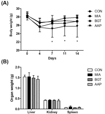
5. The weights of body and organs
The mice were recorded the changes of body weights at intervals of 3–4 days for 18 days after intra-articular injection of MIA and oral administration. When the mice were sacrificed, the organs (liver, kidney, and spleen) were harvested and their wet masses were determined.
6. Timeline of experiments
We performed a pilot experiment on a small batch of mice to determine w hich analyses to use. In the fi- nal experiment from which we collected data, the be- havioral and functional analyses were performed in the order of least stress to most stress for the mice. To ac- count for habituation formation, all mice were tested 2 times after MIA-induction OA at specific time points.
The scheme of the study is illustrated in Figure 1.
Fig. 1. Scheme of this experimental process. Osteoarthritis (OA) was induced by monoiodoacetate (MIA) injection into the joint of mice. Body weight (BW) was measured at regular intervals after intra-articular injection of MIA. Mice were orally administrated with Bogol-tang extract (BGT) or acetaminophen (AAP) once a day for 18 days. We performed two kinds of functional assessments and postmortem assessment after the final functional analysis. H&E: hematoxylin-eosin.
7. Animal behavioral tests
1) Hot plate analysis
The analgesic response was the latency observed from the time the mouse was placed on the heated surface until the first overt behavioral sign of nociception. Mice were transferred to the room for analysis at least 30 min before the experiment. Then, the mouse was placed on the hot plate at 55°C one at a time (Columbus Instruments, Columbus, OH, USA). The latency period for hind limb response (e.g., shaking, jumping, or licking) was recorded as response time. Each trial has a maximum time of 45 s.
The mouse was removed from the hot plate immediately after a response was observed.
2) Gait analysis
The paws of the mice were brushed with ink by an examiner blinded to the MIA-induced OA procedures.
Right and left hind paws were colored with ink of dif- ferent color. Immediately after ink was applied, the mice were allowed to run on a 60 cm long, 6 cm wide Plexiglas track with white paper on the bottom. A dark chamber was present in the end of the track to entice the mice. Upon completion of the test, paper was scan- ned at 300 dpi. The distance of the same paw between
two steps were defined as stride. The areas of third to the fifth set of footsteps were quantified by image J software (U.S. National Institutes of Health, Bethesda, MD, USA; https://imagej.nih.gov/ij/, 1997-2018) N=8 in each group.
8. Histopathology
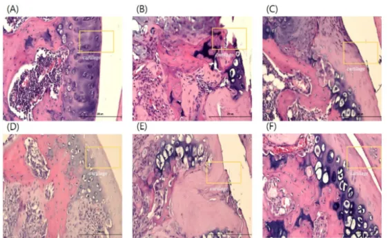
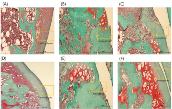
Mice were sacrificed at 18 days following BGT administration. The knee joint of each mouse was de- calcified in 14% ethylenediaminetetraacetic acid for 5 days at room temperature. After dehydration and em- bedding in paraffin, the sagittal section (5 μm) of the knee joint were stained with hematoxylin-eosin (H&E) and Safranin-O Fast green method. The stained sections were photographed under a light microscope using a Leica microscope (Leica Microsystems, Wetzlar, Germany).
9. Cell examination
1) Culture of primary rat chondrocytes
The tibial and femoral cartilages of 4–5-week old Sprague Dawley rats were dissected, added to 10 mL of 0.2% trypsin at 37°C for 45 min, and then 0.2% (w/v) collagenase Type II and digested for 45 min at 37°C,
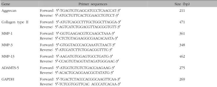
Gene Primer sequences Size (bp) Aggrecan Forward: 5ʹ-TGAGTGTGAGCATCCCTCAACCAT-3ʹ
Reverse: 5ʹ-ATGCTGTTCACTCGAACCTGTCCT-3ʹ 211
Collagen type II Forward: 5ʹ-ATGTCAGCCTTTGCTGGCTTAGGA-3ʹ
Reverse: 5ʹ-AGTCATCTGGACGTTAGCGGTGTT-3ʹ 471
MMP-1 Forward: 5ʹ-GGTGAAGACGTCCAAGCTAAA-3ʹ
Reverse: 5ʹ-CTCTGTAGAAGGCGAACACAATA-3ʹ 361
MMP-3 Forward: 5ʹ-GTGGTACCCACCAAATCTAACT-3ʹ
Reverse: 5ʹ-ATCGATCTTCTGGACGGTTTC-3ʹ 348
MMP-13 Forward: 5ʹ-AAGATGTGGAGTGCCTGATG-3ʹ
Reverse: 5ʹ-CCAGTGTAGGTATAGATGGGAAC-3ʹ 462
ADAMTS-5 Forward: 5ʹ-ATGGTGTGTCTGACCAAGAAG-3ʹ
Reverse: 5ʹ-ACACTGCAGGAACGGTATATG-3ʹ 275
GAPDH Forward: 5ʹ-TGACTCTACCCACGGCAAGTTCAA-3ʹ
Reverse: 5ʹ-TCTCGTGGTTCAC ACCCATCACAA-3ʹ 269
MMP: matrix metalloproteinase, ADAMTS: a disintegrin and metalloproteinase with thrombospondin motifs, GAPDH: glyceraldehyde 3-phosphate dehydrogenase.
Table II. The Primer Sequences Used in This Study
two times. The cartilage pieces were retrieved and placed in 10 mL of 0.2% collagenase Type II solution at 0.5 mg/mL overnight at 37°C. Primary rat chondrocytes were cultured at 37°C in a 5% CO2 incubator in Dulbeco's Modified Eagle's Medium containing 10% fetal bovine serum and 1% penicillin/streptomycin.
2) Cell viability assay
Cell proliferation were determined using a 3-(4,5- dimethylthiazol-2-yl)-2,5-diphenyltetrazolium bromide (MTT) assay. For cell proliferation assay, primary rat chon- drocytes were seeded at 1.5×10⁵ cells/mL, incubated for 5 days, and treated with varying concentrations of BGT for 24 h. The crystallized formazan was dissolved in di- methyl sulphoxide, and the absorbance was measured at 570 nm on a SpectraMax M2 Microplate reader (Molecular Devices, Sunnyvale, CA, USA).
3) Nitric oxide assay
Primary rat chondrocytes were seeded at 1.5×10⁵ cells/mL in 24-well plates. Primary rat chondrocytes were pretreated with varying concentrations (0.25, 0.5, and 1 mg/mL) of BGT for 24 h and subsequently cultured with IL-1β (10 ng/mL) for 24 h. Nitrite (NO2) accumu-
lation in the culture medium was measured as an in- dicator of NO production, based on the Griess reaction.
In brief, 100 μL of each supernatant from BGT-treated samples was mixed with an equal volume of Griess re- agent (1% [w/v] sulfanilamide in 5% [v/v] phosphoric acid and 0.1% [w/v] naphthylethylenediamine) in a dark room for 10 min. Absorbance was then measured at 540 nm on a SpectraMax M2 Microplate reader (Molecular Devices, Sunnyvale, CA, USA). The nitrite concentration was determined by comparison with a standard curve of sodium nitrite.
4) Prostaglandin E2 assay
To measure prostaglandin E2 (PGE2), primary rat chondrocytes were plated at 1.5×10⁵ cells/mL in a 24-well plate. Primary rat chondrocytes were pretreated with varying concentrations (0.25, 0.5, and 1 mg/mL) of BGT for 24 h and subsequently cultured with IL-1β (10 ng/mL) for 24 h. The concentration of PGE2 w as measured using the PGE2 Parameter Assay Kit (R&D system Inc., Minneapolis, MN, USA), according to the manufacturer’s instructions.

![Fig. 1. Effects of Pyrola japonica extract (NJ) on the formation of tartarate-resistant acid phosphatase positive (TRAP[+] multinucleated cells [MNCs])](https://thumb-ap.123doks.com/thumbv2/123dokinfo/5446796.236727/52.892.146.744.633.986/effects-japonica-formation-tartarate-resistant-phosphatase-positive-multinucleated.webp)
