pISSN 2288-9272 eISSN 2383-8493 J Oral Med Pain 2019;44(2):45-53 https://doi.org/10.14476/jomp.2019.44.2.45
Could Crepitus Be an Indication for Early Temporomandibular Joint Osteoarthritis?
Hye-Min Ju 1 , Sun-Hee Lee 1 , Hye-Mi Jeon 2 , Kyung-Hee Kim 3 , Yong-Woo Ahn 4 , Soo-Min Ok 4 , Sung-Hee Jeong 4
1 Department of Oral Medicine, Dental Research Institute, Pusan National University Dental Hospital, Yangsan, Korea
2 Department of Oral Medicine, Pusan National University Hospital, Busan, Korea
3 Department of Oral Medicine, Inje University Busan Paik Hospital, Busan, Korea
4 Department of Oral Medicine, Dental Research Institute, School of Dentistry, Pusan National University, Yangsan, Korea
Received March 25, 2019 Revised April 8, 2019 Accepted April 8, 2019
Purpose: To determine whether crepitus may be a clinical indication for early temporoman- dibular joint (TMJ) osteoarthritis (OA) and to investigate the correlation between crepitus and the occurrence of TMJ OA with respect to factors, such as patient sex, age, chewing habits, and diagnosis.
Methods: This is retrospective analysis of clinical data for 162 TMJs. The criteria for a joint to be included in this study was a minimum of two cone-beam computed tomography (CBCT) scans performed with no OA observed during the initial scan. The Diagnostic Criteria for Temporomandibular Disorders was used for OA diagnosis. Crepitus was recorded when it was objectively palpated during the follow-up period. Correlations between various patient factors and progression to TMJ OA were calculated using the Pearson’s chi-square test. A linear-by-linear association was used to analyze trends of OA progression with increasing age.
Results: Among the 162 joints, 101 progressed to OA and 61 did not. In the joints where crepitus had been present before OA was confirmed at next or last CBCT, OA progressed at a high rate, and especially higher in female and older patients (p<0.01). Patients in the pain- related disorder group with crepitus were observed to have higher rates of OA progression compared to patients in the intra-articular disorder group (p<0.01).
Conclusions: If a patient experiences pain in the TMJs and crepitus, close monitoring through regular CBCT scans is necessary even if there is no evidence of radiologically con- firmed OA after the first CBCT.
Key Words: Crepitus; Early osteoarthritis; Temporomandibular joint; The Diagnostic Criteria for Temporomandibular Disorders
Correspondence to:
Sung-Hee Jeong
Department of Oral Medicine, Dental Research Institute, School of Dentistry, Pusan National University, 49
Busandaehak-ro, Mulgeum-eup, Yangsan 50612, Korea
Tel: +82-55-360-5242 Fax: +82-55-360-5238 E-mail: [email protected]
https://orcid.org/0000-0002-6296-4775 This study was supported by 2019 Clinical Research Grant, Pusan National University Dental Hospital.
JOMP Journal of Oral Medicine and Pain
Copyright Ⓒ 2019 Korean Academy of Orofacial Pain and Oral Medicine. All rights reserved.
CC
This is an open-access article distributed under the terms of the Creative Commons Attribution Non-Commercial License (http://creativecommons.org/licenses/by-nc/4.0/), which permits unrestricted non-commercial use, distribution, and reproduction in any medium, provided the original work is properly cited.
INTRODUCTION
Osteoarthritis (OA) is one of the most common diseases that affect the temporomandibular joint (TMJ), causing de- structive changes in the bone. In general, TMJ OA is con- firmed when subchondral cyst, erosion, generalized scle- rosis, and osteophyte are detected on radiographic images
[1]. However, computed tomography (CT) or cone-beam CT
(CBCT) cannot detect tissue-related changes, such as dam-
ages to articular cartilage. Only when the disease has ad-
vanced to the extent that the subcortical layer has been
destroyed would it be recognized as OA [2]. Once a bone is
damaged, it does not easily recover; therefore, early diag-
nosis and treatment of TMJ OA are essential in reducing the
degree of joint injury and lowering the risk of possible fu- ture OA changes. Early detection of OA is much more com- plicated than the diagnosis of established OA [3,4]. Patients with early OA who show no radiological signs may go un- diagnosed and have a lack of follow-up, thereby leading to continuing deterioration.
TMJ contains fibrocartilage, unlike most joints that con- tain hyaline cartilage; however, TMJ is similar to other joints in terms of structure and function [5]. Considering the fact that knee OA is often used as an example when describing TMJ OA, it can be argued that using magnetic resonance imaging (MRI) to evaluate cartilage damage and bone marrow lesion may be considered as an appropriate method for early diagnosis of TMJ OA. There have been various studies on the early diagnosis of knee OA using MRI; however, MRIs may be prohibitively expensive despite its possible usefulness [3,6,7].
There are many studies on the clinical features and ra- diological findings of advanced TMJ OA or temporoman- dibular disorder (TMD), but there is a lack of research on the clinical manifestations of early TMJ OA. Crepitus, a characteristic sound clinically detected in patients with TMJ OA, is generally thought to occur in the later stages of OA.
However, some previous studies on knee OA and TMJ rheu- matoid arthritis (RA) have reported that crepitus may be an early clinical indicator of them [8-10]. Therefore, this study aims to determine whether crepitus may be an early clini- cal indicator of TMJ OA and investigate the association be- tween crepitus and the occurrence of OA, considering vari- ous factors about patients, such as sex, age, chewing habits, and diagnosis.
MATERIALS AND METHODS
1. Subjects
All clinical and radiographic data of patients who vis- ited the Department of Oral Medicine at Pusan National University Hospital between June 2013 and December 2016 were retrospectively reviewed. Among the 1,206 patients who underwent at least two CBCT scans, patients with ra- diographic findings of skeletal deformity, condyle frac- ture, TMJ tumor, or bilateral TMJ OA at the first CBCT were excluded. The remaining 100 patients did not have TMJ
abnormalities on at least one side.
Among 200 joints (100 patients), 162 joints were investi- gated–38 joints that showed OA at the first CBCT scan were excluded–and, they were either the ones that progressed into OA on the next or last CBCT scan, or others that did not progress until last CBCT scan. The diagnosis of each joint at the first visit was done by an orofacial pain spe- cialist, and the reading of CBCT was conducted by an oral radiologist.
All patients received conservative treatments, such as medication, behavioral conditioning, and physical therapy.
During the follow-up period, CBCT was performed at in- tervals of a minimum of 6 months and a maximum of 36 months, and the average of intervals is 9.79±5.3. At first visit, sex, age, diagnosis and chewing habits of each patient were investigated, and the presence or absence of crepi- tus on charts was studied before being confirmed by next or last CBCT. Among them, 124 were females (76.5%) and 38 were males (23.5%), and the ages ranged from 13 to 83 years (mean age, 29.40±15.46 years). This study was ap- proved by the Institutional Review Board of Pusan National University Dental Hospital (IRB no. PNUDH-2017-027).
Written informed consent was obtained from all patients at the first visit.
2. Methods
1) Clinical data collection
Clinical data were studied based on the initial record of
age, sex, presence of crepitus, unilateral chewing habit,
and diagnosis. The patients were divided into the following
three age groups; Group I (11 to 20 years), Group II (21 to
30 years), and Group III (>30 years). The data of crepitus on
two groups were collected, and the specific criteria of the
data collection are as follows; In Progression-to-OA Group,
the existence or non-existence of the detection of crepitus
on charts was studied before the OA was observed in the
CBCT reading. In No-Progression-to-OA Group, the exis-
tence or non-existence of the detection of crepitus on charts
was studied until the last visit. Crepitus was expressed as
present (P) or absent (A). Crepitus was considered as present
when it was objectively palpated at least one time during
opening, closing, or right or left lateral or protrusive move-
ment. Subjective reports of crunching, grating, or grinding
sound by patients were not collected while patients were not in the hospital. The diagnosis at first visit was conduct- ed according to Diagnostic Criteria for TMD (DC/TMD). The patients who had visited hospital before DC/TMD was pub- lished were diagnosed again, according to DC/TMD based on their chart.
2) OA diagnostic criteria
The CBCT images were taken using Pax-Zenith 3D (Vatech, Hwaseong, Korea). At the time of the first CBCT imaging, TMJ OA was not diagnosed if the images were interpreted as normal, and considered to have no abnormal finding and posterior or anterior position of condyle by experienced oral radiologists. TMJ OA was confirmed when subchondral cyst(s), erosion(s), generalized sclerosis, or osteophyte(s) was observed during CBCT image reading, according to the cri- teria of OA diagnosis based on DC/TMD [1]. The joints that progressed to OA from the second to the last CBCT were ex- pressed as progression (Pr), and joints not progressed to OA were expressed as no progression (N).
3) Statistical analysis
All statistical analyses were performed using IBM SPSS
Statistics ver. 21.0 software (IBM Co., Armonk, NY, USA).
The association between crepitus and the occurrence of OA, considering various factors about patients, such as sex, age, chewing habits, and diagnosis–was calculated using the Pearson’s chi-square test. If the expected frequency of the cell was lower than 5, Fisher’s exact test was used. A linear by linear association was used to analyze trends of OA pro- gression rate as the age of the patient increases. A p-value of <0.05 was considered statistically significant.
RESULTS
Among the 162 TMJs, 61 (37.7%) progressed to OA and 101 (62.3%) did not.
1. Association between Crepitus and TMJ OA Progression Rate
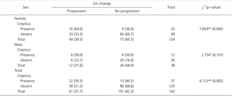
There was a statistically significant difference in the pres- ence or absence of crepitus between OA-progressed joints and non-OA-progressed joints, with a higher OA progres- sion rate when crepitus was present (p<0.01; Table 1).
Table 1. Correlation between sex and TMJ OA progression
Sex OA change
Total χ
2(p-value)
Progression No progression
Female Crepitus
Presence 16 (64.0) 9 (36.0) 25 7.854** (0.005)
Absent 33 (33.3) 66 (66.7) 99
Total 49 (39.5) 75 (60.5) 124
Male Crepitus
Presence 6 (50.0) 6 (50.0) 12 2.754
a(0.101)
Absent 6 (23.1) 20 (76.9) 26
Total 12 (31.6) 26 (68.4) 38
Total Crepitus
Presence 22 (59.5) 15 (40.5) 37 9.712** (0.002)
Absent 39 (31.2) 86 (68.8) 125
Total 61 (37.7) 101 (62.3) 162
TMJ, temporomandibular joint; OA, osteoarthritis.
Values are presented as number (%).
The p-value was determined using chi-square test.
a
Using fisher’ s exact test.
**p<0.01.
2. Association between Sex, Crepitus and Progression to TMJ OA
Among 162 TMJs, there were approximately three times more female TMJs (124) than male TMJs (38). Among the 38 male TMJs, the rate of joints that progressed to OA was 68.4% and that in females was 65.3%, which showed no statistically significant difference (p>0.05). In males, there was no statistically significant correlation (p>0.05) between OA occurrence and existence and nonexistence of crepitus;
however, in females, there was a higher probability of OA progression when crepitus was present (p<0.01; Table 1).
3. Association between Age, Presence or Absence of Crepitus, and Rate of Progression to TMJ OA
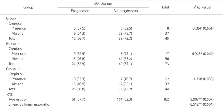
The age distribution of the 162 joints is as follows: Group I (11-20 years) had 45 joints (27.8%), Group II (21-30 years) had 73 (45.0%; the largest group), and Group III (>30 years) had 44 (27.2%). The risk of OA increased as age increased, from 26.7% in Group I to 32.9% in Group II, and 56.8% in Group III (p<0.01; Table 2).
There was no statistically significant difference in the rate of progression of OA according to existence and nonexis- tence of crepitus in Group I; however, there was a statisti- cally significant difference in Group II (p=0.044), and in Group III, when crepitus was present, a higher rate of OA progression was seen (p=0.030).
4. Analysis of OA Progression Rate with Crepitus in Intra- Articular Disorders, and Pain Related Disorders Initial diagnosis included myalgia, arthralgia, disc dis- placement with reduction (DD/wR), disc displacement with- out reduction (DD/woR), and RA according to DC/TMD (in- cluding multiple diagnoses; a total n=269). Myalgia was the most common (33.5%; n=90); however, DD/wR was the most common (n=52) among the intra-articular disorders.
When all patients were divided into two groups according to existence or absence of OA progression and relative rate of diagnosis names at initial diagnosis was studied, divided into two groups, diagnosis names (myalgia, arthralgia, and normal) showed almost similar distribution regardless of
Table 2. Correlation between age, the presence or absence of crepitus and progression to TMJ OA
Group OA change
Total χ
2(p-value)
Progression No progression
Group I Crepitus
Presence 3 (37.5) 5 (62.5) 8 0.584
a(0.661)
Absent 9 (24.3) 28 (75.7) 37
Total 12 (26.7) 33 (73.3) 45
Group II Crepitus
Presence 9 (52.9) 8 (47.1) 17 4.043* (0.044)
Absent 15 (26.8) 41 (73.2) 56
Total 24 (32.9) 49 (67.1) 73
Group III Crepitus
Presence 10 (83.3) 2 (16.7) 12 4.728 (0.030)
Absent 15 (46.9) 17 (53.1) 32
Total 25 (56.8) 19 (43.2) 44
Total
Age group 61 (37.7) 101 (62.3) 162 9.907** (0.007)
Linear by linear association 8.512** (0.004)
TMJ, temporomandibular joint; OA, osteoarthritis.
Values are presented as number (%).
Group I, 11-20 years; Group II, 21-30 years; Group III, >30 years.
The p-value was determined using the chi-square test.
a