大짧放射옳톨훌훌융誌 第 25 5/양 第5 없 pp. 799 - 806, 1989 Joumal of Korean Radiological Society, 25(5) 799-806, 1989
- Abstract-
자궁경부암의 방사선치료후 골반강내 변화의 전산화단층촬영 소견
인제대학 부속 서울 백병원 진단방사선과학교실
박 준 상
• 전 우 기 • 한 창 열 • 이 관 세.Computed Tomographic Findings in pelvic Cavity Mter Radiation Therapy for Uterine Cervical Carcinoma
Jun Sang Park , M.D., Woo Ki Jeon ,
M.D., Chang Yul Han ,
M.D., Kwan Seh Lee ,
M.D:
Department of Radiology, School of Medicine, Inje University, Paik Hospital, Seoul
The effects of pelivic irradiation may be seen mainly in the perirectal area related with the irradiating field
It is important to differentiate the postirradiation change from the recurrency or residual tumor of cervical cancer in the clinical prognostic view of the patient.
The prominent postirradiation changes may be difficult to differentiate from the recurrency or residual tumor of cervical cancer
The author analysed the computed tomographic findings of 35 patients who had received radiation therapy of cervical cancer at Inje University Seoul Paik Hospital
The radiation changes were compared with pre. and postirradiative CT findings in sequential follow.up course
The results were as follows‘
l. Significant changes could be observed at least 3 months after irradiation 2. Postirradiation changes include:
Symmetrical thickening of perirectal fascia 28 cases 77 % Increased perirectal fat 26 cases 75 %
Fibrous connection between sacrum and rectum 20 cases 57 % widening of presacral space 14 cases 42 %
Irregular thickening of rectal wall 5 cases 14 %
• 중앙대 학교 의 과대 학 방사선과학교실
• Department of Radlology, College of AfedlClIIe, Chung-Ang Unlverslty 이 논문은 1989년 7월 14 일 접수하여 1989년 8월 21 일에 채택되었음
- 799 -
- 大韓放射線뿔學會픔 : 第 25卷 第5 號 1989 -
1. 서 론
CT의 해상능력이 점차 개선됨에따라 각 장기에 발 생하는 악성종양의 뱀위 내지는 그 뱅기에 판한 결정 이 점차 정확해져 가고 있우며 특히 우리냐라 여성에 있어서 가장 많은 반도를 보이는 자궁경부암 환자에 있어서도 그 종양의 자궁땅결합조직의 첨융 유우에 대 한 정확성이 더욱 높아지고 있는 실정이마.
일단 임상적인연파 방사선파학석으로 영기를 판정 딸은 자궁경부암 환자중 수술의 적응증이 되지 않은 환자에게는 방사선치료요업이 적용되는데 이 치료선 량의 방사선 작용으로 인해 자궁경부암병소자체의 변 화와 더불어 골반강내의 쿠조물, 주로 직장과 직장 주 위부위의 성유조직의 변화가수반되는데 이런 변화들 이 심한경우에 혜혜로 방사선치료중이나치료후에 발 생하는 자궁경부암의 재딸이나 남아 있는 자궁경부암 종양의 직장주위로의 첨윤과 강벨하기 어려운 경우가 발생 할 수 있는데 두 경우를 감별 하는것은 환자의 예 상되는 예후의 결정과 앞으로의 치료 방법의 선택에 중요한 문제 점 이 라 하겠 다. 따라서 저 자들은 1984 년 1 월부터 1989년 3월까지 인제대학 부속 서울 백명원을 내원한 자궁경부암 환자중 방사선치료가 적용된 환자 중에 치료천파 치료후에 추석검사를 옥적 A로 CT 가 실시 되 었던 35 명의 환자를 대상A로 치료전파 치료후 의 사진을 후향석으로 비 교하여 방사선치료후에 발생 할 수 있는 골반강내의 변화의 종류와 이러한 변화들 이 말생 되 는 시 기 를 판찰함」프로써 방사선치 료후의 변 화와 자궁경부암 종양의 챔 윤을 보다 용이 하게 쿠별 할 수 있도록 하기 위해서 본 연구를 시행하였다.
2.
연구대상 및 방법1984 년 1월부터 1989년 3월까지 자궁경부암 환자중 수술의 석응증이 되지 않아(Table 1) 치료선량의 체외 조사를 받은후 2회 이 상. 3개 월에 서 2 년에 걸쳐 CT를 통해 추석 검 사를 받은 환자를 대 상A로 골반강내 쿠조 물의 변화를 후향적으로 조사하였다. 이 혜 방사선 치 료중 자궁경부암종양의 크기 가 더욱 증가 했거 냐 치 료 전부터 이미 종양의 첨범이 직장 주위까지 있어서 방 사선 치료후에 발생 하는 골반강내 의 변화와 혼돈될 여 지가 있는 경우는 대상에서 제외하였다.
3.
걸 과방사선치 료후 골반강내 구조물의 변화는 마음파 같다(Table 2)
1. 직장주위조직섬유의 대칭척 비후가 35 영중 28예 (77 % )(Fig. 2)
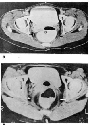
2. 직장주위지방의 증가가 35 영중 26예 (75 % )(Fig 1).
3. 천골파 직장사이의 연결 성유성결합조직의 증가 가 35 영중 20여](57 %)(Fig. 2)
4. 천골파 직장사이의 거리의 증가가 35 명중 14예 (42 % )(Fig. 3)
5. 직장백의 불규칙석인 비후가 35 명중 5예(14
%)
였마(Fig. 4) 또한 시간석 변화를 판창해 보연 (Table 3) 방사선치료후 1-2개월 사이에는 35 영중 2예를 제
A
B
Fig. 1. A. Preirradiation CT scan. B. Postirradiation CT scan, 5 months
CT scan reveals typical “Halo" appearance due to sym- metrically increased perirectal fat surrounded by thick-
ened perirectal fascia(arrows)
- 800 -