대한소화기학회지 2007;49:245-250
INTRODUCTION
Gastric adenocarcinoma is one of the most common malig- nancies in human, whereas primary gastric lymphoma is un- common, and accounts for less than 5% of primary gastric neoplasm.1,2 Moreover, the occurrence of malignant gastric lymp- homa and gastric cancer in the same patient, whether sync- hronous3-5 or metachronous is rare.6-10Little is known regarding the clinicopathologic characteristics of such cases, although Helicobacter pylori (H. pylori) is believed to play a causative
role in chronic active gastritis, peptic ulcer, gastric cancer, and primary gastric lymphoma, especially in the mucosa-associated lymphoid tissue (MALT) type.11 Here, we describe a case of metachronous early gastric cancer that followed after regression of MALT lymphoma.
CASE REPORT
A 53-year old male patient was admitted due to melena complaining of epigastric pain and weight loss in December
접수: 2007년 2월 27일, 승인: 2007년 3월 5일
연락처: 권계숙, 400-711, 인천시 중구 신흥동 3가 7-206 인하대학교병원 내시경센터
Tel: (032) 890-2548, Fax: (032) 890-2549 E-mail: [email protected]
위 점막연관림프조직 림프종 관해 후 발생한 조기 위암 1예
인하대학교 의과대학 내과학교실, 두뇌한국21사업단*, 해부병리학교실†
서동범·권계숙·박현신·이돈행*·김형길·신용운·김영수·김준미
†Metachronous Gastric MAL T L ymphoma and Early Gastric Cancer: A Case Report
Dong Beom Seo, M.D., Kye Sook Kwon, M.D., Hyun Shin Park, M.D., Don Haeng L ee, M.D.*, Hyung Gil Kim, M.D., Yong Woon Shin, M.D.,
Young Soo Kim, M.D., and J oon Mi Kim, M.D.†
Departments of Internal Medicine and Pathology†, Inha University College of Medicine, Center for Advanced Medical Education, Inha University Collage of Medicine by BK-21 Project*, Incheon, Korea
Metachronous association between gastric lymphoma and early gastric cancer is a rare event. Recent studies have suggested that a relationship exists between gastric mucosa-associated lymphoid tissue (MALT) lymphoma and gastric carcinoma although the mechanism is unknown. Herein, we report a 53-year-old man who visited to our hospital due to melena. Esophagogastroduodenoscopy (EGD) revealed a MALT lymphoma on the greater curvature of lower body. The patient received anti-Helicobacter pylori eradication therapy, followed by 6 cycles of chemotherapy and radiation therapy, and achieved complete remission 12 months after the therapy. Three years later, he revisited our hospital with epigastric pain. EGD revealed an early gastric cancer on the anterior wall of proximal antrum, nearly opposite to the previous lymphoma site, and a partial gastrectomy was performed. To the best of our knowledge, this is the first case report of metachronous MALT lymphoma and subsequent gastric carcinoma in Korea. (Korean J Gastroenterol 2007;49:245-250)
Key Words: Gastric cancer; Mucosa-associated lymphoid tissue; lymphoma; Helicobacter pylori
Correspondence to: Kye Sook Kwon, M.D.
Department of Internal Medicine, Inha University Hospital 7-206, Sinheung-dong 3-ga, Jung-gu, Incheon 400-711, Korea Tel: + 82-32-890-2548, Fax: + 82-32-890-2549
E-mail: [email protected]
246 대한소화기학회지: 제49권 제4호, 2007
2001. There was no evidence of superficial lymphadenopathy, hepatosplenomegaly or abdominal mass upon physical exami- nation. He had no history of fever and denied any history of gastric cancer in his family.
Initial laboratory studies showed hemoglobin 10.3 g/dL, LDH 443 IU/L (normal range 313-618 Iu/L). Other laboratory studi- es including liver and renal function tests and urinalysis were normal. Chest radiograph showed no abnormality. Esophago- gastroduodenoscopy (EGD) revealed a huge ulcerative lesion on the greater curvature side of lower body (Fig. 1). Pathologic evaluation of biopsy specimens revealed a diffuse large B cell lymphoma with low grade B cell MALT type lymphoma component (Fig. 2). Rapid urease test for H. pylori infection produced a positive result. Abdominal computed tomography (CT) revealed enlarged left gastric and suprapancreatic lymph nodes (Fig. 3). The bone marrow was not involved. The patient underwent H. pylori eradication (omeprazole, metronidazole,
clarithromycin) followed by 6 cycles of CHOP chemotherapy (cyclophosphamide 700 mg, doxorubicin 40 mg, vincristine 2 mg and prednisone 20 mg). After H. pylori eradication and chemotherapy, abdominal CT showed that the left gastric and suprapancreatic lymph node had decreased in size. EGD re- vealed a healing ulcer with an irregular margin and an uneven base on the greater curvature of lower body (the previous lymphoma site). Pathologic evaluation of a biopsy specimen revealed no evidence of tumorous cells or H. pylori like organisms. Thereafter, he recieved radiation therapy (3,960 cGy). In December 2002, abdominal CT revealed that the multiple small lymph nodes in the left gastric area and suprapancreatic area are disappeared. The patient was con- sidered to achieve complete remission after 12 months of therapy, and did not show up until 2005.
In April 2005, he revisited our hospital with epigastric pain.
Physical examination was non-specific and all laboratory tests were normal. Abdominal CT revealed no evidence of abnormal node enlargement, but EGD revealed a 0.8 cm sized shallow ulcer on the anterior wall of proximal antrum, nearly opposite to the previous lymphoma site, which was shown as an ulcer scar on the greater curvature of lower body (Fig. 4). Rapid urease test was negative. Biopsy of the ulcer revealed poorly differentiated adenocarcinoma with signet ring cell component.
Therefore, partial gastrectomy was performed in May 2005. The final pathologic evaluation showed early gastric cancer (type IIc) with signet ring cell component (Fig. 5). The patient was discharged without complication. There was no evidence of recurrence of either neoplasm during his subsequent visits.
DISCUSSION
Adenocarcinoma is the most common tumorous condition
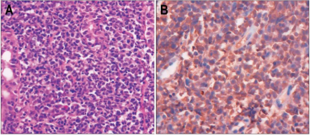
Fig. 2. Microscopic findings. (A) Biopsy specimens demonstrate in- filtration of diffuse small- to me- dium-sized lymphoid cells, with in- dented nuclei and clear cytoplasm in the lamina propria forming a lymp- hoepithelial lesion (H&E stain, × 200. (B) Immunohistochemical stain- ing for CD20 reveals positive cyto- plasmic reaction (× 400).
Fig. 1. Endoscopic finding. It shows a huge ulcerative lesion on the greater curvature side of lower body.
서동범 외 7인. 위 점막연관림프조직 림프종 관해 후 발생한 조기 위암 1예 247
arising from the stomach, and accounts for approximately 95%
of gastric neoplasms. The majority of the remaining tumors are lymphomas, and mesenchymal tumors are rare.2
MALT lymphoma was first introduced by Issacson and Wright in 1983.12 It was a low-grade primary B-cell lymphoma, which was difficult to diagnose earlier due to its variable gas- trointestinal symptoms and non-specific, variable endoscopic findings.
A few reports published over the last three decades had suggested a possible association between MALT lymphoma and gastric adenocarcinoma, and the synchronous or metachronous occurrence of these two neoplasms.3-10There had been a larger number of reports on synchronous rather than metachronous occurrences; i.e., four in Korea13-15 and more than 30 abroad.
The observation of concurrent or metachronous MALT lymp- homa and gastric adenocarcinoma is of interest as both are etiologically related to H. pylori. H. pylori infection has an established causal relationship with histological gastritis, gastric atrophy, gastric cancer, and gastric MALT lymphoma.11 More- over, H. pylori eradication has been reported to lead a complete remission of early stage gastric MALT lymphoma in about 70% of cases.16 Serological investigations have demonstrated a high prevalence of anti-H. pylori IgG antibodies within the areas associated with high incidence of gastric cancer,17 and many case controlld studies have reported 3-6 folds increased risk of gastric cancer in H. pylori positive individuals.11 Moreover, epidemiological studies and animal models have demonstrated a clear causal link between H. pylori infection
Fig. 5. Pathologic findings of resect- ed stomach. (A) The short arrow indicates the site of EGC and the long arrow indicates the site of pre- vious MALT lymphoma. (B) Micro- scopic finding of the ulcer lesion reveals a signet ring cell carcinoma confined to the mucosa (H&E stain,
× 200).
EGC, early gastric cancer; MALT, mucosa-associated lymphoid tissue.
Fig. 3. Abdominal CT finding. It shows enlarged left gastric and suprapancreatic lymph node.
Fig. 4. Endoscopic finding after three years. The short arrow in- dicates a newly developed ulcer (EGC) on the anterior wall of proximal antrum. The long arrow indicates the ulcer scar (previous MALT lymphoma) on the greater curvature of lower body.
EGC, early gastric cancer; MALT, mucosa-associated lymphoid tissue.
248 The Korean Journal of Gastroenterology: Vol. 49, No. 4, 2007
and gastric carcinogenesis.11 Ioachim and co-workers suggested the possibility of immunological containment of dysplastic epithelial cells by lymphocytic infiltrate,18 and raised the question as to whether an immunological imbalance resulting from lymphocytic infiltrate regression could contribute to the rapid development of gastric cancer. Thus, it appears that H.
pylori infection may play a significant role in the pathogenesis of both tumors.
The development of metachronous gastric adenocarcinoma following gastric MALT lymphoma has been reported in 10 patients (Table 1) overseas,6-10but has not yet been reported in Korea.
One previous case report found that H. pylori infection was associated with the pathogenesis of both tumors.10 Gastric adenocarcinoma developed with H. pylori re-infection after 17 months of remission following eradication and surgical resection of gastric MALT lymphoma. However, H. pylori infection was not found in most of other cases after subsequent adenocarcinoma development.
It was supposed that prolonged residual MALT lymphoma could constitute an additional risk factor for the development of
gastric carcinoma.9 Three patients in their report who were diagnosed as gastric MALT lymphoma received eradication and alkylating agent, and then achieved complete remission. After long-term endoscopic follow-up, three patients developed gastric adenocarcinoma at the same locations of previous MALT lymphomas and were treated by gastrectomy. Gastrectomy specimens showed residual MALT lymphoma in all three cases.9
Patients with Hodgkin's disease or nodal non-Hodgkin's lymphomas appear to be at higher risk of developing other cancers. A high incidence of other cancers has also been found in some gastric MALT lymphoma series.19 Montalban and co-workers were able to show that other cancers occur in patients with gastric MALT lymphoma. Among 136 patients with gastric MALT lymphoma, other cancers were detected in 16 patients (11.7%), either prior to the diagnosis of MALT lymphoma, concomitantly, or following diagnosis.20 Lymphoid neoplasms and gastric carcinomas develop more frequently than other malignancies in MALT lymphoma patients and are more frequent than in the normal population, but without statistical significance. Prospective study is required to validate these Table 1. Literature Review of Metachronous MALT Lymphoma and EGC
Location of H. MALT Duration† Location of H.
Authers Age/sex Stage lymphoma Type
MALT pylori* (month) gastric cancer pylori‡
treatment
Morgner et al6 74/M Antrum I E + Antibiotics 49 Same location IIa -
77/F Angle I E + Antibiotics 48 Opposite location IIc -
70/M Upper body I E + Antibiotics 60 Prepyloric anturm IIa+IIc -
Nakamura et al7 82/F Body I E + Resection 13 Antrum IIa +
Hasegawa et al8 72/F Mid body to angle I E + Antibiotics 6 Angle IIc NM
Copie-Bergman et al9 46/M Antrum I E + Antibiotics 30 Same location NM -
65/F Body II E + Alkylating agent 66 Same location NM -
and antibiotics
40/M Body IV + Alkylating agent 156 Same location NM -
and antibiotics
44/M Body IV - Alkylating agent 204 Antrum NM -
Ghoshal et al10 32/M Body I E + Antibiotics and 17 Body NM +
gastrectomy
This Case 52/M Lower body II EA + Antibiotics, CHOP 40 Proximal antrum IIc - and radiotherapy
NM, not mentioned.
* H. pylori, detected at the time of MALT lymphoma diagnosis.
†Duration, duration between the development of MALT lymphoma and EGC.
‡H. pylori, detected at the time of EGC diagnosis.
EGC, early gastric cancer; MALT mucosa-associated lymphoid tissue; H. pylori, Helicobacter pylori; CHOP, cyclophosphamide+ doxorubicin + vincristine+ prednisone.
Seo DB, et al. Metachronous Gastric MALT Lymphoma and Early Gastric Cancer: A Case Report 249
results with a large number of patients.
In conclusion, we experienced a case of early gastric cancer in a patient who had been diagnosed and treated for primary gastric MALT lymphoma. Although the association between gastric MALT lymphoma and subsequent gastric adenocarcino- ma developement has not been fully delineated, we suggest that the patient population treated for primary gastric lymphoma should be more frequently surveyed for the development of gastric adenocarcinoma, because it appears that the occurrence of gastric adenocarcinoma showed higher tendency in patients treated for primary gastric lymphoma as compared to general population.
요 약
위림프종 관해 후 위선암종이 발생하는 것은 매우 드문 일이다. 최근 위 점막연관림프조직(mucosa-associated lymp- hoid tissue, MALT) 림프종과 위선암종과의 연관성에 대한 논문들이 발표되고 있으나, 정확한 기전은 알려진 바 없다.
53세 남자 환자가 흑색변을 주소로 내원하여 시행한 상부위 내시경 검사에서 위체하부의 대만에 Helicobacter pylori (H.
pylori) 양성 MALT 림프종을 진단 받았다. 환자는 H. pylori 제균 치료 후에 항암화학요법 및 방사선요법을 시행 받았 다. 환자는 12개월 후에 완전 관해 진단을 받았다. 3년 후 환자는 상복부 통증을 주소로 다시 내원하였다. 상부위내시 경 검사에서 이전의 MALT 림프종 반대쪽인 위전정부 근위 부에 조기 위암이 발견되어 위부분절제를 시행하였다. 저자 들은 이전에 국내에서 보고된 바 없는 위 MALT 림프종 관 해 후 발생한 조기 위암 1예를 경험하였기에 문헌고찰과 더 불어 보고한다.
REFERENCES
1. Aozasa K, Tsujimoto M, Inoue A, et al. Primary gastro- intestinal lymphoma. A clinicopathologic study of 102 pa- tients. Oncology 1985;42:97-103.
2. Nakamura S, Akazawa K, Yao T, Tsuneyoshi M. A clini- copathological study of 233 cases with special reference to evaluation with the MIB-1 index. Cancer 1995;76:1313-1324.
3. Wotherspoon AC, Isaacson PG. Synchronous adenocarcinoma and low grade B-cell lymphoma of mucosa associated lymp- hoid tissue (MALT) of the stomach. Histopathology 1995;27:
325-331.
4. Chan AO, Chu KM, Yuen ST, Leung SY, Lam SK, Wong J.
Synchronous gastric adenocarcinoma and mucosa-associated
lymphoid tissue lymphoma in association with Helicobacter pylori infection: comparing reported cases between the East and West. Am J Gastroenterol 2001;96:1922-1924.
5. Kelly SM, Geraghty JM, Neale G. H pylori, gastric car- cinoma, and MALT lymphoma. Lancet 1994;343:418.
6. Morgner A, Miehlke S, Stolte M, et al. Development of early gastric cancer 4 and 5 years after complete remission of Heli- cobacter pylori associated gastric low grade marginal zone B cell lymphoma of MALT type. World J Gastroenterol 2001;
7:248-253.
7. Nakamura S, Aoyagi K, Iwanaga S, Yao T, Tsuneyoshi M, Fujishima M. Synchronous and metachronous primary gastric lymphoma and adenocarcinoma: a clinicopathological study of 12 patients. Cancer 1997;79:1077-1085.
8. Hasegawa N, Kato K, Yamada K, et al. Early-stage gastric adenocarcinoma: revealed after anti-Helicobacter pylori therapy of MALT lymphoma. Gastrointest Endosc 2001;53:495.
9. Copie-Bergman C, Locher C, Levy M, et al. Metachronous gastric MALT lymphoma and early gastric cancer: is residual lymphoma a risk factor for the development of gastric car- cinoma? Ann Oncol 2005;16:1232-1236.
10. Ghoshal UC, Guha D, Bandyopadhyay S, et al. Gastric ade- nocarcinoma in a patient re-infected with H. pylori after regression of MALT lymphoma with successful anti-H. pylori therapy and gastric resection: a case report. BMC Gastro- enterol 2002;2:6.
11. Sugiyama T, Asaka M. Helicobacter pylori infection and gastric cancer. Med Electron Microsc 2004;37:149-157.
12. Issacson P, Wright DH. Malignant lymphoma of mucosa- associated lymphoid tissue. a distinctive type of B-cell lymp- homa. Cancer 1983;52:1410-1416.
13. Min BH, Choi KD, Im JP, et al. A case of synchronous early gastric cancer and low-grade MALT lymphoma. Korean J Gastrointest Endosc 2002;25:203-207.
14. Lee KH, Kim HS, Baik SK, et al. A case of synchronous mucosa-associated lymphoid tissue lymphoma and early carci- noma of the stomach. Korean J Gastroenterol 2001;37:455-460.
15. Park SH, Rha SY, Shim DK, et al. An unusual case of gastric carcinoma with synchronous non-Hodgkin's lymp- homa. Yonsei Med J 1998;39:463-467.
16. Bayerdorffer E, Neubauer A, Rudolph B, et al. Regression of primary gastric lymphoma of mucosa associated lymphoid tis- sue type after cure of Helicobacter pylori infection. MALT Lymphoma Study Group. Lancet 1995;345:1591-1594.
17. Correa P, Fox J, Fontham E, et al. Helicobacter pylori and gastric carcinoma. Serum antibody prevalence in populations with contrasting cancer risks. Cancer 1990;66:2569-2574.
250 The Korean Journal of Gastroenterology: Vol. 49, No. 4, 2007
18. Ioachim HL, Hajdu C, Giancotti FR, Dorsett B. Lymphoid proliferations and lymphomas associated with gastric meta- plasia, dysplasia, and carcinoma. Hum Pathol 1999;30:833-842.
19. Zucca E, Pinotti G, Roggero E, et al. High incidence of other neoplasms in patients with low-grade gastric MALT lymp
homa. Ann Oncol 1995;6:726-728.
20. Montalban C, Castrillo JM, Lopez-Abente G, et al. Other cancers in patients with gastric MALT lymphoma. Leuk Lymphoma 1999;33:161-168.