I. 서론
최근 10여년의 연구 결과 신경계가 면역계 에 대하여 면역 반응을 증진 혹은 억제시키 는 조정 기능을 하고 있는 것이 밝혀졌다1, 2). 이와 관련된 신경 전달 물질인 neuropeptide 들 중에서 가장 많은 연구가 진행되었던 Substance P(SP)는 여러 자극에 의하여 지각 신경 C 섬유 말단으로부터 유리되어, 혈관 투 과성, 점액 분비, 백혈구에 대한 화학 주성, 기관지 수축과 같은 다양한 염증 및 면역 반 응을 유도한다는 사실이 보고되었다3-6). SP와 같은 neuropeptide 들이 신경계와 면역계 사이 의 연결 역할을 하는 물질임이 알려져 있다.
즉, 임파 기관에 peptide성 신경이 와 있어, 지 각 신경과 면역 세포 사이에 근접한 관계가 가능한 점7-11)과, 대식세포는 물론 T, B 임파 구의 특정 아군집에 SP수용체가 존재한다는
점1, 2, 12)도 이와 무관하지 않다. 또한 SP와 관
련된 면역 조절 기능들로는, in vitro에서 T 임파구의 증식 촉진 작용13, 14), 면역 글로불린 합성 증진 효과15, 16), 비만세포 탈과립과 histamine 분비 촉진 작용17-19), 대식세포와 호 중구 활성 효과20-23) 및 임파구 기능의 조정
작용24-27)등이 있으며, 이러한 사실들은 SP가
신경성 면역 반응의 매개 역할을 담당하고 있음을 시사하고 있다.
신경계의 면역 반응 조절은 치주질환의 진 행 양상에도 peptide성 신경이 영향을 미칠 수 있음을 의미한다. 실제 치주염과 그 발병 기전이 유사하다고 알려져 있는 류마치스성 관절염에 있어, 그 질병 진행 양상과 SP 사이 에 밀접한 상관 관계가 있음이 밝혀졌다. 즉, 흰쥐에서 실험적으로 일으킨 관절염을 SP가 더욱 악화시켰으며28), 대식세포를 활성화시켜 prostaglandin과 다른 염증성 매개체 및 IL-1 분비를 촉진시켰고20, 29, 30), 활막세포에서 PGE2와 collagenase 생산을 증가시켰다고 Lotz등31)은 보고하였다. SP의 면역 및 염증 조절기능을 파악하여 이를 치주염의 원인 규 명과 예방, 치료에 이용할 수 있는 가능성이 매우 높으나, 아직까지 이 방면에 대한 연구 가 미흡한 실정이다.
Interleukin(IL)은 면역 세포가 분비하는 수 용성 산물로서 면역 반응의 중요한 조절 인 자로 작용하는데, 이들 IL의 발현에 SP와 같 은 neuropeptide 들이 영향을 줌으로써 면역 조절 작용이 매개 될 가능성이 매우 크다. 이 러한 예로, SP는 다양한 형의 세포와 조직들 에서 IL-1, TNF-α, IL-6, IFN-γ및 MIP-1α의 대한치주과학회지 : Vol. 27, No. 4, 1997
Substance P가 T 임파구의 세포증식과 IL-2 생산에 미치는 영향
문진균·최병선·이석초·김형섭
전북대학교 치과대학 치주과학교실 전북대학교 치의학 연구소
805
분비를 유도한다고 밝혀졌다30, 32-37). 또한 SP 는 골수의 단핵세포에서 IL-3나 GM-CSF를 유도함으로써 조혈 기능을 촉진하는 작용도 가지고 있다38). 또한 SP는 사람의 T 임파구 와 T 임파구주(T cell line)의 성장을 촉진하 며14), murine splenocyte와 murine T 임파구주 인 EL-4 및 LBRM-T6G로부터 IL-2 분비를 촉진할 뿐 아니라38, 39), mitogen으로 자극된 사람의 T 임파구로부터 IL-2 분비를 유도한 다고 보고되었다40).
본 연구에서는 첫째 균질한 사람의 T 임파 구주를 이용하여, 생리적 농도의 SP가 세포 증식과 IL-2 분비로서 대표되는 T 임파구의 기능에 대하여 어떠한 영향을 미치는지 알고 자 하였고, 둘째 T 임파구의 기능 변화를 SP 가 유도(induce) 하는 것인지, 아니면 cosignal 로서 증폭 작용을 하는 것인지 판별하고저 하였으며, 셋째 만일 T 임파구가 IL-2 분비를 증진시킨다면 이것이 세포 증식에 따른 결과 로서 나타난 현상인지를 판별하고자 하였다.
이를 위하여 mitogen으로 자극한 혹은 자극하 지 않은 Jurkat과 HuT78 세포에 대하여 [3H]-thymidine 편입 실험, 및 IL-2 의존형 세포를 이용한 IL-2 bioassay를 시행함으로써, 세포 증식 속도와 IL-2 분비량에 미치는 SP의 영향을 관찰하여, 면역계 특히 세포매개 면역 반응에서의 SP의 역할과 신경성 염증 현상의 치주질환에 대한 영향을 밝히고자 하였다.
II. 실험재료 및 방법
1. 세포 배양사람의 T세포주인 Jurkat과 HuT78은 10%
fetal calf serum(FCS)이 포함된 RPMI1640 (Gibco)에 서 배 양 하 였 고 , 생 쥐 의 살 상 (cytolytic) T 세포주인 CTLL-2는 이틀에 한 번씩 동일 배양액에 100 unit/ml의 rIL-2를 첨가하면서 배양하였다. 모든 배양 조건은 37
℃, 5% CO2 하에서 시행하였다.
2. [3H]-thymidine 편입 실험에 의한 세포 성장 속도 검사
Jurkat과 HuT78 세포(1.5×10³ cell/well)를 96 microwell plate에 넣고, substance P(SP, Sigma), concanavalin A(Con A, Sigma), phetohemagglutinin(PHA, Sigma), 및 phorbol 12-myristate 13-acetate(PMA, Sigma) 등의 시약을 적정 농도 가한 후 18시간 자극하였 고, 자극 마지막 6시간 동안 [3H]-thymidine 0.5 μCi/well(sp. act. 2 Ci/mmol, New England Nuclear, Dupont)로 pulse 처리하였다. 그 후, cell harvester로 harvest하여 세포내로 편입된 [3H]-thymidine을 liquid scintillation counter (Beckman)로 측정하였다. 이상의 모든 실험 은 3선 반복 시행하였다.
3. IL-2 bioassay
IL-2 bioassay는 IL-2 의존형 세포주인 CTLL-2를 이용하여 시행하였다41). Jurkat 혹 은 HuT78 세포를 24 well plate에 1×10⁶ cell/well씩 넣어 SP, Con A, 혹은 PHA+
PMA 적당 농도를 첨가한 후 SP 단독 투여 시에는 12시간, mitogen과 SP를 동시에 처리 하였을 경우에는 3시간 배양하였다. 그 후, 각 각을 microtube에 넣고 1000 rpm으로 5분간 원심 분리하여 상청액만 IL-2 bioassay에 이 용하였다. 100 μl의 상청액을 96 microwell plate에 넣고 CTLL-2 세포를 Dulbecco's phosphate buffered saline으로 5회 세척하여 100 μl 씩 5×10³ cell/well 되도록 well에 넣고, Jurkat 및 HuT78 세포에서 얻었던 배양 상청 액을 일정량씩 첨가하여 18시간 배양하고, 마 지막 6시간 동안 0.5 μCi/well의 [3H]- thymidine으로 pulse 처리하였다. 그 후, 세포 를 cell harvester로 harvest하여 liquid
scintillation counter로 측정하였다. 모든 실험 은 3선 반복 시행하였다. 세포를 배제하여 각 실험군과 동일한 배양 혼합물을 24 well에서 병행하여 배양하고, 이를 IL-2 bioassay시에 각 대조군으로 하여 사용하였으며, 각 대조군 의 cpm값을 각 실험군 값에서 제하여 줌으로 써 CTLL-2의 성장에 PHA, PMA 혹은 SP 가 줄지도 모를 영향을 배제하였다. 또한, IL- 2 여러 농도를 CTLL-2에 가하여 [3H]- thymidine 편입 실험을 시행함으로써 표준 곡 선을 그리고, 이 좌표를 이용해 cpm 값으로 부터 IL-2 unit를 계산해 내었다. IL-2 측정의 예민도는 0.05 unit/ml 였다.
4. 통계 분석
Student's t-test에 의하여 차이에 대한 통계 적 유의성을 검정하였다.
III. 결과
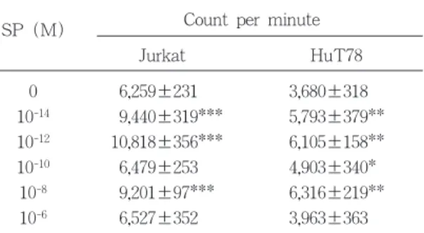
T 세포의 성장에 미치는 SP 단독의 효과를
각 농도별로 측정한 결과, Jurkat과 HuT78 모 두 SP 10-12M과 10-8M 두 농도에서 정점을 나타내는 종 형태(bell-shape)의 좌표 모양을 나타내었으나, Jurkat에 대한 SP의 효과가 통 계적으로 더 큰 유의성을 나타내었다(표 1, 그림 1).
그림 2는 CTLL-2 세포를 이용한 IL-2
807
표 1 Effect of substance P on the cell proliferation of T lymphocyte
SP (M) Count per minute
Jurkat HuT78
0 6,259±231 3,680±318
10-14 9,440±319*** 5,793±379**
10-12 10,818±356*** 6,105±158**
10-10 6,479±253 4,903±340*
10-8 9,201±97*** 6,316±219**
10-6 6,527±352 3,963±363
Mean±SD(n=3)
* : P < 0.05 when compared to control
** : P < 0.01 when compared to control
*** : P < 0.001 when compared to control
그림 1 Biphasic bell-shape dose-dependent effect of substance P on the cell proliferation of Jurkat and Hut 78
* P<0.05 when compared to control
** P<0.01 when compared to control
*** P<0.001 when compared to control
0 10-14 10-12 10-10 10-8 10-6 0 10-14 10-12 10-10 10-8 10-6 14
12 10 8 4 2 0
8 7 6 5 4 3 2 1 0
cpm×10
-3cpm×10
-3(A) Jurkat
Substance P(M) Substance P(M)
(B) Hut78
표 2 Effect of substance P alone on the IL-2 production of T lymphocyte
Mean±SD(n=3)
All data were not significant compared to control group Count per minute
SP (M)
Jurkat HuT78
0 1,830±192 1,886±163
10-14 1,874±170 1,820±178
10-12 2,078±218 1,663±218
10-10 1,675±151 1,846±176
10-8 1,722±129 1,789±97
10-6 1,764±63 1,827±120
0 1 2 4 8 16 32
cpm×10
-38 7 6 5 4 3 2 1 0
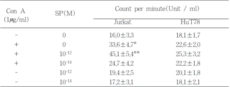
그림 2 IL-2 standard curve 표 3 Effect of substance P on the IL-2 production of con A-activated T lymphocytes
Con A SP(M) Count per minute(Unit / ml)
(1μg/ml) Jurkat HuT78
- 0 16.0±3.3 18.1±1.7
+ 0 33.6±4.7* 22.6±2.0
+ 10-12 45.1±5.4** 25.3±3.2
+ 10-14 24.7±4.2 22.2±1.8
- 10-12 19.4±2.5 20.1±1.8
- 10-14 17.2±3.1 18.1±2.1
Mean ± SD(n=3)
* : P < 0.05 when compared to control
** : P < 0.01 when compared to control
그림 3 Costimulatory effect of substance P on the IL-2 production of Con A activated Jurkat and HuT78
* P<0.05 when compared to control
** P<0.01 when compared to control
- + + + - -
0 0 10-12 10-14 10-12 10-14
- + + + - - Con A(1㎍/ml)
0 0 10-12 10-14 10-12 10-14SP(M)
IL-2(unit/ml) IL-2(unit/ml)
60 50
40 30
20 10
0
30
25 20
15 10
5 0
(A) Jurkat (B) HuT78
bioassay 표준 곡선의 대표적인 한 예이다. 0 에서 32 unit/ml 사이의 IL-2 농도에서 비교 적 정확한 농도 비례적 반응을 나타내었다. T 세포의 IL-2 분비에 미치는 SP 단독의 효과 를 각 농도 별로 측정한 결과, 어느 농도에서 도 SP 단독으로는 IL-2의 분비를 유도하지 못하였다(표 2).
반면 con A로 활성화시킨 T 세포의 IL-2 분비에 대하여 SP는 뚜렷한 효과를 나타내었 다. 즉, Jurkat 세포의 경우, con A 1μg/ml 자 극에 의하여 16.0±3.3에서 33.6±4.7 unit/ml로
IL-2 분비량이 약 2배 증가했으며(P<0.05), 여 기에 SP 10-12M의 투여시 45.1±5.4 unit/ml로 더욱 IL-2 분비량이 증가하여 통계적으로 더 욱 유의한 차이(P<0.01)를 나타내었으나, Con A 단독 효과에 비하여서는 통계적으로 유의 한 차이가 관찰되지 않았다(표 3, 그림 3A).
그러나 HuT78의 경우, 전체적인 분비 유도 양상은 Jurkat과 유사하였으나, 통계적으로 유 의한 분비량의 차이가 관찰되지 않았다(표 3, 그림 3B). Con A가 T 세포의 성장 자체에 미치는 영향을 관찰한 결과, IL-2 분비량이
809
표 4 Effect of Con A on the cell proliferation of T lymphocytes
ConA Count per minute
(μg/ml) Jurkat HuT78
0 1,095±115 1,025±92
1 3,626±51*** 889±68
Mean ± SD (n=3)
*** : P < 0.001 when compared to control
(A) Jurkat (B) HuT 78
IL-2(unit/ml) IL-2(unit/ml)
- + + + - -
0 0 10-12 10-14 10-12 10-14
- + + + - -
0 0 10-12 10-14 10-12 10-14
PHA(3 ug/ml) +
PMA(5 ng/ml) SP(M) 80
70 60 50 40 30 20 10 0
30
25
20
15
10
5
0
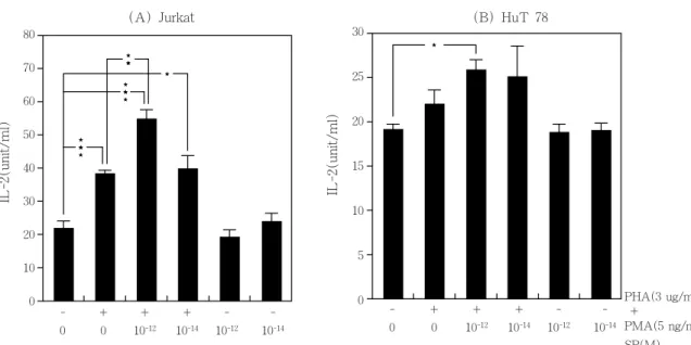
그림 4 Costimulatory effect of substance P on the IL-2 production of PHA+PMA activated Jurkat and HuT 78
* P<0.05 when compared to control
** P<0.05 when compared to PHA+PMA activated Jurkat
*** P<0.001 when compared to control
증가하였던 Jurkat에서는 3배 이상 성장을 촉 진하였고(P<0.001), HuT78에서는 오히려 약 간 감소시키는 경향을 나타내었다(표 4).
PHA와 PMA로 활성화시킨 T 세포의 IL-2 분비에 대하여 SP는 con A로 활성화시킨 T 세포에서와 비슷한 양상의 효과를 나타내었 다. 즉, Jurkat의 경우, PHA+PMA 자극으로
인해 21.3±2.4에서 38.2±1.9 unit/ml로, 약 2 배 가까운 IL-2 분비 증가를 보였다(P<0.001).
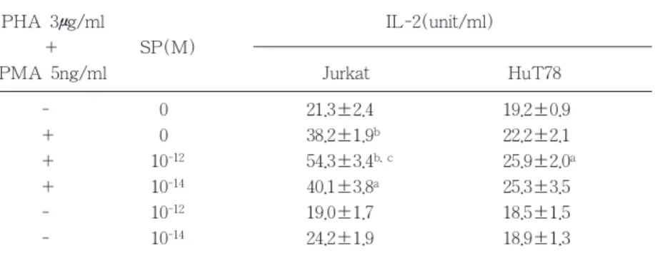
여기에 SP 10-12M을 첨가 투여함으로써, IL-2 분비량이 54.3±3.4 unit/ml로 더욱 증가 하였 으며(P<0.001), 이 값은 PHA+PMA로만 자 극한 경우와도 통계적으로 유의한 차이를 보 였고(P<0.01), SP 10-14M 투여시 이러한 SP의 표 5 Effect of substance P on the IL-2 production of PHA+PMA activated T lymphocyte
PHA 3μg/ml IL-2(unit/ml)
+ SP(M)
PMA 5ng/ml Jurkat HuT78
- 0 21.3±2.4 19.2±0.9
+ 0 38.2±1.9b 22.2±2.1
+ 10-12 54.3±3.4b, c 25.9±2.0a
+ 10-14 40.1±3.8a 25.3±3.5
- 10-12 19.0±1.7 18.5±1.5
- 10-14 24.2±1.9 18.9±1.3
Mean±SD(n=3)
a : P < 0.05 when compared to control b : P < 0.001 when compared to control
c : P < 0.05 when compared to PHA+PMA activated Jurkat
표 6 Effect of PHA+PMA on the cell proliferation of T lymphocytes
Mitogen Count per minute
Jurkat HuT78
PHA 0 1,966±211 1,414±83
(μg/ml) 3 981±138* 1,403±115
0 995±92 3,035±182
PMA 3 4,919±359*** 2,924±133
(ng/ml) 5 4,714±278*** 1,072±88***
10 980±110 872±81***
PHA 0, 0 1,101±109 4,215±288
+ 3, 5 7,963±412*** 2,574±194**
PMA 3, 10 1,718±202* 1,182±47***
Mean±SD(n=3)
* : P < 0.05 when compared to control
** : P < 0.01 when compared to control
*** : P < 0.001 when compared to control
costimulation 효과가 나타나지 않았다. HuT78 에서도 전체적인 양상은 Jurkat과 비슷하였으 나, PHA+PMA 자극 및 SP 10-12M을 투여한 군에서만 통계적으로 유의한 차이의 IL-2 분 비 증가를 나타내었고(P<0.05), PHA+PMA 자극만 한 경우에 비하여 SP 10-12M 첨가 투 여로 인한 IL-2 분비의 증가량은 통계적 유의 성을 나타내지 않았다(표 5, 그림 4).
표 6은 본 실험에서 사용한 PHA와 PMA 농도가 T 세포의 성장 속도에 미친 영향을 나타낸 것으로서, PHA 3μg/ml은 Jurkat의 성 장 속도를 반 이상 감소 시켰으며, HuT78은 변화가 없었다. PMA는 3, 5 ng/ml 농도에서 Jurkat의 성장 속도를 5배 가까이 증가시켰으 나 HuT78에서는 용량에 비례하여 감소되었 다. PHA 3 μg/ml과 PMA 5 ng/ml 농도에서 IL-2 분비의 큰 증가를 보였던 Jurkat은 1101
±109에서 7936±412 cpm으로 거의 8배 가까 운 성장 속도 상승을 보였으나(P<0.001), 그렇 지 못했던 HuT78은 4215±288에서 2574±194 cpm으로 오히려 성장 속도가 감소하였다 (P<0.01). Con A 자극의 경우와 매우 유사한 결과를 보였으며, 따라서 IL-2 분비량과 세포 의 성장 속도 간에는 연관이 있는 것으로 나 타났다.
IV. 고찰
본 연구의 결과 SP는 그 자체로서 사람의 T 세포주의 성장 속도를 증가시켰으며, mitogen과 동시에 작용하여 T 세포주의 IL-2 분비를 증진 시킴으로써, T 세포의 기능적 변 화를 유도하여 면역계를 조정할 수 있는 것 으로 나타났다. 이미 SP와 같은 tachykinin이 T 세포 증식을 유도하고, 어떤 T 세포 아군 집은 SP에 특이적인 수용체를 발현하는 것으 로 보고 되었으며42, 13, 14), SP의 성장 촉진 작 용은 IL-2와 같은 T 세포 성장 인자를 SP가 유도함으로써 매개되는 것으로 생각되어졌다.
실제 SP는 murine T 세포주와 비세포에서 IL-2의 분비를 증진 시켰다고 Rameshwar 등
38, 39)은 보고하였고, Nio 등46)은 항원이나 con
A로 자극된 T 세포주에서 SP가 IL-2 분비를 증가시켰다고 하였으며, Calvo 등40)은 PHA와 PMA로 자극된 사람의 T 세포주와 말초 혈 액 T 세포에서 SP가 IL-2 mRNA 발현을 증 가시켰다고 보고하였다. Scicchitano 등16)의 보고에 의하면, in vivo로 SP를 지속적으로 투여했을 때, murine Peyer's patch 임파구의 세포 증식이 증가되었을 뿐 아니라, 면역 글 로불린의 합성도 증진되었다고 하였다.
본 실험의 결과 T 세포의 성장에 대하여 생리적 농도 범위43, 44)에서의 SP는 최대 용량 에서 오히려 성장의 촉진 효과가 감소되는 형태를 보여 용량에 따라 2개의 정점을 가진 종 모양(bell-shape)의 곡선을 나타내었다. 종 모양(bell-shape)의 용량 반응 곡선은 이미 다 른 여러 model에서 SP에 의해 처리된 세포에 서 기술되었던 형태의 좌표이다. 즉, mitogen 으로 자극된 T 세포의 성장14, 45), 말초 혈액 단핵구에서 IFN-γ의 분비37), T 세포의 IL-2 mRNA 발현40), splenocyte의 IL-2 분비38), T 세포주의 IL-2 분비46)에 대하여 SP는 일원적 (monophasic), 또는 이원적(biphasic) 형태의 bell 모양 용량 반응 곡선을 나타내었다. 또한 본 연구에서 세포 성장 촉진 효과의 peak를 보였던 SP의 농도는 각각 10-12과 10-8M로서, 동일 세포주에서 IL-2 mRNA 발현에 대한 효과를 분석하였던 Calvo 등40)의 보고에서도 동일한 농도에서 peak가 나타났다고 하였다.
고농도에서 효과가 감소하는 것이 세포 독성 때문인지 아닌지는 아직은 확실히 밝혀져 있 지 않지만, SP수용체의 탈감작, 혹은 감소와 관련 가능성 있는 기전으로 추측되고 있다.
최근의 연구 보고에 의하면 10-6M의 SP를 적 용하였을 때 IM-9 lymphoblastoid 세포주의 SP수용체 숫자가 감소하였고47), 여러 연구에 서 SP수용체에 대한 ligand의 결합에 있어
811
desensitization 현상이 나타난다는 사실48)이 이러한 가설을 뒷받침 하고 있다.
SP 자체의 IL-2 분비에 대한 본 실험의 연 구 결과, 세포 증식에 관한 결과와는 달리, SP가 T 세포주의 IL-2 분비량에 아무런 영 향도 나타내지 못하였다. 그러나 con A, 혹은 PHA와 PMA 같 은 mitogen과 SP가 costimulator로써 T 세포주의 IL-2 분비를 촉 진하는 것으로 관찰되었다. PHA와 PMA를 Jurkat, 혹은 HuT78에 적용하였을 때, IL-2 mRNA 발현의 kinetics에서 보면 4 내지 6 시 간에 peak를 나타낸다고 알려져 있으므로40), mitogen에 의한 IL-2 분비가 시작될 초기인 3 시간 만에 T 세포주에서 분비된 IL-2의 양을 측정함으로써 mitogen 단독 작용과, mitogen 및 SP의 cosignal 효과를 차별되게 관찰할 수 있었다. SP의 mitogen에 대한 cosignal 효과가 SP에 의한 세포 증식 때문에 나타난 것이 아 니다는 것은, SP 단독으로 IL-2 분비 촉진이 일어나지 않은 것으로부터 확실히 설명할 수 있다. Con A나 PHA+PMA가 T 세포 성장 을 크게 증진시켰던 것은 사실이나, 본 실험 에서는 Jurkat과 HuT78을 3 시간만 mitogen 혹은 SP를 처리하여 IL-2 bioassay를 하였기 때문에, 배가 시간이 16-24 시간에 이르는 이 들 세포주들에 있어 각 군간에 세포 수의 차 이는 매우 미미할 것으로 보이므로, 세포 수 가 IL-2 분비량의 차이에 미치는 영향은 극히 적을 것으로 사료된다. 또한 본 실험에 사용 된 농도의 mitogen 처리시 Jurkat과는 달리 HuT78에서는 세포 증식 속도에서 변화가 거 의 없거나 오히려 감소되는 양상을 나타내었 다. 그러나 세포 수 자체가 IL-2 bioassay에서 는 영향을 줄 수 없었다는 앞에서와 같은 이 유로, IL-2 분비 촉진 효과가 비교적 적게 나 타난 것에 대한 설명으로서 세포 증식 감소 효과를 들 수 없다고 본다. 오히려 반대로, 분 비된 IL-2가 결과적으로 세포의 성장에 영향 을 미쳤으리라고 판단된다. 따라서 IL-2 분비
증가의 폭이 컸던 Jurkat의 성장 속도 증가가 크게 나타났던 것으로 보인다. HuT78 세포주 는 생산된 IL-2 중, 많은 양이 분비되지 않고 세포막에 존재하는 것으로 알려져 있으므로, 이 때문에 전체적인 반응 양상은 비슷하지만 Jurkat에 비하여 HuT78에 있어 IL-2 분비가 현저히 미약했다고 볼 수도 있다. T 세포의 IL-2 분비에 있어 SP가 inducer가 아닌 cosignal로서 작용하리라는 가설에는 여러 일 치된 보고가 있다. Rameshwar 등39)은 EL-4 및 LBRM-T6G와 같은 murine T 세포주에 최적 이하, 혹은 이상 농도의 PMA와 SP를 동시에 처리하였을 때, IL-2 분비가 증가 되 었다고 하였고, Calvo 등40)은 말초 혈액 T 세 포와 Jurkat, HuT78 세포주의 IL-2 mRNA 발현에 대하여 PHA 및 PMA와 함께 SP가 cosignal로 서 작 용 한 다 고 하 였 다 . 또 한 Rameshwar 등38)은 murine splenocyte에서 con A 혹은 anti-CD3 항체 등과 함께 SP가 cosignal로 작용하여 IL-2 분비를 증진시킨다 고 보고하였다. 사람 말초 혈액의 T 세포 및 T 세포주의 SP수용체는 mitogen 처리로 인 하여 그 수가 현저하게 증가한다는 사실42)은 SP의 cosignal 증폭 효과를 설명할 수 있는 가능한 기전이다. Mitogen 처리 후 이를 완전 히 제거하여 mitogen 자체에 의한 IL-2 mRNA 발현 효과가 사라진 상황에서 SP를 단독처리 하였을 때, SP 투여로 IL-2 mRNA 발현이 일어났던 사실40)도 SP수용체에 대한 이러한 가설과 유관한 결과라고 추측된다.
SP의 고친화도 수용체는 NK-1 수용체로서 G-protein coupled receptor family에 속한다49-
52). 임파구의 SP수용체 역시 NK-1형 수용체 이며 IL-2 생산도 이 수용체를 매개로 해서
일어난다1, 2). IL-2 생산에서 peak를 보이는
SP의 농도가 10-12, 10-8M 둘 인것은 고친화 도, 및 저친화도의 두가지 종류 SP수용체가 T 세포에 존재하기 때문인 것으로 추측되지 만, 아직 SP수용체가 완전히 cloning 되지 않
은 단계이므로 속단하기는 이르다.
여러 조직에서 SP의 세포내 신호 전달 경 로에 phosphatidylinositol 가수분해가 관련되어 있다고 보고 되었다53-55). Mitogen에 의하여 T 세포가 활성화되어 IL-2 생산이 일어나는데 는, 적어도 두가지 이상의 서로 다른 세포내 신호 전달 경로를 밟는 것으로 알려져 있다.
즉, tyrosine kinases와 phosphatidylinositol 가수 분해의 활성화가 그것이다56-58). 따라서 SP는 T 세포의 IL-2 생산과 활성화에 필요한 세포 내 신호 전달 경로 활성화에 기여함으로써, 면역 체계에서 조정 역할을 수행하는 것으로 추측된다. 이러한 가설을 확인하기 위하여 SP의 세포내 신호 전달 체계에 대한 분자 수 준의 연구가 앞으로 진행되어야 할 것으로 사료된다.
V. 결론
T 세포매개 면역반응에서의 Substance P(SP)의 역할을 규명하기 위하여, mitogen으 로 자극한 혹은 자극하지 않은 T 임파구주, Jurkat과 HuT78 세포에 대하여 [3H]- thymidine 편입 실험, IL-2 의존형 세포를 이 용한 IL-2 bioassay를 시행함으로써, T 임파구 주의 세포 증식 속도와 IL-2 생산에 미치는 SP의 영향을 관찰하여 다음의 결과를 얻었다.
1. SP는 T 임파구주의 세포 증식 속도를 증가시켰으며, 10-12M과 10-8M, 두가지 농 도에서 정점을 보이는 2원적 종 형태 (bell-shape)의 용량 반응 곡선을 나타내 었으나, SP 단독으로는 IL-2 분비를 증 가시키지 못하였다.
2. Con A로 자극한 T 임파구주의 IL-2 분 비에 대하여 10-12M의 SP는 costimulation 효과를 보였으나, 차이의 통계적 유의성 은 관찰되지 않았다.
3. Con A 투여시 IL-2 분비 촉진 효과가
크게 나타났던 Jurkat에서만, 세포 증식 속도가 Con A 로 인하여 3배 이상 증가 하였다(P<0.001).
4. PHA와 PMA로 자극한 T 임파구주의 IL-2 분비에 대하여 10-12M의 SP는 costimulation 효과를 보였으나, Jurkat에 서만 차이의 통계적 유의성이 관찰되었 다(P<0.01).
5. PHA와 PMA 투여시에도 Jurkat에서는 8배의 세포 증식 속도 상승을 나타내었 으나(P<0.001), HuT78에서는 오히려 감 소되었다(P<0.01).
이상의 결과로부터, SP는 T 임파구주의 세 포 증식 속도를 증가시키며, mitogen에 의한 IL-2 분비 유도를 costimulation 함으로써, T 임파구의 활성을 증가시킨다고 결론지을 수 있었다.
VI. 참고문헌
1. Payan, D. G., McGillis, J. P., and Organist, M. L. : Binding characteristics and affinity labeling of protein constituents of the human IM-9 lymphoblast receptor for substance P. J.
Biol. Chem., 261 : 14321, 1986.
2. Bost, K. L. : Hormone and neuropeptide receptors on mononuclear leukocytes.
Prog. Allergy, 43 : 68, 1988.
3. Pernow, B. : Substance P. Pharmacol.
Rev., 35 : 85, 1983.
4. Lundberg, J. M., Saria, A., Brodin, E., Rosell, S., and Folkers, K, : A substance P antagonist inhibits vagally induced increase in vascular permeability and bronchial smooth muscle contraction in the guinea pig. Proc. Nat. Acad. Sci.
USA, 80 : 1120, 1983.
813
5. Gashi, A. A., Borson, D. B., Finkbeiner, W. E., and Basbaum, C. B. : Neuropeptides degranulate serous cells of ferret tracheal glands. Am. J. Physiol., 251 : 223, 1986.
6. Goetzl, E. J., Adelman, D. C., and Sreedharan, S. P. : Neuroimmunology.
Adv. Immunol., 48 : 161, 1990.
7. Weihe, E., Muller, S., Fink, T., and Zentel, H. J. : Tachykinins, calcitonin- gene related peptide and neuropeptide Y in nerves of the mammalian thymus : interactions with mast cells in autonomic and sensory neuroimmunomodulation ?.
Neurosci. Lett., 100 : 77, 1989.
8. Weihe, E., Nohr, D., Michel, S., Muller, S., Zentel, H. J., Fink, T., and Krekel, J.
: Molecular anatomy of the neuro- immune connection. Intern. J. Neurosci., 59 : 1, 1991.
9. Bellinger, D. L., Lorton, D., Romano, T.
D., Oischowka, J. A., Felten, S. Y., and Felten, D. L. : Neuropeptide innervation of lymphoid organs. Ann. NY Acad.
Sci., 254 : 17, 1990.
10. Kurkowski, R., Kummer, W., and Heym, C. : Substance P immunoreactive nerve fibers in tracheobronchial lymph nodes of the guinea pig : origin, ultrastructure and coexistence with other peptides. Peptides, 11 : 13, 1990.
11. Zentel, H. J., Nohr, D., Albrecht, R., Jeurissen, S. H. M., Vainio, O., and Weihe, E. : Peptidergic innervation of the bursa fibrichii : interrelation with T-lymphocyte subsets. Int. J. Neurosci., 59 : 177, 1991.
12. Stanisz, M. A., Scicchitano, R., Payan, D., and Bienenstock, J. : In vitro studies
of immunoregulation by substance P and somatostatin. Ann. NY Acad. Sci., 496 : 217, 1987.
13. Payan, D. G., and Goetzl, E. J. : Modulation of lymphocyte function by sensory neuropeptides. J. Immunol., 135 : 783, 1985.
14. Payan, D. G., Brewster, D. R., and Goetzl, F. J. : Specific stimulation of human T lymphocytes by substance P.
J. Immunol., 131 : 1613, 1983.
15. Stanisz, A. M., Befus, D., and Bienenstock, J. : Differential effects of vasoactive intestinal peptides, substance P, and somatostatin on immunoglobulin synthesis and proliferation by lymphocytes from Peyer's patches, mesenteric lymph nodes, and spleen. J.
Immunol., 136 : 152, 1986.
16. Scicchitano, R., Bienenstock, J., and Stanisz, A. M. : In vivo immunomodulation by the neuropeptide substance P. Immunology, 63 : 733, 1988.
17. Shanahan, F., Denburg, J. A., Fox, J., Bienenstock, J., and Befus, D. : Mast cell heterogeneity : effects of neuroenteric peptides on histamine release. J. Immunol., 135 : 1331, 1985.
18. Mazurek, N., Pecht, I., Teichberg, V. I., Blumberg, S. : The role of N-terminal tetrapeptide in the histamine releasing action of substance P. Neuropharmacology, 20 : 1025, 1981.
19. Fewtrell, C. M. S., Foreman, J. C., Jordan, C. C., OEhme, P., Renner, H., and Stewan, J. M. : The effects of substance P on histamine and 5- hydroxytryptamine release in the rat. J.
Physiol., 330 : 393, 1982.
20. Hartung, H., Wolters, K., and Toyka, K.
V. : Substance P binding properties and studies on cellular responses in guinea pig macrophages. J. Immunol., 136 : 3856, 1986.
21. Bar-Shavit, Z., Goldman, R., Stabinsky, Y., Gottlieb, P., Fridkin, M., Teichberg, V. I., and Blumberg, S. : Enhancement of phagocytosis. A newly found activity of substance P residing in N-terminal tetrapeptide sequence. Biochem. Biophys.
Res. Commun., 94 : 1445, 1980.
22. Wozniak, A., McLennan, G., Betts, W.
H., Murphy, G. A., and Scicchitano, R.
: Activation of human neutrophils by substance P : effect of FMLP- stimulated oxydative and arachidonic metabolism on antibody-dependent cell- mediated cytotoxicity. Immunology, 68 : 359, 1989.
23. Serra, M. C., Bazzoni, F., Della Bianca, V., Greskowiak, M., and Rossi, F. : Activation of human neutrophils by substance P. J. Immunol., 141 : 2118, 1988.
24. Perianin, A., Snyderman, R., and Malfroy, B. : Substance P primes human neutrophil activation : a mechanism for neurological regulation of inflammation. Biochem. Biophys. Res.
Commun., 161 : 520, 1989.
25. Marasco, W. A. H., Showell, J., and Becker, E. L. : Substance P binds to the formylpeptide chemotaxis receptor on the rabbit neutrophil. Biochem.
Biophys. Res. Commun., 99 : 1065, 1981.
26. Matsuda, H., Kawakita, K., Kiso, Y., Nakano, T., and Kitamura, Y. :
Substance P induces granulocyte infiltration through degranulation of mast cells. J. Immunol., 142 : 927, 1989.
27. Moore, J. C., Lami, J. L., and Spruck, C. H. : Substance P increases lymphocytes traffic and lymph flow through peripheral lymph nodes of sheep. Immunology, 67 : 109, 1989.
28. Levine, J. D., Clark, R., Devor, M., Helms, C., Moskowitz, M. A., and Busbaum, A. I. : Intraneuronal substance P contributes to severity of experimental arthritis. Science, 226 : 547, 1984.
29. Hartung, H. P., and Toyka, K. V. : Activation of macrophages by substance P induction of oxidative burst and thromboxane release. Eur. J. Pharmacol., 89 : 301, 1983.
30. Kimball, E., Persico, F., and Vaught, J.
L. : Substance P, neurokinin A, and neurokinin B induce generation of IL-1- like activity in P388D1 cells. J.
Immunol., 141 : 3564, 1988.
31. Lotz, M., Carson, D. A., and Vaughan, J. H. : Substance P activation of rheumatoid synoviocytes : neural pathway in pathogenesis of arthritis.
Science, 235 : 893, 1987.
32. Lee, J. Y., Kim, H. S. : Effects of Substance P on the Release of Cytokines from Immune Cell Lines. J.
Korean Academy Periodontol., 1997. (in press)
33. Okamoto, Y., Shirotori, K., Kudo, K., Ishikawa, K., Ito, E., Togawa, K., and Saito, I. : Cytokine expression after the topical administration of substance P to human nasal mucosa. The role of
815
substance P in nasal allergy. J.
Immunol., 151 : 4391, 1993.
34. Hart, R., Dancygier, H., Wagner, F., Lersch, C., and Classen, M. : Effect of substance P on immunoglobulin and interferon-gamma secretion by cultured human duodenal mucosa. Immunol.
Lett., 23 : 199, 1989/1990.
35. Laurenzi, M. A., Persson, M. A. A., Dalsgaard, C., and Hoegerstrand, A. : The neuropeptide substance P stimulates production of IL-1 in human blood monocytes : activated cells are preferentially influenced by the neuropeptide. Scand. J. Immunol., 31 : 529, 1990.
36. Lotz, M., Vaughan, J. H., and Carson, D. A. : Effect of neuropeptides on the production of inflammatory cytokines by human monocytes. Science, 241 : 1218, 1988.
37. Wagner, F., Fink, R., Hart, R., and Dancygier, H. : Substance P enhances interferon-γ production by human peripheral blood mononuclear cells.
Regul. Pept., 19 : 355, 1987.
38. Rameshwar, P., Garea, D., and Gascon, D. : In vitro stimulatory effect of hematopoiesis. Blood, 81 : 391, 1993.
39. Rameshwar, P., Gascon, P., and Garea, D. : Immunoregulatory effects of neuropeptides : stimulation of interleukin 2 production by substance P. J.
Neuroimmunol., 37 : 65, 1992.
40. Calvo, C. F., Chavanel, G., and Senik, A. : Substance P enhances IL-2 expression in activated human T cells. J.
Immunol., 148 : 3498, 1992.
41. Gillis, S., and Smith, K. A. : Long term
culture of tumour-specific cytotoxic T cells. Nature, 268 : 154, 1977.
42. Payan, D. G., Brewster, D. R., Missirian- Bastian, A., and Goetzl, E. J. : Substance P recognition by a subset of human T lymphocytes. J. Clin. Invest., 14 : 1532, 1984.
43. Skrabanek, P., Cannon, D., Kirrane, J., Legge, D., and Powell, D. : Circulating immunoreactive substance P in man.
Irish J. Med. Sci., 145 : 399, 1976.
44. Pearse, A. G. E., Polak, J. M., and Bloom, S. R. : The newer gut hormones. Gastroenterology, 72 : 746, 1977.
45. Paegelow, I., and Werner, H. : Immunomodulation by some oligopeptides. Methods Find. Exp. Clin.
Pharmacol., 8 : 91, 1986.
46. Nio, D. A., Moylan, R. N., and Roche, J.
K. : Modulation of T lymphocyte function by neuropeptides : evidence for their role as local immunoregulatory elements. J. Immunol., 150 : 5281, 1993.
47. Parnet, P., Payan, D. G., Kerdelhue, B., and Mitsuhashi, M. : Neuroendocrine interaction on lymphocyte : testosterone-induced modulation of the lymphocyte substance P receptor. J.
Neuroimmunol., 28 : 185, 1990.
48. Harada, Y., Takahashi, T., Kuno, M., Nakayama, K., Masu, Y., and Nakanishi, S. : Expression of two different tachykinin receptors in Xenopus oocytes by exogenous mRNAs. J.
Neurosci., 10 : 3265, 1987.
49. Shigemoto, R., Yokota, Y., Tsuchida, K., and Nakanishi, S. : Cloning and expression of a rat neuromedin K
receptor cDNA. J. Biol. Chem., 265 : 623, 1990.
50. Yokota, Y., Sasai, Y., Tanaka, K., Fujiwara, T., Tsuchida, K., Shigemoto, R., Kakizuka, A., Ohkubo, H., and Nakanishi, S. : Molecular characterization of a functional cDNA for rat substance P receptor. J. Biol.
Chem., 264 : 17649, 1989.
51. Nakanishi, S. : Substance P precursor and kininogen : their structures, gene organizations, and regulation. Physiol.
Rev., 67 : 1117, 1987.
52. Takeda, Y., Chou, K. B., Sachais, B. S., Krause, J. E. : Molecular cloning, structural characterization and functional expression of the human substance P receptor. Biochem. Biophys. Res.
Commun., 179 : 1232, 1991.
53. Maggio, J. E. : Tachykinins. Annu.
Rev. Neurosci., 11 : 13, 1988.
54. MacDonald, S. G., and Boyd, N. D. :
Regulation of substance P receptor affinity by guanine nucleotide binding proteins. J. Neurochem., 53 : 264, 1989.
55. McGillis, J. P., Mitsuhashi, M., and Payan, D. G. : Immunomodulation by tachykinin neuropeptides. Ann. NY Acad. Sci., 294 : 85, 1990.
56. Gallagher, R. B., and Cambier, J. C. : Signal transmission pathways and lymphocyte function. Immunol. Today, 11 : 187, 1990.
57. Stanley, J. B., Gorczynski, R. D., Huang, C. K., Love, J., and Mills, G. B. : Tyrosine phosphorylation is an obligatory event in IL-2 secretion. J. Immunol., 145 : 2189, 1990.
58. Desai, D. M., Newton, M. E., Kadlacek, T., and Weiss, A. : Stimulation of the phosphatidyl-inositol pathway can induce T cell activation. Science, 243 : 355, 1990.
817
-Abstract-
Effects of Substance P on the Cell Proliferation and IL-2 Production of T Lymphocyte
Jin-Kyun Moon, Byung-Son Choi, Seok-Cho Lee, Hyung-Seop Kim Department of Periodontology, College of Dentistry, Chon-buk National University
Immune responses of periodontal tissue may be regulated by products of sensory afferent nerve endings such as neuropeptides. Substance P(SP), a tachykinin neuropeptide, has been previously reported to stimulate the activities of T lymphocyte. Therefore, I examined the role of SP in IL- 2 production and cell proliferation by using a homogeneous line of T lymphocytes(Jurkat and HuT78). Cell proliferation rate was determined by [3H]-thymidine incorporation test, and IL-2 was quantitated by the growth rate of CD4+ IL-2-dependent T lymphocyte line CTLL-2.
SP stimulated cell proliferation of T lymphocytes at the concentration of 10-12 and 10-8M in a biphasic bell-shape dose-dependent manner. However, SP alone did not induce IL-2 release at the concentration range of 10-6 to 10-14M. The upregulation of IL-2 release was observed when 10-12M SP was applied together with mitogens such as Con A or PHA+PMA on T cell lines, especially on Jurkat. Con A or PHA+PMA demonstrated to increase the rate of cell proliferation of Jurkat, which had shown to produce much amount of IL-2 indicating that mitogen-induced cell proliferation might be partially influenced by released IL-2. It was concluded that regulatory effects of SP on the immune/inflammatory response could be mediated through the costimulatory upregulation of IL-2 production and increase of cell proliferation of T lymphocyte.