I. 서 론
신경계가 면역 및 염증반응을 조정하며, substance P(SP)등과 같은 neuropeptide가 신 경성 염증반응(neurogenic inflammation)의 매 개체 역할을 한다는 것이 알려지기 시작했다.
Bost와 Stead1~3) 등의 연구에 의하면 면역세 포들에 neuropeptide의 수용체가 존재하며, lymphoid organ에 peptide성 신경이 분포되어 있어서 지각신경 말단과 면역세포 간에 긴밀 한 접촉이 이루어져 있다는 사실들은 이들 neuropeptide들이 면역체계와 신경계의 연결 기능을 하고 있음을 시사하고 있다.
치주질환의 발생에 있어 치태의 존재가 필 수적인 조건이기는 하나, 치주질환의 병인론 이 다양하게 나타나는 현상에 대하여는 충분 한 조건이 되지 못한다. 따라서 숙주반응의 본질적인 차이점들에서 치주질환 병인론의 다양성에 대한 원인을 찾는 것이 좋은 것으 로 사료된다. 동일한 염증 자극에 대하여 개 체마다 상이한 반응을 나타내는 중요한 이유 중 하나일 것으로 추측되는 것으로서, 면역체 계에 대한 지각신경계의 조정역할을 들 수 있다. Bartold4) 등이 언급한 치주조직의 혈관 주위 및 염증세포 침윤부위에 지각섬유의 신
경전달물질로 알려져 있는 neuropeptide인 SP 가 다량 분포되어 있으며, 치은섬유아세포의 성장을 SP가 촉진한다는 사실들이 이에 대한 간 접 적 인 증 거 이 다 . 또 한 Freeman과 Green5~8) 등에 의한 stress가 치주조직에 변화 를 초래하고 치주질환의 정도와도 깊은 상관 관계가 있으며, 실험동물에서는 골조직의 변 화와도 관련이 있다는 보고와도 이러한 사실 과 무관하지 않다. 치주질환과 유사한 질병 진행양상을 보이는 류마치스성 관절염에서 SP의 역할에 관한 연구 결과는, 치주질환에 서도 이 neuropeptide가 관련되어 있으리라는 추론을 더욱 강하게 한다. 즉, Levine9) 등은 흰쥐에서 실험적으로 일으킨 관절염을 SP가 더욱 악화시켰다고 하였고, Kimball 등10~12)은 대식세포를 활성화시켜 prostaglandin(PG)과 다른 염증 매개체, 및 IL-1 유리를 촉진 시켰 으며 Lotz 등13)은 synoviocyte에서 PGE2와 collagenase 생산을 증가 시켰다고 보고하였다.
SP는 dorsal root ganglia에서 합성되어, 지 각신경의 C fiber에 존재하면서 동통과 관련 된 신경전달물질 기능을 하기도 하며14~16)한 편으로는 말초의 신경말단으로 이동하여, 역 방향성(antidromic) 유해성 자극이 있을 때 유리되어 신경성 염증반응을 일으키기도 한 대한치주과학회지 : Vol. 27, No. 2, 1997
면역세포의 cytokine 유리에 미치는 substance P의 영향
이진용·김수아·서석란·김형섭 전북대학교 치과대학 치주과학교실
전북대학교 치의학 연구소
다17~19). 유리된 SP는 동통, 평활근 수축, 혈 관 이완 및 혈관 투과성 증가 등 국소 염증반 응에 기여하는 여러 작용을 하며, 시험관 내 에서 T임파구의 세포성장을 촉진하고20~21) B 임파구의 분화와 면역글로불린 합성을 유도
하며22~24) 비만세포의 탈과립과 histamine 분
비 를 유 발 시 키 고25~26) 다 형 핵 백 혈 구 의 respiratory burst, exocytosis, antibody- dependent cell-mediated cytotoxicity 및 priming 을 촉진한다고 보고되었다27~32).
또한 SP는 면역계에서 각종 cytokine의 분 비를 조정하는 것으로 최근 알려졌다. 즉, 단 핵구/대식세포에서 IL-133~36), IL-6,35) 및 TNF-α35~36)의 분비를 유발시켰고, 사람의 T 임파구 및 T 세포클론에서 IL-237~38)를, mitogen으로 자극한 말초혈액 단핵세포와 항 원으로 자극한 murine 비장세포에서 IFN-γ39-
40)를, synoviocyte에서 GM-CSF41)를, 다형핵백 혈구에서 IL-842)을 각각 분비 유도하였다고 보고 되었다.
SP의 작용은 대상세포에 따라 매우 상이하 다. Lim33)과 Fewtrell 등43)에 의하면 비만 세 포의 histamine분비 및, 다형핵백혈구의 활성 화와 같은 작용들은 micromole 단위를 필요로 한다고 하였는데 Bartold4)과 Calvo 등38)은 대 식세포, 임파구 및 섬유아세포의 활성에는 nanomole 내지 picomole 단위의 저농도를 필요 로 한다고 했으며 Mantyh44)은 동시에 존재하 는 다른 화학적 signal의 종류가 무엇이냐에 따라 동일 세포일지라도 촉진, 혹은 억제의 전 혀 반대되는 효과를 나타내기도 한다고 하였 다. 따라서 SP는 다른 mitogen, 매개체, 혹은 cytokine들 과 동 시 에 작 용 했 을 때 , 즉 costimulator로 작용하였을 때의 신호전달체계 가 경우에 따라 매우 상이한 결과를 나타내므 로, 치주염과 같이 주기적이고 episodic한 염증 반응을 나타내는 질환에서는 상황에 따라 이 화성(catabolic)과 동화성(anabolic) 의 양쪽 기 능을 모두 나타낼 수 있으리라 추측된다.
본 실험에서는 균질적(homogeneous)인 각 종 면역세포주를 이용하여, 이들 세포주의 cytokine 분비에 미치는 SP의 cosignal로서의 영향을 관찰하여 SP의 면역 조절기전을 밝힘 으로서, 향후 치주질환의 진단, 예방 및 치료 에 응용하고자 하였다.
II. 실험재료 및 방법
1. 세포배양본 실험에 사용한 세포주의 종류는 단핵구/
대식세포주인 U937, RAW264.7 및 WR19m.1 과 T임파구세포주인 CTLL-R8, HuT78, CCRF-CEM 및 Jurkat, B 임파구세포주인 Daudi, A20, SKW6.4, Namulwa 및 Raji, erythroid/promyeloid 세포주인 K562, KG1a, 및 HL-60등 이었다. 이중 RAW264.7과 WR19m.1은 10% fetal calf serum(FCS)이 포 함된 Dulbecco's modified Eagle medium (DMEM, Gibco)에서 배양하였고, KG1a는 20% FCS가 포함된 Iscove's medium(Gibco) 에서 배양하였으며, 나머지 세포주들은 모두 10% FCS가 포함된 RPMI1640(Gibco)에서 배 양하였다. 모든 경우, 5% CO2, 100% 습도 및 37℃ 온도의 동일한 조건으로 배양하였다.
2. 세포자극 및 배양상청액 시료 준비
각 세포주를 Dulbecco's phosphate buffered saline(DPBS, Gibco)으로 세척하여 FCS가 포 함 되 지 않 은 무 혈 청 DMEM, Iscove's medium, 혹은 RPMI1640에 각 자극제를 적당 농도로 첨가하여 16 시간 내지 18시간 배양하 였다. 자극제로 사용한 lipopolysaccharide (LPS, from E.
coli
0127:B8, Sigma)는 0.1 혹 은 1μg/ml의 농 도 를 사 용 하 였 으 며 , concanavalin A(ConA, Sigma)는 5μg/ml를, 12-O-tetradecanoylphorbol-13-acetate(TPA,Sigma)는 10ng/ml를, phytohemagglutinin (PHA, Sigma)는 3μg/ml 를, N6,O2-dibutyryl adenosine 3'5' cyclic monophosphate (dibutyl cAMP, Sigma)는 500μM를, SP(Sigma)는 10-6 내지 10-10M의 농도를 각각 사용하였다.
무혈청 상태로 16시간 내지 18시간 배양이 끝난 후, 배양액을 3000rpm으로 10분간 원침 시켜 배양상청액을 얻었다. 이를 농축 시키기 위하여, 1ml의 상청액에 300μl의 100%
trichloroacetic acid(TCA, Sigma)를 첨가한 후, 얼음 속에서 25분간 단백질을 침전시키고, 12,000rpm, 4℃의 조건으로 25분간 단백질을 원침시켜 상청액을 제거 하였다. 단백질 침전 물에 10μl의 전기영동 시료완충액(50mM Tris, pH 6.8, 2% SDS, 0.1% bromophenol blue, 10%
glycerol, 5% mercaptoethanol)을 첨가하여 용 해 시킨 다음, 100℃로 5분간 denature 시켰다.
3. Western blot 분석
각 시료와 순수한 recombinant(r) MIP-1α, rIL-1β, 및 rIL-6등의 표준시료를 Laemmli방 법(1970)에 따라, 15%(IL-1β, IL-6)와 20%
(MIP-1α) gel에서 sodium dodecyl sulfate- polyacrylamide gel electrophoresis(SDS- PAGE)한 후, PVDF membrane(Millipore)에 transfer하였다. Membrane을 tris buffered saline(TBS)/tween 20(TBST, 10 mM Tris, pH 7.4, 150 mM NaCl, 0.05% tween 20)로 세 척하고, blocking 용액(5% 탈지분유/0.02%
NaN3/TBST)에 넣어 실온에서 30분간 반응 시켰다. TBST로 membrane을 2회 세척하고, rocker로 잘 섞어주면서 primary antibody희석 액으로 1시간동안 실온에서 반응 시켰다. 이 때 사 용 한 primary antibody는 rabbit polyclonal anti-MIP-1αantibody/TBST 1:200, rabbit polyclonal anti-IL-1β antibody (Genzyme)/TBST 1:200, 및 mouse monoclonal anti-IL-6 antibody/TBST 1:500
등이었다. 반응이 끝난 후 membrane을 TBST로 격렬하게 흔들어 10분씩 두차례 세 척하고, rocker로 잘 섞어주면서 secondary antibody/TBST 1:3,000 희석액으로 1 시간 동안 실온에서 반응시켰다. 이때 사용한 secondary antibody는 alkaline phosphatase (AP) conjugated goat anti-rabbit IgG antibody(Bio-Rad), 혹은 AP conjugated anti- mouse IgG antibody(R & D)였다. 반응이 끝 난 후 격렬하게 흔들면서 membrane을 TBST로 10분씩 두차례 세척한 다음, alkaline phosphatase 의 불용성 substrate인 bromochloroindolyl phosphate(BCIP)/nitroblue tetrazolium(NBT)을 alkaline phosphatase buffer(100 mM NaCl, 5 mM MgCl2, 100 mM Tris, pH 9.5)에 희석하여 membrane의 단백질 band가 보일때까지 실온 에서 반응 시켰다.
III. 결 과
본 실험에서 사용한 여러 면역세포주 중에 서 western blot 분석법으로 cytokine 분비량 을 측정할 수 있는 high producer들을 찾아내 기 위하여, 각 세포주들에 대하여 전형적인 자극제들을 처리하고, 배양상청액에서 MIP-1 α, IL-1β및 IL-6의 유리 정도를 western blot 으로 관찰하여 임의적으로 그 정도를 비교함 으로써 표 1의 결과를 얻었다.
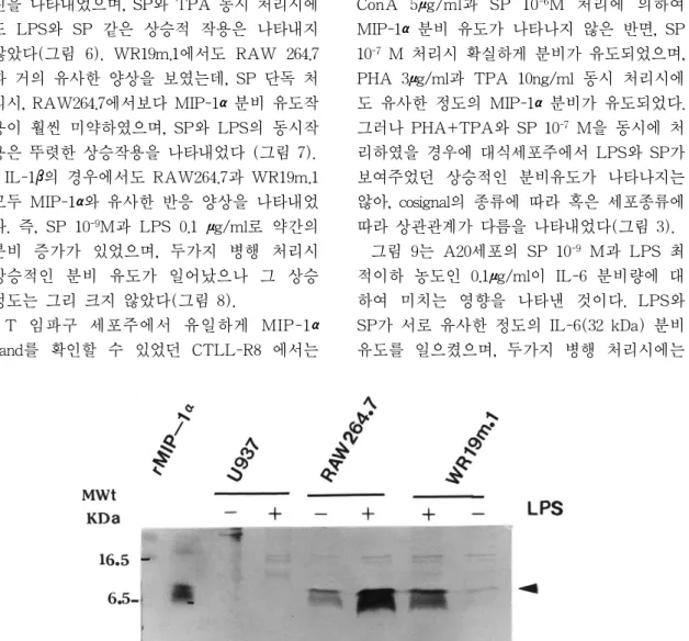
단핵세포/대식세포주는 LPS 1μg/ml로 자 극하였을 때, RAW264.7과 WR19m.1에서는 농축하지 않은 상청액에서도 상당히 진한 8 kDa의 MIP-1αband가 관찰되었으나, U937에 서는 농축된 시료임에도 불구하고 MIP-1α band가 관찰되지 않았다(그림 1, 그림 4, 표 1). IL-1β의 경우는 LPS로 자극하였을 경우, 단핵세포/대식세포주 모두 약간의 유리량 증 가를 보였으나(17 kDa band), U937은 비교적 희미한 IL-1βband를 나타내었다(그림 2, 표 1). IL-6는 모든 실험대상 단핵구/대식세포주
에서 western blot으로 관찰할 수 있는 정도의 유리량을 나타내지 못하였다(표 1).
T 임파구세포주에서는 조사된 cytokine 중, CTLL-R8을 PHA 3μg/ml과 TPA 10ng/ml로 함께 자극하였을 경우에 MIP-1α의 옅은 band만이 관찰되었을 뿐이며, HuT78, CCRF- CEM, Jurkat에서는 western blot상에서 어떠 한 cytokine band도 관찰되지 못하였다(그림 3, 그림 5A & 5B, 표 1).
B 임파구 세포주는 Daudi의 경우만 약간의 IL-1β분비가 관찰되었고(그림 4, 표 1), MIP-1 α는 모든 조사 대상세포에서 western blot으로 관찰할 수 있는 유리량을 나타내지 못하였으 며(표 1), IL-6는 A20, Daudi, SKW6.4 세포에 서만 진한 band가 관찰되었으나, 표준시료 IL-6의 분자량이 24 kDa인데 비하여, A20는 32 kDa, Daudi는 32와 35 kDa, SKW6.4는 29 kDa으로, 서로 상이한 IL-6 분자량을 나타내 었다. 이들 세포주들에서 IL-6는 일정하게 유
리되었으며, Daudi 와 SKW6.4는 LPS나 TPA 로 자극하여도 더 이상의 분비 증가가 나타나 지 않았다(그림 5B). Erythroid/Promyeloid 세 포주에서는 K562세포를 LPS로 자극할 경우에 IL-1βband만 희미하게 관찰되었을 뿐이었다.
특히 HL-60의 경우 TPA와 dibutyl cAMP로 대식세포, 혹은 다형핵백혈구로 분화를 유도 하여 자극해 보았으나 어느 cytokine도 western blot으로 관찰할 수 있는 유리량을 나 타내지 못하였다(그림 4, 그림 5A, 표 1).
그림 6은 대식세포주인 RAW264.7에 LPS, TPA 및 SP를 처리하였을 때의 MIP-1α분비 량을 비교한 것이다. SP 10-9 M 단독 처리시, 대조군에 비하여 약간의 MIP-1α분비 증가를 나타내었고, LPS 최적이하 농도인 0.1μg/ml처 리시에도 비슷한 결과를 보였으며, SP와 LPS 동시 처리시 비교적 적은 폭의 상승적 (synergistic) 분비 증가를 나타내었다. TPA는 LPS 최적이하 농도보다 매우 미약한 분비 촉 표 1 Secretion of inflammatory cytokines from immune cell lines induced by various chemical signals.
Cell Line Chemical Signal MIP-1α IL-1β IL-6
Monocyte/ U937 LPS - ± -
Macrophage RAW264.7 ++++a + -
WR19m.1 ++++a + -
T lymphocyte CTLL-R8 ConA/IL2/ + - -
HuT78 PHA+TPA - - -
CCRF-CEM - - -
Jurkat
B lymphocyte Daudi LPs/TPA - ± +
A20 - - ++
SKW6.4 - - ++
Namulwa - - -
Raji - - -
Erythroid/ K562 LPS/TPA - ± -
Promyeloid
KGla dbcAMP - - -
HL-60 - - -
Arbitrary scores are deduced from band density of western blot analysis.
a : Cell supernatant needs not to be concentrated for western blot analysis.
진을 나타내었으며, SP와 TPA 동시 처리시에 도 LPS와 SP 같은 상승적 작용은 나타내지 않았다(그림 6). WR19m.1에서도 RAW 264.7 과 거의 유사한 양상을 보였는데, SP 단독 처 리시, RAW264.7에서보다 MIP-1α분비 유도작 용이 훨씬 미약하였으며, SP와 LPS의 동시작 용은 뚜렷한 상승작용을 나타내었다 (그림 7).
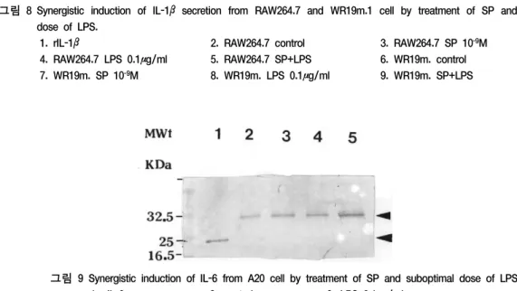
IL-1β의 경우에서도 RAW264.7과 WR19m.1 모두 MIP-1α와 유사한 반응 양상을 나타내었 다. 즉, SP 10-9M과 LPS 0.1 μg/ml로 약간의 분비 증가가 있었으며, 두가지 병행 처리시 상승적인 분비 유도가 일어났으나 그 상승 정도는 그리 크지 않았다(그림 8).
T 임파구 세포주에서 유일하게 MIP-1α band를 확인할 수 있었던 CTLL-R8 에서는
ConA 5μg/ml과 SP 10-6M 처리에 의하여 MIP-1α분비 유도가 나타나지 않은 반면, SP 10-7 M 처리시 확실하게 분비가 유도되었으며, PHA 3μg/ml과 TPA 10ng/ml 동시 처리시에 도 유사한 정도의 MIP-1α분비가 유도되었다.
그러나 PHA+TPA와 SP 10-7 M을 동시에 처 리하였을 경우에 대식세포주에서 LPS와 SP가 보여주었던 상승적인 분비유도가 나타나지는 않아, cosignal의 종류에 따라 혹은 세포종류에 따라 상관관계가 다름을 나타내었다(그림 3).
그림 9는 A20세포의 SP 10-9 M과 LPS 최 적이하 농도인 0.1μg/ml이 IL-6 분비량에 대 하여 미치는 영향을 나타낸 것이다. LPS와 SP가 서로 유사한 정도의 IL-6(32 kDa) 분비 유도를 일으켰으며, 두가지 병행 처리시에는
그림 1 Induction of MIP-1αsecretion from monocyte/macrophage cell lines by LPS stimulation. 10μl of cell supernatants were fractionated with 20% SDS-PAGE and subjected into western blot analysis.
그림 2 Induction of IL-1βsecretion from monocyte/macrophage cell lines by LPS stimulation. 1 ml of cell supernatants were TCA concentrated and fractionated with 15% SDS-PAGE and subjected into western bot analysis.
그림3 Induction of MIP-1α secretion from CTLL-R8 cell by SP or PHA+TPA. Concentrated cell supernatants were fractionated with 20% SDS-PAGE and subjected into western blot analysis.
1. rMIP-1α 2. control 3. ConA 5 ㎍/ml 4. SP 10-6 M
5. SP 10-7M 6. ConA+SP 10-7 M 7. PHA 3㎍/ml+TPA 10 ng/ml 8. PHA+TPA+SP 10-7 M
그림 4 Induction of IL-1β secretion from various cell lines by LPS stimulation. Concentrated cell supernatants were fractionated with 15% SDS-PAGE and subjected into western blot analysis.
그림 5 Induction of IL-6 secretion from various immune cell lines by stimulation
A) 1. rIL-6 2. HL-60 3. HL-60 LPS 1㎍/ml
4. HL-60 TPA 10ng/ml 5. HL-60 TPA+LPS 6. Jurkat
7. Jurkat ConA 5㎍/ml 8. CCRF-CEM 9. CCRF-CEM ConA 5㎍/ml
B) 1. rIL-6 2. A20 3. Daudi
4. Daudi TPA 10ng/nl 5. SKW 6.4 6. SKW6.4 LPS 1㎍/ml 7. SKW6.4 TPA 10ng/ml 8. HuT78 9. HuT78 ConA 5㎍/ml
그림 6 Synergistic induction of MIP-1αsecretion from RAW264.7 cell by treatment of SP and suboptimal dose of LPS.
1. rMIP-1α 2. control 3. SP 10-9 M 4. LPS 10-6 M 5. LPS+SP 6. TPA 10ng/ml 7. TPA+SP
그림 7 Synergistic induction of MIP-1αsecretion from WR19m.1 cell by treatment of SP and suboptimal dose of LPS.
1. rMIP-1α 2. control 3. SP 109M 4. LPS 0.1μg/ml 5. LPS+SP 6. TPA 10ng/ml 7. TPA+SP
그림 8 Synergistic induction of IL-1βsecretion from RAW264.7 and WR19m.1 cell by treatment of SP and suboptimal dose of LPS.
1. rIL-1β 2. RAW264.7 control 3. RAW264.7 SP 10-9M
4. RAW264.7 LPS 0.1μg/ml 5. RAW264.7 SP+LPS 6. WR19m. control 7. WR19m. SP 10-9M 8. WR19m. LPS 0.1μg/ml 9. WR19m. SP+LPS
그림 9 Synergistic induction of IL-6 from A20 cell by treatment of SP and suboptimal dose of LPS.
1. rIL-6 2. control 3. LPS 0.1μg/ml
4. SP 10-9M 5. LPS+SP
상승적인 분비 촉진 현상이 나타났다.
IV. 고 찰
지각신경계가 염증질환의 진행양상을 조정 한다고 하는 여러 실험적, 임상적 증거들에 의하여, 말초의 지각정보를 중추신경계로 전 달하는 역할을 하는 것으로만 알려졌던 neuropeptide들이 구심성(afferent) 자극전달 외에 말초조직으로의 원심성(efferent) 조절에 도 관련되어 있다는 사실이 알려지기 시작했
다45~47). Levine48)이 언급한 관절염 환자에서,
관절부 지각신경의 예민성이 증가하고, 염증 의 정도를 지각신경이 조절한다는 보고가 있 었다. 또 정상인보다 관절염 환자의 관절에서 SP를 함유한 신경섬유가 더욱 고농도로 발견 되었고9) 더 많은 대식세포가 모여 있으며 이 들 세포에는 SP수용체가 존재한다는 보고도
있었다11~12). 치주질환에서의 신경성 염증의
역할에 관하여 아직 알려진 바는 없으나, 최 근 에 치 주 조 직 의 혈 관 주 위 에 서 SP- immunoreactive fiber가 다량 발견되었고, 치 은섬유아세포의 성장을 SP가 촉진하였다고 한 Bartold 등4)의 보고에 따르면 치주질환의 진행양상에 신경성 조절이 중요한 역할을 하 리라고 추정된다. SP가 신경성 염증에서 매 개체 역할을 하리라고 추측할만한 근거는 매 우 풍부하다. 즉, SP는 강력한 혈관 이완제로 서 혈관주위에 많이 분포하고 있고 여러 염증 및 면역세포들에 기염증성 작용을 지니고 있 다고 보고되었다44), 49~55). 또한, capsaicin에 예 민한 지각신경을 비가역적으로 파괴시킨 실험 동물에서 신경성 염증반응이 유도되지 않았다
는 사실56~57) 및 상치조신경이나 하치조신경을
외과적으로 절제시킨 실험동물에서 치수, 치 주인대, 치은, 및 구강점막의 혈행역학적 (hemodynamic) 반응에 변화가 초래되었다는
보고들이50), 58~62) 이러한 추측을 뒷받침한다.
SP는 각종 cytokine의 분비를 조정하는 것
으로 알려져 있다13), 34), 36~39), 41~42). MIP-1α, IL-1β 및 IL-6와 같은 cytokine은 대표적인 기염증성 cytokine들로서, 각종 면역반응에 대 하여 다양한 조절기능을 나타내며, SP의 염 증조절기능은 상당 부분이 이들 cytokine의 발현에 영향을 미침으로서 매개된다고 볼 수 있으므로, 이들의 분비에 미치는 SP의 영향 은 신경성 염증조절 기전을 밝히는데 중요한 단서가 된다. 본 실험에서 cytokine의 분비를 측정하기 위해 사용한 western blot 분석방법 은 단백질 nanogram 단위에서 측정 가능한 예민도를 나타내므로 ELISA방법의 1/100에 미치지 못하는 예민도이기는 하나, 특이성 (specificity)에 있어서는 ELISA방법이 따라갈 수 없을 뿐 아니라, cytokine에 대한 항체 한 가지 만으로도 특이적으로 각 cytokine을 측 정할 수 있어 경제적인 측면에서 유리하다.
따라서 여러 종류의 cytokine에 대한 SP의 작 용을 측정하고저한 본 실험의 목적에는 western blot 방법이 합당하였다. Western blot 의 단점인 예민도의 미흡을 보완하기 위하여 가능한 한 많은 양의 cytokine을 분비하는 세 포주를 찾는 일이 우선되어야 했다. 15종의 면역세포주들에 대하여 3가지 cytokine의 유 리량을 통상적인 자극제를 사용하여 측정한 결과, MIP-1α는 RAW264.7과 WR19m.1에서 LPS 자극에 의하여 상당히 많은 양이 분비됨 을 확인하였고, IL-6는 A20, Daudi, SKW6.4 등 B임파구 세포주에서 다량 분비가 확인되 었으나, IL-1β는 western blot 방법으로 만족 하게 측정할 수 있는 양을 분비하는 세포를 찾지 못하였다. Promyelocyte 세포주 중 HL- 60와 같은 세포는 Lim 등33)의 보고에서 dibutyl cAMP나 TPA에 의하여 분화된 경우, SP에 의하여 항균작용이 상승되었다고 하였 으나, 본 실험의 cytokine 분비에 관한 실험에 서는 측정할 수 없었다.
MIP-1α는 대식세포를 LPS로 자극하였을 때 TNF-α와 더불어 가장 많이 분비되는 8
kDa의 cytokine으로서, 국소 염증작용, PG과 무관한 발열작용, T 임파구에 대한 화학주성 효과, stem세포 성장억제작용 등을 나타내는 기염증성 C-C chemokine중의 하나이다63-67). IL-1β는 17kDa 크기의 기염증성 cytokine으로 서, 자극이나 감염시에 유리되어 다양한 생물 학적 활성을 나타내므로, 질병의 병인론에 큰 영향을 미치는 것으로 알려져 있어, 관절염이 나 치주염과 같은 이화적 염증성 질환에서 매우 중요한 역할을 한다. IL-6는 면역반응, 급성반응, 조혈기능을 조절하는 다기능성 cytokine으로서68~70) glycosylation 정도에 따라 세포마다 다른 분자량의 IL-6를 생산한다. 본 실험에서 표준시료 IL-6 분자량이 24 kDa 이 었던데 반하여 세포에 따라 29, 32, 35 kDa의 여 러 band가 관 찰 된 것 은 이 러 한 glycosylation 차이에 기인한 것으로 사료된다.
CTLL-R8을 제외한 나머지 세포주는 SP 농도 10-9 M에서 가장 큰 작용을 나타내었는 데, 이는 다른 여러 보고들의 결과와 일치한 다. Rameshwar과 Bartold 등4), 13), 38), 73)에 의 하면 치은섬유아세포 성장촉진 작용의 경우 는 10-9 M, T 세포의 IL-2 mRNA 발현작용 은 10-12 M, 피부 섬유아세포는 10-7 M, synovial cell 은 10-8 M, 골수에서의 cytokine 발현은 10-9 M 등, 대체적으로 10-6 M 에서 10-13 M 사이에서 그 활성을 나타낸다고 한 다. 그러나 Lim과 Lowman 등 25), 30), 33)은 비 만세포의 histamine 유리에는 10-5 M 내지 10-6 M, 다형핵백혈구에 대한 활성화에는 10-5 M 등 높은 SP농도를 필요로 한다고 하였고 Kroegel 등74)은 호산구의 탈과립도 비슷한 유 효농도를 요구한다고 보고하였다. 이러한 고 농도는 초기 과염증상태에서 쉽게 도달할 수 있는 SP 농도이며, 비만세포, 호중구, 호산구 등의 세포는 초기 염증단계에서 신속하고 짧 은 기간동안만 주로 그 역할을 담당하여 염 증반응을 즉각 증폭시키는 기능을 하므로, 초 기 염증단계에서 나타나는 SP 고농도에 대하
여만 이들 세포가 반응을 하는 것은 지각신 경에 의하여 평상시에도 용이하게 활성화 되 는것을 차단하기 위하여 마련된 생체 기전일 것으로 사료된다42). SP 용량-반응 곡선의 특 징은 2개의 peak를 나타내는 biphasic activity 가 관찰된다는 것과, bell 모양의 형태를 나타 낸다는 것이다. Bell 모양의 용량-반응 곡선은 T세포 성장 촉진46), 75) 과 말초혈액 단핵세포 에 의한 IFN-γ의 분비40) 및 T세포에서의 IL- 2 분비38)에서 관찰되었지만 그 기전은 아직 알 수 없다. 본 실험에서는 이 중, peak를 나 타내는 농도를 선택해서 사용하였고, CTLL- R8에서 10-6 M 농도에서보다 10-7 M에서 더 MIP-1α유리가 촉진되었던것은 이러한 사실 의 단편적인 예이다. Biphasic 한 용량-반응 곡선의 원인은 SP의 특이적 수용체인 NK-1 외의 NK-2 혹은 NK-3와 같은 낮은 친화력 의 수용체와 SP가 결합하여 반응을 일으키기 때문인 것으로 추측된다. 한편 단일세포에 두 가지 이상의 subset들이 존재하여 SP 수용체 의 3차원적인 구조가 약간 차이가 있어 결합 친화력이 달라질 수도 있을 것으로 추측할수 있지만, 현재로서는 결론을 짓기가 어렵다.
본 실험에서 SP는 단독으로는 뚜렷한 cytokine 유리를 촉진하지 못하였으나, 최적이 하 농도의 LPS와 동시에 처리하였을 때 상승 적인 작용을 나타내는 것으로 관찰되었다. 이 러한 결과는 SP가 T세포의 IL-2 분비유도제 로서 작용하기보다는, 반응 증폭제 역할을 하 는 cosignal로서 작용한다고 보고한 Calvo 등
38)의 결과와 일치한다. 또한 Bartold 등4)도 치 은섬유아세포의 성장에 대하여 SP가 촉진효 과를 나타내기 위하여는 다른 co-stimulation 인자가 필수적으로 있어야 한다고 보고하였 고, Nilsson 등76)도 최적이하농도의 platelet derived growth factor를 첨가하였을 때 SP가 섬유아세포의 성장을 촉진한다고 하여, SP 단독보다는 주위의 다른 화학물질의 존재에 의하여 SP 작용이 결정 되어짐을 추측할 수
있다. SP는 주위에 어떠한 종류의 화학적 signal이 존재하느냐에 따라 동일한 세포에 대 하여 촉진, 혹은 억제적으로 작용할 수 있다
44). 예를 들어, SP는 다형핵백혈구를 priming 시켜 fMLP로 자극하였을 때 superoxide anion의 생산을 증가시키지만, TNF가 또다른 자 극 제 로 사 용 된 경 우 , SP는 오 히 려 superoxide anion 생산을 감소시킨다고 Serra 등42)은 보고하였다. 본 실험의 결과에서도 대 식세포의 MIP-1α유리에 대하여 최적이하농도 의 LPS와 SP사이에는 상승적 작용이 관찰되 었으나, TPA와 SP사이에서는 일어나지 않았 고, T임파구에서도 PHA+TPA와 SP 사이에 MIP-1α유리에 대한 상승작용이 관찰되지 않 았다. 그러므로 SP는 염증부위의 다른 인자 들의 존재에 따라서, 이화반응의 결과를 유발 하는 기염증성 작용을 나타낼 수도 있고, 아 니면 동화반응의 결과를 유발하는 조직 재생 작용을 나타낼 수도 있다4).
SP가 어떠한 기전을 통하여 복잡한 염증조 절반응을 나타내는지 확실히 알려져 있지는 않으나, tachykinin 수용체 중 NK-1수용체와 특이적 결합을 함으로써 활성을 나타내게 된 다46), 77~78). SP수용체는 G-protein coupled receptor family에 속하며79~81), 수용체가 자극 을 받으면 G-protein은 adenylate cyclase 활성 을 변화시켜 세포내 cAMP농도의 변화나 phosphatidylinositol 의 가수분해를 유발시킴으 로써 세포내 신호전달체계가 활성화된다고
한다30), 82~83). SP의 수용체를 통한 신호전달
체계가 다른 cosignal의 신호전달계와 만나 어 떠한 경로로 세포반응의 증폭, 또는 억제 등 의 방향으로 전개되는지를 밝히기 위해서는 세포내 분자수준에서의 연구가 뒤따라야할 것으로 사료된다.
V. 결 론
본 연구는 각종 면역세포주의 기염증성
cytokine 분비에 미치는 SP의 cosignal로서의 영향을 관찰함으로써 SP의 면역 조절기전을 밝히고자 하였다. 세포 배양상청액내로 유리 된 MIP-1α, IL-1β및 IL-6를 western blot 분 석법으로 측정하여 다음의 결과를 얻었다.
1. 조사대상 15종의 면역세포주에 자극을 가했을 때 MIP-1α의 분비량이 가장 많았 던 것은 대식세포주인 RAW264.7과 WR19m.1 이었고, T 임파구세포주인 CTLL-R8도 약간의 분비를 나타냈으며, IL-6는 B 임파구세포주인 A20(32kDa), Daudi(32, 35kDa) 및 SKW6.4(29kDa)에 서 비교적 많은양이 분비되었으며, IL-1β 는 RAW264.7과 WR19m.1 및 erythroid/
promyeloid 세포주인 K562에서 매우 적은 양이 관찰되었을 뿐, 그외의 어느세포주 에서도 westerm blot 법으로 측정 가능한 수준의 분비량이 관찰되지 않았다.
2 최적이하 농도의 LPS(0.1μg/ml) 혹은 SP 10-9 M을 처리하였을 때, RAW264.7과 WR19m.1에서 MIP-1α와 IL-1β의 분비가 약간 증가되었으나, LPS와 SP를 동시에 처리하였을 때, 두가지 cytokine에서 모두 상승적인 분비증가가 관찰되었으며, TPA 와 SP 사이에는 이러한 상승작용이 관찰 되지 않았다.
3. CTLL-R8을 SP 10-7M 혹은 PHA+
TPA로 처리한 경우 MIP-1α분비의 유도 가 일어났으나, 이들 사이의 상승작용은 관찰되지 않았다.
4. A20, Daudi 및 SKW6.4에서는 일정하게 IL-6분비가 일어났으며, A20 에서 최적 이하농도의 LPS와 SP 10-9 M에 의하여 IL-6 분비량이 약간 증가하였고, LPS와 SP 사에에 상승적 분비유도작용이 관찰 되었다.
이 상 의 결 과 에 서 , SP는 염 증 부 위 의 cytokine 유리를 유발하는 cosignal의 작용을
조절함으로써 면역 기능을 조정하는 것으로 추론된다.
참고문헌
1. Bost, K.L.: Hormone and neuropeptide receptors on mononuclear leukocytes.
Prog. Allergy., 43:68, 1988.
2. Stead, R.H., Tomioka, M., Quinonez, G., Simon, G.T., Felten, S.Y., and Bienenstock, J.: Intestinal mucosal mast cells in normal and nematode-infected rat intestines are in intimate contact with peptidergic nerves. Proc. Natl.
Acad. Sci. USA., 84:2975-2979, 1987.
3. Felten, D.L., Felten, S.Y., Carlson, S.L., Olschowka, J.A., and Livnat, S.:
Noradrenergic and peptidergic innervation of lymphoid tissue. J.
Immunol., 135:755, 1985.
4. Bartold, P.M., Kylstra, A., and Lawson, R.:
Substance P: An immunohistochemical and biochemical study in human gingival tissues.
A role for neurogenic inflammation? J.
Periodontol., 65:1113, 1994.
5. Freeman, R. and Goss, S.: Stress measures as predictors of periodontal disease-a preliminary communication.
Comm. Dent. Oral Epidemiol.., 21:176- 177, 1993.
6. Green, L.W., Tryan, W.W., Marks, B., and Huryn, N.: Periodontal disease as a function of life events stress. J. Human Stress., 12:32-36, 1986.
7. DeMarco, T.J.: Periodontal emotional stress syndrome. J. Periodontol., 37:67- 68, 1976.
8. Fedi, P.F.: The effects of stress on the periodontium of the Syrian hamster. J.
Periodontol., 29:293-299, 1958.
9. Levine, J.D., Clark, R., Devor, M., Helms, C., Moskowitz, M.A., and Busbaum, A.I.: Intraneuronal substance P contributes to severity of experimental arthritis. Science., 226:547-549, 1984.
10. Kimball, E.S., Perisco, F.J., and Vaught, J.L.: Substance P, neurokinin A, and neurokinin B induce generation of IL-1 like activity in P388D1 cells. J.
Immunol., 141: 3564-3569, 1988.
11. Hartung, H.P., Wolters, K., and Toyka, K.V.: Substance P: Binding properties and studies on cellular responses in guinea pig macrophages. J. Immunol., 136:3856-3863, 1986.
12. Hartung, H.P. and Toyoka, K.V.:
Activation of macrophages by substance P induction of oxidative burst and thromboxane release. Eur. J. Pharmacol., 89:301-305, 1983.
13. Lotz, M., Carson, D.A., and Vaughan, J.H.: Substance P activation of rheumatoid synoviocytes: Neural pathway in pathogenesis of arthritis.
Science., 235:893-895, 1987.
14. Pernow, B.: Substance P. Pharmacol.
Rev., 35:85, 1983.
15. Jiang, Z., Dun, N.J., and Karczmar, A.G.: Substance P: a putative sensory transmitter in mammalian autonomic ganglia. Science., 217:739, 1982.
16. Ho..
felt, T., Kellerth, J.O., Nilsson, G., and Pernow, B.: Substance P: localization in the central nervous system and in some primary sensory neurons. Science., 190:889-890, 1975.
17. Goetzl, E.J., Adelman, D.C., and Sreedharan, S.P.: Neuroimmunology.
Adv. Immunol., 48:161, 1990.
18. Holzer, P.: Local effector functions of capsaicin-sensitive sensory nerve endings: involvement of tachykinins, calcitonin gene-related peptide and other neuropeptides. Neuroscience., 24:39, 1988.
19. Lembeck, F. and Holzer, P.: Substance P as neurogenic mediator of antidromic vasodilation and neurogenic plasma extravasation. Naunyn-Schmiedebergs Arch. Pharmacol., 310:175-183, 1979.
20. Payan, D.G. and Goetzl, E.J.:
Modulation of lymphocyte function by sensory neuropeptides. J. Immunol., 135:783, 1985.
21. Payan, D.G., Breqster, D.R., and Goetzl, E.J.: Specific stimulation of human T lymphocytes by substance P. J.
Immunol., 131:613, 1983.
22. Bost, K.L. and Pascual, D.W.:
Substance P: a late-acting B lymphocyte differentiation cofactor. Am.
J. Physiol., 262:C537-C545, 1992.
23. Scicchitano, R., Bienenstock, J., and Stanisz, A.M.: In vivo immunomodulation by the neuropeptide substance P. Immunology., 63:733-735, 1988.
24. Stanisz, A.M., Befus, D., and Bienenstock, J.: Differential effects of vasoactive intestinal peptide, substance P, and somatostatin on immunoglobulin synthesis and proliferation by lymphocytes from Peyer's patches, mesenteric lymph nodes, and spleen. J.
Immunol., 136:152-156, 1986.
25. Lowman, M.A., Benyon, R.C., and Church, M.K.: Characterization of neuropeptide-induced histamine release from human dispersed skin mast cells.
Br. J. Pharmacol., 95:121, 1988.
26. Shanahan, F., Denburg, J.A., Fox, J., Bienenstock, J., and Befus, D.: Mast cell heterogeneity: effects of neuroenteric peptides on histamine release. J.
Immunol., 135:1331-1337, 1985.
27. Wozniak, A., Betts, W.H., McLennan, G., and Scicchitano, R.: Activation of human neutrophils by tachykinins.
Effect on formyl-ethionyl-leucyl- phenylalanine and platelet-activating factor-stimulated superoxide anion production and antibody-dependent cell- mediated cytotoxicity. Biochem. Biophys.
Res. Commun., 78:629, 1993.
28. Brunelleschi, S., Tarli, S., Giotti, A., and Fantozzi, R.: Priming effects of mammalian tachykinin on human neutrophils. Life Sci., 48:1, 1990.
29. Wozniak, A., McLennan, G., Betts, W.H., Murphy, G.A., and Scicchitano, R.: Activation of human neutrophils by substance P: effect of FMLP- stimulated oxidative and arachidonic acid metabolism and on antibody-dependent cell-mediated cytotoxicity. Immunology., 68:359-364, 1989.
30. Serra, M.C., Bazzoni, F., Della Bianca, V., Greskowiak, M., and Rossi, F.:
Activation of human neutrophils by substance P. Effect on oxidative metabolism, exocytosis, cytosolic Ca2+
concentration and inositol phosphate formation. J. Immunol., 141:2118, 1988.
31. Marasco, W.A., Showell, H.J., and Becker, E.L.: Substance P binds to the formylpeptide chemotaxis receptor on the rabbit neutrophil. Biochem. Biophys.
Res. Commun., 99:1065, 1981.
32. Bar-Shavit, Z., Goldman, R., Stabinsky, Y., Gottlieb, P., Fridkin, M., Teiberg, V.I., and Blumberg, S.: Enhancement of phagocytosis: a newly found activity of substance P residing in its N-terminal tetrapeptide sequence. Biochem. Biophys.
Res. Commun., 94:1445, 1980.
33. Lim, J.D., Oh, K.O., and Kim, H.S.:
Effects of substance P on the activities of immune cells. J. Korean academy of Periodontol., 1996; 26(2): 376-395 34. Laurenzi, M.A., Persson, M.A.A.,
Dalsgaard, C.J., and Haegerstrand, A.:
The neuropeptide substance P stimulates production of interleukin-1 in human blood monocytes: Activated cells are preferentially influenced by the neuropeptide. Scand. J. Immunol., 31:529-533, 1990.
35. Lotz, M., Vaughan, J.H., and Carson, D.A.: Effect of neuropeptides on production of inflammatory cytokines by human monocytes. Science., 241:1218- 1221, 1988.
36. Cozens, P.J. and Rowe, F.M.: Substance P is a potent inducer of TNF and IL-1 secretion by macrophages. A potent role for TNF in the pathogenesis of asthma.
Immunobiology., 175:7, 1987.
37. Nio, D.A., Moylan, R.N., and Roche, J.K.: Modulation of T lymphocyte function by neuropeptides: evidence for their role as local immunoregulatory elements. J. Immunol., 150:5281, 1993.
38. Calvo, C.F., Chavanel, G., and Senik, A.: Substance P enhances IL-2 expression in activated human T cells. J.
Immunol., 148:3498-3504, 1992.
39. Blum, A.M., Metwali, A., Cook, G.,
Mathew, R.C., Elliott, D., and Weinstock, J.V.: Substance P modulates antigen-induced, IFN-γ production in murine Schistosomiasis mansoni. J.
Immunol., 151: 225-233, 1993.
40. Wagner, F., Fink, R., Hart, R., and Dancygier, H.: Substance P enhances interferon-γ production by human peripheral blood mononuclear cells.
Regul. Pept., 19:355, 1987.
41. Agro, A., and Stanisz, A.M.: Are lymphocytes a target for substance P modulation in arthritis? Semin. Arthritis Rheum., 21:252, 1992.
42. Serra, M.C., Calzetti, F., Ceska, M., and Cassatella, M.A.: Effect of substance P on superoxide anion and IL-8 production by human PMNL. Immunology., 82:63, 1994.
43. Fewtrell, C.M.S., Foreman, J.C., Jordan, C.C., CEhme, P., Renner, H., and Stewart, J.M.: The effects of substance P on histamine and 5-hydroxytryptamine release in the rat. J. Physiol., 330:393- 411, 1982.
44. Mantyh, P.W.: Substance P and the inflammatory and immune response.
Ann. New York Acad. Sci., 632:263- 271, 1991.
45. Weinstock, J.V.: Neuropeptides and the regulation of granulomatous inflammation.
Clin. Immunol. Immunopathol., 64:17-22, 1992.
46. Payan, D.G. and Goetzl, E.J.:
Neuropeptide regulation of immediate and delayed hypersensitivity. Int. J.
Neurosci., 38:211-221, 1988.
47. Johnson, H.M. and Torres, B.A.:
Immunoregulatory properties of
neuroendocrine peptide hormones. Prog.
Allergy., 43:37-67, 1988.
48. Levine, J.P., Goetzl, E.J., and Basbaum, A.T.: Contribution of nervous system to pathophysiology of rheumatoid arthritis and other polyarthropathies. Rheum. Dis.
Clinic North Am., 13:369-383, 1987.
49. Baluk, P., Nadel, J.A., and McDonald, D.M.: Substance P immunoreactive sensory axons in the rat respiratory tract: A quantitative study of their distribution and role in neurogenic inflammation. J. Comp. Neurology., 319:586-598, 1992.
50. Gyorti, A., Fazekas, A., and Rosivall, L.: Neurogenic inflammation and the oral mucosa. J. Clin. Periodontol., 19:731-736, 1992.
51. Nagata, E., Ohura, K., Shinohara, M., et al.: Distribution of nerve fibers with substance P- or calcitonin gene-related peptide-like immunoreactivity in ODU plaque susceptible rats with experimental gingivitis. Acta. Histochem. Cytochem., 25:161-168, 1992.
52. Scott, D.T., Lam, F.Y., and Ferrell, W.R.: Acute inflammation enhances substance P-induced plasma protein extravasation in the rat knee joint.
Regulatory Peptides., 39:227-235, 1992.
53. Ballieux, R.E.: Impact of mental stress on the immune response. J. Clin.
Periodontol., 18:427-430, 1991.
54. Farber, E.M., Lanigan, S.W., and Rein, G: The role of psychoneuroimmunology in the pathogenesis of psoriasis. Cutis., 46:314-316, 1990.
55. Ferrel, W.R. and Russel, N.J.W.:
Extravasation in the knee induced by
antidromic stimulation of articular C fiber afferents of the anesthetized cat. J.
Physiol., 379:407-416, 1986.
56. Gamse, R., Holzer, P., and Lembeck, F.: Decrease of substance P in primary afferent neurons and impairment of neurogenic plasma extravasation by capsaicin. Br. J. Pharmacol., 68:207-213, 1980.
57. Jansco, G., Kiraly, E., and Jansco-Gabor, A.: Pharmacologically induced selective degeneration of chemosensitive primary neurons. Nature., 270:741-743, 1977.
58. Olgart, L.M.: Involvement of sensory nerves in hemodynamic reactions. Proc.
Finn. Dent. Soc., 88(Suppl. 1):403-410, 1992.
59. Fazekas, A., Gyorfi, A., Posch, E., Jakab, G., Bartafai, Z., and Rosivall, L.:
Effect of denervation on the neurogenic inflammation of the rat mandibular mucosa. Naunyn Schmiedenbergs Arch.
Pharmacol., 343:393-398, 1991.
60. Olgart, L., Edwell, L., and Gazelius, B:
Involvement of afferent nerves in pulpal blood-flow reactions in response to clinical and experimental procedures in the cat. Arch. Oral Biol., 36:575-581, 1991.
61. Izumi, H. and Karita, K: The effects of capsaicin applied topically to inferior alveolar nerve on antidromic vasodilatation in cat gingiva. Neuroscience Lett., 112:65-69, 1990.
62. Karita, K., Izumi, H., Tabata, T., Kuriwada, S., Sasano, T., and Sanjo, D.:
The blood flow in the periodontal ligament regulated by the sympathetic and sensory nerves in the cat. Proc.
Finn. Dent. Soc., 85:289-294, 1989.
63. Graham, C.J., Zhou, L., Weatherbee, J.A., Tsang, M.L., Napolitano, M., Leonard, W.J., and Pragnell, I.B.: Cell Growth Differ. 4:137, 1993.
64. Kwon, B.S., Zhou, Z., Pollok, K., and Kim, Y.: FASEB., 6:A1147, 1992.
65. Davatelis, G., Wolpe, S., Sherry, B., Dayer, J., Chicheportiche, R., and Cerami, A.: Macrophage inflammatory protein-1: a prostaglandin-independent endogenous pyrogen. Science., 243:1066, 1989.
66. Wolpe, S., Davatelis, G., Sherry, B., Beutler, B., Hesse, D., Nguyen, H., Moldawer, L., Nathan, C., Lowry, S., and Cerami, A.: Macrophage secret a novel heparin-binding protein with inflammatory and neutrophil chemokinetic properties. J.
Exp. Med., 167:570, 1988.
67. Oh, K.O., Zhou, Z., Kim, K.K., Samanta, H., Fraser, M., Kim Y.J., Broxmeyer, H.E., and Kwon, B.S.: Identification of cell surface receptors for murine macrophage inflammatory protein-1α. J.
Immunol., 147:2987, 1991.
68. Hirano, T.: Chem. Immunol., 51:153, 1992.
69. Hirano, T. and Kishimoto, T.: In Handbook of Experimental Pharmacology Peptide Growth Factors and Their Receptors. vol. 95/1(ed. M.B. Sporn and A.B. Roberts), Springer, Berlin, p 633- 665, 1990.
70. Sehgal, P.B., Grienger, G., and Tosata, G.: Ann. NY Acad. Sci., 557:1, 1989.
71. Santhanam, U., Ghrayeb, J., Sehgal, P.B., and May, L.T.: Arch. Biochem.
Biophys., 274:161, 1989.
72. May, L.T., Grayeb, J., Santhanam, U., Tatter, S.B., Sthoger, Z., Helfgott, D.C., Chiorazzi, N., Grieninger, G., and Sehgal, P.B.: J. Biol. Chem., 263:7760, 1988.
73. Rameshwar, P., Ganea, D., and Gascon, P.: Induction of IL-3 and granulocyte- macrophage colony stimulating factor by substance P in bone marrow cell partially mediated through the release of IL-1 and IL-6. J. Immunol., 152:4044, 1994.
74. Kroegel, C., Giembycz, M.A., and Barnes, P.J.: Characterization of eosinophil cell activation by peptides.
Differential effects of substance P, milittin, and FMEY-Leu-Phe. J.
Immunol., 145:2581, 1990.
75. Paegelow, I. and Werner, H.:
Immunomodulation by some oligopeptides.
Methods Find. Exp. Clin. Pharmacol., 8:91, 1986.
76. Nilsson, J., Von Euler, A.M., and Dalsgaard, C.J.: Stimulation of connective tissue cell growth by substance P and substance K. Nature., 315:61-63, 1992.
77. Quirion, R.: Multiple tachykinin receptors. Trends Neurosci., 8:183, 1985.
78. Lee, C.M., Iversen, L.L., Hanley, M.R., and Sandberg, B.E.B.: The possible existence of multiple receptors for substance P. Arch. Pharmacol., 318:281, 1982.
79. Takeda, Y., Chou, K.B., Sachais, B.S., and Krause, J.E.: Molecular cloning, structural characterization and functional expression of the human substance P receptor. Biochem. Biophys. Res.
Commun., 179:1232, 1991.
80. Shigemoto, R., Yokota, Y., Tsuchida, K., and Nakanishi, S.: Cloning and expression of a rat neuromedin K receptor cDNA. J. Biol. Chem., 265:623, 1990.
81. Yokota, Y., Sasai, Y., Tanaka, K., Fujiwara, T., Tsuchida, K., Shigemoto, R., Kakizuka, A.,Ohkubo, H., and Nakanishi, S.: Molecular characterization of functional cDNA for rat substance P receptor. J. Biol. Chem., 264:17649, 1989.P
82. Catalan, R.E., Martinez, A.M., Aragones,
M.D., Miguel, B.G., Robles, A., and Hernandez, F.: Dual mechanism of phosphatidylinositol hydrolysis by substance P in brain. Eur. J. Biochem., 172:547, 1988.
83. Laniyonu, A., Sliwinski-Lis, E., and Fleming, N.: Different tachykinin receptor subtypes are coupled to the phosphoinositide or cyclic AMP signal transduction pathways in rat submandibular cells. FEBS, Lett., 240:186, 1988.
-Abstract-
Effects of Substance P on the Release of Cytokines from Immune Cell Lines
Jin-Yong Lee, Soo-Ah Kim, Seok-Ran Seo, Hyong-Seop Kim
Department of Periodontology, College of Dentistry, Chon-buk National University
The neuropeptide substance P(SP) has been implicated in the mediation of inflammation and immune-mediated disease such as arthritis. Recently, it was reported that SP was markedly increased around the blood vessels in inflamed gingiva as well as in close association with the inflammatory cell infiltrate. These results support that SP may contribute to the pathophysiology of neuronal inflammation in human periodontal tissues. SP may regulate inflammatory/immune responses by stimulating the proliferation of human T cells, differentiation and antibody-secreting potential of B cells, macrophage respiratory burst, connective tissue proliferation, and the secretion of cytokines from monocytes and T cells. Here, I studied potential role of SP as a costimulatory chemical signal in inflammatory/immune responses, by determining the released proinflammatory cytokines such as MIP-1α, IL-1β, and IL-6 from culture supernatants of homogeneous immune cell lines. Serum free cell supernatants were concentrated with TCA precipitation, fractionated with SDS-PAGE, and subjected into western blot analysis. Among 15 cell lines tested, macrophage/monocyte cell line RAW264.7 and WR19m.1 showed the highest level of induction of MIP-1αwhen stimulated with LPS. Discrete IL-6 bands with multiple forms of molecular mass were detected from supernatants of B cell lines A20(32kDa), Daudi(32, 35kDa), and SKW6.4(29kDa), which were expressed constitutively. IL-1β could not be detected by the method of western blot analysis from supernatants of all cell lines tested except RAW264.7, WR19m.1, and erythroid cell line K562 which showed the least amount of IL-1βsecretion. SP 10-9M with suboptimal dose of LPS treatment showed synergistic induction of MIP-1αrelease from RAW264.7 or WR19m.1, and also IL-6 release from A20, but this synergism is not the case in costimulation of RAW264.7 or WR19m.1 with SP 10-9M and TPA. Although treatment of T cell line CTLL-R8 with SP 10-7M or PHA+TPA induced modest level of MIP-1α secretion, synergism was not observed when they are applied together. These findings all together suggest the possibility of a regulatory role of SP in inflammatory/immune reaction through differential modulation of bioactivities of other chemical cosignals.
Keywords : Substance P, Inflammatory/immune response, Proinflammtory cytokines.