INTRODUCTION
The association of pancreatic disease with fat necrosis at distant foci was first described by Chiari in 1883 (1). The most common pancreatic disorders associated with pancre- atic panniculitis are acute or chronic pancreatitis, especially the alcohol-related types, and pancreatic carcinoma. Other pancreatic disorders have been infrequently reported to be associated with pancreatic panniculitis, and these include post-traumatic pancreatitis, pancreatic pseudocyst, pancreas divisum, and hemophagocytic syndrome (2). To the best of our knowledge, only three cases of pancreatic panniculitis have been reported in the Korean literature. One case was associated with pancreatic adenocarcinoma, and the other two were associated with acute and chronic pancreatitis, respectively (1, 3, 4).
We report here on a case of pancreatic panniculitis associ- ated with acute pancreatitis that had a fatal outcome. Physi- cians should be aware of this disease entity because this rare cutaneous manifestation may be associated with major mor- bidity and significant mortality.
CASE REPORT
A 50-yr-old man with a history of alcohol abuse present- ed with increasing fatigue, generalized weakness, decreased appetite, and abdominal distension and discomfort for the
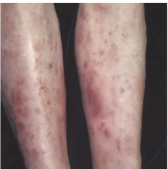
past 2 weeks. He was admitted to the Department of Inter- nal Medicine under the impression of acute pancreatitis, and he was referred to the Department of Dermatology for the mul- tiple painful subcutaneous nodules on his legs, which had suddenly developed 3 weeks before (Fig. 1).
On admission, his hemoglobin value was 11.4 g/dL (refer- ence range: 13-18 g/dL), the total count of white blood cells was 31,300/ L (reference range: 4,000-10,000/ L) with 90.7%
segment neutrophils, but the coagulation profiles and platelet counts were normal. The serum amylase was 1,909 U/L (ref- erence range: 20-120 U/L), and the lipase was 2,306 U/L (ref- erence range: 5-51 U/L). Liver function testing revealed an aspartate aminotrans terase level of 104 U/L (reference range:
13-40 U/L), an alanine aminotransferase level of 24 U/L (ref- erence range: 7-40 U/L), and a lactate dehydrogenase level of 665 U/L (reference range: 200-400 U/L). The fasting glucose level was 133.9 mg/dL (reference range of 70-110 mg/dL) and the electrolytes were unbalanced. The calcium level was 7 mg/
dL (reference range: 8.6-10 mg/dL), and the sodium level was 125 mEq/L (reference range: 136-145 mEq/L). The blood urea nitrogen and creatinine were 40.1 mg/dL and 2.1 mg/dL, respectively. After 48 hr, the blood urea nitrogen was increased to 62.7 mg/dL after intravenous fluid administration. As his leukocytosis, elevated serum LDH at admission, and hypocal- cemia, hypoalbuminemia, increase in blood urea nitrogen during initial 48 hr were poor prognostic factors in Ranson criteria, increased risk of complications was predicted. The chest radiograpy showed mild pleural effusion. On the abdomi-
Woo Sun Lee, Mi Yeon Kim, Sang Woo Kim*, Chang Nyol Paik*, Hyung Ok Kim, Young Min Park
Departments of Dermatology and Internal Medicine*, College of Medicine, The Catholic University of Korea, Seoul, Korea
Address for correspondence Young Min Park, M.D.
Department of Dermatology, The Catholic University of Korea, Kangnam St. Mary’s Hospital, 505 Banpo-dong, Seocho-gu, Seoul 137-701, Korea
Tel : +82.2-590-1351, Fax : +82.2-599-9950 E-mail : [email protected]
914 J Korean Med Sci 2007; 22: 914-7
ISSN 1011-8934
Copyright � The Korean Academy of Medical Sciences
Fatal Pancreatic Panniculitis Associated with Acute Pancreatitis:
A Case Report
Pancreatic panniculitis is a rare disease in which necrosis of fat in the panniculus and other distant foci occurs in the setting of pancreatic diseases; these diseases include acute and chronic pancreatitis, pancreatic carcinoma, pseudocyst, and other pancreatic diseases. This malady is manifested as tender erythematous nodules on the legs, buttock, or trunk. Histopathologically, it shows the pathognomonic findings of focal subcutaneous fat necrosis and ghost-like anucleated cells with a thick shadowy wall. We herein report a case of fatal pancreatic panniculitis that was associated with acute pancreatitis in a 50-yr-old man. He presented with a 3-week history of multiple tender skin nodules, abdominal pain and distension. Laboratory and radiologic findings revealed acute pancreatitis, and skin biopsy showed pan- creatic panniculitis. Despite intensive medical care, he died of multi-organ failure 3 weeks after presentation.
Key Words : Pancreatitis; Fat Necrosis; Panniculitis
Received : 18 May 2006 Accepted : 11 August 2006
A Case of Pancreatic Panniculitis Complicated in Acute Pancreatitis 915
nal computed tomography scan and magnetic resonance (MR) imaging taken on the second day of admission, a swollen pan- creas with an dilated pancreatic duct, a loculated fluid collec- tion in the left anterior perirenal space, multiple hepatic cysts, and massive ascites were noted (Fig. 2). On abdominal para- centesis, the amylase level was 14,696 U/L, the serum-ascites albumin gradient was calculated to be 1.07, and there was no evidence of malignancy.
A skin biopsy performed on the 4th day of admission from the nodule on the left lower leg showed a diffuse subcutane-
ous fat necrosis and ghost-like cells with thick shadowy walls and no nuclei. There was a fine granular basophilic material deposited within and around the necrotic fat cells (Fig. 3).
These findings were consistent with pancreatic panniculitis.
After 10 days of intensive medical care for the pancreatic disease, the patient’s condition began to worsen; he and his family began to refuse any further treatment. Despite a strong warning by physicians, he insisted on the discharge and died at home 1 week later.
DISCUSSION
The pathogenesis of pancreatic panniculitis is still unknown, but the released pancreatic enzymes such as trypsin may in- crease the permeability of the microcirculation. Lipase or amylase is then involved in the process of fat degradation, which results in the liberation of free fatty acids that com- bine with calcium to form soap (2). Although elevation of the pancreatic enzymes is common in pancreatitis patients, pancreatic panniculitis is a very rare malady. Mullin et al.
(5) identified only one case in a retrospective review of 893 patients who had suffered with pancreatic disease from vari- ous causes. Furthermore, well documented cases of fat necro- sis with normal serum lipase levels have also been described (6). These reports, suggest that there would be some other factors that allow the pancreatic enzymes to escape from the circulation and act on the subcutaneous fat. Zellman (7) sug- gested that some damage to the blood vessels via inflamma- tion, edema, or altered immunity may act as the initiating factor.
As for the two cases in the Korean literature, one case was
Fig. 1.Multiple erythematous to brownish nodules are shown on both lower legs.
Fig. 2.(A) Contrast-enhanced computed tomography showing an edematous pancreas with dilated pancreatic duct. (B) Loculated fluid at left anterior perirenal space with massive ascites was noted (arrow).
A B
916 W.S. Lee, M.Y. Kim, S.W. Kim, et al.
associated with chronic pancreatisis, in which the cutaneous lesions had occurred 3 months later than the other systemic symptoms, and they involuted as the underlying pancreati- tis was ameliorated (4). The other case was associated with acute pancreatitis with pancreas divisum, in which his skin lesions had appeared 2-3 days after his admission due to acute pancreatitis; the skin lesions also resolved as the under- lying pancratitis subsided with conservative treatment (1).
On the contrary, in our patient, the skin lesions preceded the other systemic symptoms.
Although the underlying pancreatic pathologic conditions can vary, the clinical features of pancreatic panniculitis are similar. The legs were the most commonly affected area, but the lesions can also occur on the arms, thighs, and trunk. The lesions usually began as erythematous or red-brown subcu- taneous nodules with a tendency to show central softening.
In the mild form, they may involute within weeks and leave an atrophic hyperpigmented scar. If the fat necrosis is severe, individual nodules may break down and extrude necrotic material (8). Although patients with other panniculitides such as erythema nodosum, erythema induratum, lupus pan- niculitis, Weber-Christian pannicultitis, or alpha-1 antitry- psin deficiency-associated panniculitis can have similar clin- ical lesions, the diagnosis of pancreatic panniculitis is sug- gested by the presence of pancreatic disease and this is con- firmed relatively clearly by the characteristic histopatholog- ic findings. The pathognomonic findings are collections of
‘ghost cells’ and anucleated adipocytes containing intracyto- plasmic fine basophilic granular material from the saponifi- cation of fat. The resistance of the fat cell membrane to lipase produces the shadowy walls and the fatty acids combine with
calcium to form calcium soap (10).
Medical and surgical treatments of the underlying pancre- atic disease are the primary therapeutic approaches. Some- times conservative care can ameliorate the pancreatitis, and this then results in the resolution of the skin lesions. In iso- lated cases, surgical correction of anatomic ductal anomaly or pancreatic pseudocyst, or the removal of gallstones has result- ed in complete resolution (8). However, disseminated fat necrosis is associated with major morbidity and mortality.
In a review of 27 patients with pancreatic panniculitis, all 8 patients with pancreatic carcinoma and 42% of the 19 pa- tients with pancreatitis died of their disease, as was seen in our case (9).
Pancreatic panniculitis is a pathognomic finding of pan- creatic disease, and as in our case, the cutaneous lesions may precede the usual manifestations of underlying pancreatic disease by several weeks to months (11). Therefore, clinicians should be aware that panniculitis may herald serious pan- creatic disease.
REFERENCES
1. Oh YS, Kang BD, Kim IH, Kye YC, Kim SN. Case reports: a case of subcutaneous fat necrosis associated with pancreatitis. Ann Der- matol 1996; 8: 38-42.
2. Johnson MA, Kannan DG, Balachandar TG, Jeswanth S, Rajendran S, Surendran R. Acute septal panniculitis. A cutaneous marker of a very early stage of pancreatic panniculitis indicating acute pancre- atitis. JOP 2005; 6: 334-8.
3. Kim HJ, Lee KG. Subcutaneous fat necrosis associated with pan- Fig. 3.(A) Diffuse fat necrosis and fine granular basophilic material deposited in the subcutis. (B) Ghost-like fat cells with thick shadowy walls and no nuclei are noted in the necrotic area (Hematoxylin-eosin stain, A: ×40, B: ×200).
A B
A Case of Pancreatic Panniculitis Complicated in Acute Pancreatitis 917
creatic adenocarcinoma: a case report. Korean J Pathol 1996; 30:
155-60.
4. Park SW, Wang HY, Lee HJ, Wang JK. A case of nodular fat necro- sis associated with pancreatitis. a case report. Korean J Dermatol 1998; 36: 346-9.
5. Mullin GT, Caperton EM Jr, Crespin SR, Williams RC Jr. Arthritis and skin lesions resembling erythema nodosum in pancreatic disease.
Ann Intern Med 1968; 68: 75-87.
6. Berman B, Conteas C, Smith B, Leong S, Hornbeck L III. Fatal pancreatitis presenting with subcutaneous fat necrosis. J Am Acad Dermatol 1987; 17: 359-64.
7. Zellman GL. Pancreatic panniculitis. J Am Acad Dermatol 1996;
35: 282-3.
8. Dahl PR, Su WP, Cullimore KC, Dicken CH. Pancreatic panniculi- tis. J Am Acad Dermatol 1995; 33: 413-7.
9. Potts DE, Mass MF, Iseman MD. Syndrome and pancreatic disease, subcutaneous fat necrosis and polyserositis: case report and review of literature. Am J Med 1975; 58: 417-23.
10. Szymanski FJ, Bluefarb SM. Nodular fat necrosis and pancreatic diseases. Arch Dermatol 1961; 83: 224-9.
11. Hughes SH, Apisarnthanarax P, Mullins F. Subcutaneous fat necro- sis associated with pancreatic disease. Arch Dermatol 1975; 111:
506-10.