147
"J. Korean Soc. Radiol., Vol. 13, No. 2, April 2019"
Reduction of Radiation Exposure Dose of Eyeball and Thyroid for Chest and Abdomen CT Scan
Jun Seok Lee,1 Kwon Su Chon1,2*
1Department of Radiological Science, The Graduate School of Daegu Catholic University
2Department of Radiological Science, Daegu Catholic University
Received: January 22, 2019. Revised: April 26, 2019. Accepted: April 30, 2019
ABSTRACT
In chest and abdomen CT scans, the radiation exposure doses by scattering lines were measured at the eyeball and thyroid. Radiation exposure was investigated by using shielding devices. The chest and abdomen CT scan protocols used in the real examination were applied to measure and compare radiation doses before and after the use of shielding devices at the eyeball and the thyroid. The radiaton doses were measured with OSLD dosimeters.
Barium, tungsten sheets, goggles and neck shields were used to protect the scattered X-ray. The chest CT scans showed respectively 3.01 mSv and 6.21 mSv at the eyeball and the thyroid by the scattered X-ray. The abdomen CT scans showed 0.55 mSv and 3.22 mSv for the eyeball and the thyroid respectively. Barium and tungsten sheets had 11% to 13% protection rates at the eyeball and the thyroid for chest CT scan, and 34% to 49%
reduction in radiation dose for the abdomen CT scan. Because of the significant radiation dose, which causes cataracts and thyroid cancer by the repeated and continuous radiation exposure, for the chest and the abdomen CT scans, it is required to use shielding devices to reduce radiation dose for examinations.
Keywords: CT, Dose, Scattered Ray, Shielding Sheet
Ⅰ. INTRODUCTION
CT 검사에 대한 국민건강보험의 적용으로 최근 10년 사이 검사 건수가 크게 증가하였다.[1] CT의 이용으로 질환의 진단이 용이해지고, 의료 분야에 서 없어서는 안 될 검사가 되었지만, CT 검사가 증 가할수록 전리방사선의 피폭도 함께 증가하였다.
CT 검사로 수반되는 방사선 피폭이 위험한 것은 저선량의 전리방사선이 발암의 원인이기 때문이 다.[2] CT 검사로 인한 방사선량은 10~100 mSv 정 도로 원자폭탄 생존자들의 방사선 피폭선량과 비 슷한 수준으로 암 발생 위험도 비슷하다.[3,4]
CT 검사는 경우에 따라 1회 스캔으로 끝나는 것 이 아니라 조영제를 사용하면 3~4회까지 스캔이 이 루어지는 경우가 대부분이기 때문에 환자의 피폭
선량이 증가하며, 그로 인해 검사 부위 내의 장기 뿐만 아니라 검사 부위를 제외한 다른 장기의 피폭 도 함께 우려되는 상황이다. 이런 이유로 CT 검사 중 방사선에 민감한 장기를 보호하기 위해 차폐체 를 이용하여 방사선을 차폐하고자 노력 해왔다.
두부 CT 검사 시 비스무스(bismuth) 차폐체를 사 용하여 수정체의 선량을 감소시키고,[5-7] 납 앞치마 를 이용하여 복부 및 생식선을 보호하는 등의 연구 가 다수 있었다. 하지만, 대부분의 임상에서는 흉부 및 복부 CT 검사 시 방사선 민감도가 높은 안구와 갑상선에 대해 방사선 차폐없이 검사를 실시하고 있는 실정이다. 이와 관련하여 CT 검사 시 안구와 갑상선에 대한 방사선 차폐에 대한 연구가 일부 진 행되었으나, 대부분의 경우 실제를 임상에서 적용 하는 프로토콜이 아닌 1회 스캔 조건에 대해서 방
* Corresponding Author: Kwon Su Chon E-mail: [email protected] Tel: +82-53-850-2521 https://doi.org/10.7742/jksr.2019.13.2.147
사선 차폐에 대한 효과를 연구하였다. 본 연구에서 는 1회 스캔으로 피폭선량을 측정한 것이 아니라 실제 임상에서 사용하는 프로토콜을 적용하여 흉 부 및 복부 CT 검사 시 산란선에 의한 안구와 갑상 선의 방사선 피폭선량을 측정하고, 피폭선량의 감 소를 위해 차폐체를 사용함으로써 방사선피폭 저 감 정도를 알아보고자 한다.
Ⅱ. MATERIAL AND METHODS
1. 흉부 및 복부 CT 스캔
본 연구에 64절편 다중검출기 CT(MDCT;
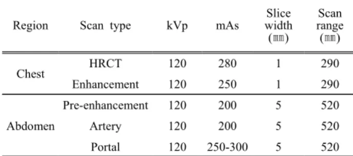
MultiDetector CT, Siemens, SOMATOM Sensation)를 이용하였고, 스캔 파라메터(Scan Parameter)와 검사 조건 등은 Table 1과 같다. 검사방법은 인체모형 팬 톰(ALDERSON RANDO)을 촬영대 위에 올려놓고, 환자 검사용 프로토콜을 적용하여 스캔을 하였다.
흉부 CT에서는 고분해능 CT(HRCT)와 조영제 주입 모드(Chest Enhancement)로 스캔 하였고, 복부 CT는 조영 주입 전과 동맥상(Artery Phase), 정맥상(Portal Phase), 지연기(Delayed Phase) 등을 가정하여 스캔 을 하였다. 팬톰을 이용한 흉부 CT와 복부 CT의 유효선량은 CT 모니터에 제시되는 선량-길이곱 (Dose-Length Product, DLP)에 유효선량전환계수를 곱하여 산출하였다. 유효선량(E)을 k × DLP (k는 전환계수이며 흉부 0.014, 복부 0.015이다)로 계산 하였다.[8]
Table 1. Scan protocols for chest and abdomen CTs.
Region Scan type kVp mAs
Slice width (㎜)
Scan range (㎜)
Chest HRCT 120 280 1 290
Enhancement 120 250 1 290
Abdomen
Pre-enhancement 120 200 5 520
Artery 120 200 5 520
Portal 120 250-300 5 520
2. 선량계 및 차폐체
광 자극 형광 선량계(OSLD; Optically Stimulated Luminescent dosimeter)를 이용하여 선량을 측정하 였다. 팬톰의 양쪽의 안구와 갑상선 위치에 OSLD
을 부착하여 CT 스캔하였으며, 차폐체를 사용하기 전과 후의 선량을 비교하였다. OSLD의 측정 가능 최소선량(Low Limit Dose)은 100 μSv이며, 에너지 범위는 5 keV ~ 20 MeV이고 정확도(Accuracy)는
±5%이다. 각 검사 당 10회 선량을 측정하여 평균값 을 구하였다.
산란선에 의한 피폭의 저감을 위해 바륨 및 텅스텐 시트, 고글(Goggle) 및 목 차폐체(Neck Protector)를 사용 하였다. 바륨 및 텅스텐 시트의 두께는 1 ㎜이고, 안구 와 갑상선 모양에 맞게 크기를 변형하여 사용하였다.
(a) (b)
Fig. 1. OSLDs attached on the human phantom (a) and shield sheet of barium covered eye balls and thyroid (b) to reduce the radiation dose.
Ⅲ. RESULT
1. 검사부위의 DLP와 유효선량
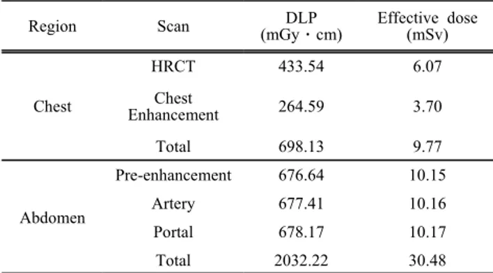
조영제 주입 전과 후로 2회 스캔한 흉부 CT에서 총 유효선량은 Table 2와 같이 9.77 mSv이었다. 스 캔당 평균 4.88 mSv였다. Axial mode로 스캔한 HRCT의 유효선량은 Helical mode로 스캔한 Chest enhancement의 유효선량 보다 1.64배 많았다. 조영 제 주입 전과 후, 지연상 총 3회 스캔에서는 복부 CT의 총 유효선량은 30.48 mSv이고 평균 스캔당 10.16 mSv였다. 이는 흉부 CT의 총 유효선량에 비 해 3.12배 많은 양이다. 복부 CT는 흉부 CT와 달리 각 촬영마다 비슷한 피폭선량을 보였다.
2. 흉부 CT에서의 갑상선과 안구의 표면선량 및 차폐효과
흉부 CT에서 안구는 3.01 mSv,갑상선은 6.21
"J. Korean Soc. Radiol., Vol. 13, No. 2, April 2019"
mSv의 피폭선량을 보었다. 갑상선은 일부 1차 엑 스선에 노출되었고 스캔 범위가 흉부와 가까워 안 구보다 2.06배 많은 선량을 보였다. 안구는 고글이 40.54%,갑상선은 목 차폐체가 55.23%로 차폐율 이 가장 높았다. 안구에서 완제품인 고글과 연구용 으로 제작한 바륨 시트의 차폐율을 비교하면 고글 이 3.2배 높다.
Table 2. DLP and Effective dose for the chest and the abdomen CT examinations.
Region Scan DLP
(mGyㆍcm)
Effective dose (mSv)
Chest
HRCT 433.54 6.07
Chest
Enhancement 264.59 3.70
Total 698.13 9.77
Abdomen
Pre-enhancement 676.64 10.15
Artery 677.41 10.16
Portal 678.17 10.17
Total 2032.22 30.48
Table 3. Dose and shield rate for the eyeball and the thyroid in the chest CT.
Region Shield Dose
(mSv)
Shield rate (%)
Eye ball
No shield 3.01±0.28
Barium sheet 2.63±0.05 12.63 Tungsten sheet 2.66±0.05 11.23
Goggle 1.79±0.22 40.54
Thyroid
No shield 6.21±0.25
Barium sheet 5.46±0.02 12.08 Tungsten sheet 5.40±0.04 13.05 Neck protector 2.78±0.06 55.23
3. 복부 CT에서의 갑상선과 안구의 표면선량 및 차폐효과
복부 CT에서 안구는 0.55 mSv,갑상선은 3.22 mSv의 피폭선량을 나타내었다. 갑상선은 스캔 범 위가 안구 보다 복부와 가까워 안구보다 5.85배 많 은 선량을 보였다. 거리에 의한 피폭선량 감쇠 효 과로 흉부 CT보다 차폐율이 2배 이상 높게 나타 났다. 갑상선에서 완제품인 목 차폐체와 연구용으 로 제작한 바륨 시트의 차폐율을 비교하면 목 차폐 체가 1.87배 높다.
Table 4. Dose and shield rate for the eyeball and the thyroid in the abdomen CT.
Region Shield Dose
(mSv)
Shield rate (%)
Eye ball
No Shield 0.55±0.01
Barium sheet 0.35±0.01 36.37 Tungsten sheet 0.36±0.01 34.55
Goggle 0.17±0.02 69.10
Thyroid
No Shield 3.22±0.05
Barium sheet 1.63±0.03 49.38 Tungsten sheet 1.62±0.05 49.69 Neck Protector 0.25±0.04 92.24
Ⅳ. DISCUSSION
흉부 CT 스캔 시 차폐를 하지 않고 스캔한 안구 는 3.01 mSv,갑상선은 6.21 mSv정도 측정 되었으 며 복부 CT 스캔 시 차폐를 하지 않고 스캔한 안구 는 0.55 mSv,갑상선은 3.22 mSv로 방사선피폭을 받았다. 이와 같은 검사가 반복, 지속적으로 이루어 진다면 방사선 피폭으로 인해 갑상선 암, 백내장 등 방사선 위해가 발생할 가능성이 높아질 수 있다.
바륨과 텅스텐 시트는 흉부 CT 검사 시 안구와 갑상선의 차폐율이 11~13%이었고, 복부 CT 검사 시에는 34~49%까지 선량 감소가 있었다. 안구와 갑상선에서의 흉부와 복부에서의 차폐율 차이는 1차선의 피폭과 거리에 의한 피폭선량 감쇠 효과 와 관계가 있어 차이가 발생하였다.
또한 차폐율이 높은 고글은 흉부 CT 검사 시 40%, 복부 CT 검사 시 69%,목 차폐체는 흉부 CT 검사 시 55%, 복부 CT 검사 시 92% 정도 선량 감소 효과가 있었다. 고글과 목 차폐체는 임상에서 사용 중인 완제품(고글의 납 성분 함유량은 0.07 mmPb, 목 차폐체는 0.5 mmPb)이기에 차폐율이 높 고 바륨 시트와 텅스텐 시트는 연구용으로 제작된 두께 1 mm이기에 차폐율이 낮다. 두께 5 mm로 제작한다면 완제품인 고글과 목 차폐체와 비슷한 차폐율을 나타낼 것이다.
본 연구의 제한점은 첫째, 흉부 및 복부 CT 검사 시 팬텀을 이용하여 AEC mode로 스캔을 하였기 때문에 실제 환자 선량은 이보다 더 높을 수 있고,
안구 및 갑상선의 선량도 더 높을 수 있다. 둘째, 64채널 MDCT 단일 기기를 이용하여 실험하였기 때문에 다른 기종의 CT 기기에 일괄적으로 동일한 차폐율을 적용할 수 없다. 셋째, 검사자에 따라 스 캔설정 범위를 다르게 하면 방사선 피폭선량 값이 달라진다.
Ⅴ. CONCLUSION
흉부 및 복부 CT 스캔 시 검사 목적부위 이외의 장기인 안구와 갑상선에 피폭되는 선량은 안구는 3.01 mSv,갑상선은 6.21 mSv 정도 측정 되었으며 복부 CT 스캔 시 차폐를 하지 않고 스캔한 안구는 0.55 mSv,갑상선은 3.22 mSv로 측정되었다.
방사선피폭 저감을 위해 바륨과 텅스텐 시트는 흉부 CT 검사 시 안구와 갑상선의 차폐율이 11~
13%이었고, 복부 CT 검사 시에는 34~49%까지 방 사선의 피폭이 저감되었다.
흉부 및 복부 CT 검사 시 방사선 피폭 정도가 상 당하기 때문에 검사가 반복, 지속적으로 이루어진 다면 방사선피폭으로 인해 갑상선 암, 백내장 등 방사선 위해가 발생할 가능성이 있어 검사 시 차폐 체를 사용하는 것이 요구된다.
Reference
[1] "Special medical device health insurance requisition present condition," Health Korea News, 19 SEP.
2010. http://www.hira.or.kr/main.do
[2] National research council of the National academies,
"Health Risks from Exposure to Low Levels of Ionizing Radiation: BEIR VII–Phase 2," Division on Earth and Life Studies, Board on Radiation Effects Research, Washington, DC: National Academy Press, 2005.
[3] D. A. Pierce, D. L. Preston, "Radiation-related cancer risks at low doses among atomic bomb survivors,"
Radiat Res, Vol. 154, No. 2, pp. 178-186, 2000.
[4] D. L. Preston, E. Ron, S. Tokuoka, S. Funamoto, N.
Nishi, M. Soda, K. Kodama, "Solid cancer incidence in atomic bomb survivors: 1958-1998," Radiat Res, Vol. 168, No. 1, pp. 58-64, 2007.
[5] D. J. McLaughlin, R. B. Mooney, "Dose reduction to radiosensitive tissues in CT. Do commercially available shields meet the users' needs?," Clinical Radiology, Vol. 59, No. 5, pp. 446-450, 2004.
[6] P. K. Cho, I. J. Choi, S. G. Chang, J. P. Chung, H.
Lee, J. S. Kim, W. H. Lee, "Assessment of the Eye Lens Dose Reduction by Bismuth Shields in Rando Phantom Undergoing CT of the Head," Journal of the Korean Society of Radiological Technology, Vol.
31, No. 2, pp. 171-175, 2008.
[7] M. Y. Jung, D. C. Kweon, S. I. Kwon, "Bismuth shielding effect of lens by using photoluminescence glass dosimeter in CT scan," Journal of the Korean Society of Radiological Technology, Vol. 32, No. 3, pp. 307-312, 2009.
[8] G. Bongartz, S. J. Golding, A.G. Jurik, M. Leonardi, EvP van Meerten, J. Geleijns, "European guidelines on quality criteria for computed tomography," EUR 16262, 2000. http://www.drs.dk/guidelines/ct/quality
"J. Korean Soc. Radiol., Vol. 13, No. 2, April 2019"
흉부 및 복부 CT 검사 시 안구와 갑상선의 방사선 피폭선량 저감
이준석,1 천권수1,2,*
1대구가톨릭대학교 일반대학원 방사선학과
2대구가톨릭대학교 방사선학과
흉부 및 복부 CT 검사 시 산란선에 의한 안구와 갑상선의 방사선 피폭선량을 측정하고, 피폭선량의 감소 를 위해 차폐체를 사용함으로써 방사선 피폭 정도를 조사하였다. 임상에서 사용되는 흉부 및 복부 CT 검사 프로토콜을 적용하여 안구와 갑상선의 차폐체 사용 전과 후의 선량을 측정하여 비교하였다. 안구와 갑상선 의 표면선량은 OSLD를 사용하여 측정하였다. 산란선을 차폐하기 위해 바륨, 텅스텐 시트와 고글과 목 차 폐체를 사용하였다. 흉부 CT 스캔 시 차폐를 하지 않고 스캔한 안구는 3.01 mSv,갑상선은 6.21 mSv로 측 정 되었고 복부 CT 스캔 시 차폐를 하지 않고 스캔한 안구는 0.55 mSv,갑상선은 3.22 mSv를 나타내었다.
바륨과 텅스텐 시트는 흉부 CT 검사 시 안구와 갑상선의 차폐율이 11~13%이었고, 복부 CT 검사 시에는 34~49%까지 방사선 피폭의 저감 효과가 있었다. 흉부 및 복부 CT 검사 시 방사선 피폭 정도가 상당하기 때문에 검사가 반복, 지속적으로 이루어진다면 방사선 피폭으로 인해 갑상선 암, 백내장 등 방사선 위해가 발생할 가능성이 있어 검사 시 차폐체를 사용하는 것이 요구된다.
중심단어: CT, 선량, 산란선, 차폐시트
성명 소속 직위
(제1저자) 이준석 대구가톨릭대학교 일반대학원 방사선학과 대학원생
(교신저자) 천권수 대구가톨릭대학교 방사선학과 교수
연구자 정보 이력