Celecoxib Enhances Susceptibility of Multidrug Resistant Cancer Cells to
17-Allylamino-17-demethoxy geldanamycin through Dual Induction of Apoptotic and Autophagic Cell Death
Hyun-Jung Moon, So-Young Park, Su-Hoon Lee, Chi-Dug Kang* and Sun-Hee Kim*
Department of Biochemistry, Pusan National University School of Medicine, Yangsan 626-870, Korea Received March 8, 2018 /Revised May 23, 2018 /Accepted May 29, 2018
Autophagy is a complex signaling process and has been implicated in tumor suppression and anti- cancer therapy resistance. Autophagy can produce tumor-suppressive effect by inducing autophagic cell death, either in collaboration with apoptosis. In this current study, we found that celecoxib (CCB), a nonsteroidal anti-inflammatory drug (NSAID) with multifaceted effects, induced autophagy includ- ing enhanced LC3 conversion (LC3-I to LC3-II) and reduced autophagy substrate protein p62 level in multidrug-resistant (MDR) cancer cells. CCB sensitized human multidrug resistant (MDR) cancer cells to the ansamycin-based HSP90 inhibitor 17-allylamino-17-demethoxygeldanamycin (17-AAG), a benzo- quinoid ansamycin, which causes the degradation of several oncogenic and signaling proteins, by in- ducing autophagic cell death and apoptosis. CCB significantly augmented 17-AAG-mediated level of LC3-II/LC-I, indicating the combined effect of 17-AAG and CCB on the induction of autophagy.
Autophagic degradation of mutant p53 (mutp53) and activation of caspase-3 in 17-AAG-treated MDR cells were accelerated by CCB. Inhibition of caspase-3-mediated apoptotic pathway by Z-DEVD-FMK, a caspase-3 inhibitor, did not completely block CCB-induced cell death in MCF7-MDR cells. In addi- tion, treatment of MDR cells with Z-DEVD-FMK failed to prevent activation of autophagy by com- bined treatment with 17-AAG and CCB. Based on our findings, the ability of clinically used drug CCB to induce autophagy has important implications for its development as a sensitizing agent in combina- tion with Hsp90 inhibitor of MDR cancer.
Key words : Apoptosis, autophagy, cancer, celecoxib, Hsp90 inhibitor
*Corresponding authors
*Tel : +82-51-510-8082, Fax : +82-51-510-8086
*E-mail : [email protected] (Chi-Dug Kang)
*Tel : +82-51-510-8081, Fax : +82-51-510-8086
*E-mail : [email protected] (Sun-Hee Kim)
This is an Open-Access article distributed under the terms of the Creative Commons Attribution Non-Commercial License (http://creativecommons.org/licenses/by-nc/3.0) which permits unrestricted non-commercial use, distribution, and reproduction in any medium, provided the original work is properly cited.
Journal of Life Science 2018 Vol. 28. No. 7. 778~785 DOI : https://doi.org/10.5352/JLS.2018.28.7.778
Introduction
Autophagy is an important cellular process involving the degradation of intracellular aggregated or misfolded pro- teins and damaged organelles through lysosomal machinery in response to stress or starvation [10]. To accurately esti- mate autophagic activity, it is essential to determine auto- phagic flux. LC3 is currently the most widely used autopha- gosome marker because the amount of LC3-II reflects the number of autophagosomes and autophagy-related structures.
Degradation of p62 is another widely used marker to mon- itor autophagic activity because p62 directly binds to LC3
and is selectively degraded by autophagy [25]. Deregulation of autophagy is implicated in several human diseases includ- ing cancers, and autophagy is a double-edged sword in tu- morigenesis, acting both as a tumor suppressor and a pro- tector of tumor cell survival, and therefore plays an im- portant role in the resistance of cancer cells to chemotherapy [1]. The pro-death or pro-survival roles of autophagy are highly dependent on the tumor type and treatment characteristics. Depending on the type of tumor and stage of disease, autophagy induces both cell survival and death during the initiation, progression, maturation and main- tenance of cancer [5]. It has been reported that autophagy is the major cellular route for the degradation of mutant p53 (mutp53) and controls mutp53 expression levels [4]. The ac- quired p53 mutations are the most common genetic alter- ations in human cancer, which are mostly missense mutations. Some p53 mutants not only result in the loss of wild-type p53 activity, but also may acquire new oncogenic properties known as gain-of-function. The mutation usually leads to the formation of mutp53 proteins, which often accu- mulate at high levels in cancer [8], especially in multi-
drug-resistant (MDR) cancer [6]. It has been reported that heat shock protein Hsp90 interacts with mutp53 and leads to its accumulation in tumors [12]. As the notion that mutp53 accumulation augments their oncogenic potential, the field of molecular modulators of mutp53 level and activity is gaining interest.
A large body of evidence has indicated that autophagy plays dual roles in MDR [11]. Autophagy protects MDR can- cer cells from apoptosis and promotes resistance to chemo- therapy treatment, and inhibition of autophagy may sensi- tize MDR cells to anticancer drugs. Conversely, autophagy can also play a pro-death role and trigger autophagic cell death in apoptosis-deficient MDR cells [11]. Under certain conditions, autophagy can be a scavenger in apoptosis- blocked signaling pathways, sensitizing MDR tumors to apoptosis. It has been reported that further stimulation of autophagy by rapamycin accelerates feroniellin A-induced apoptosis in MDR cells [7]. Coincidentally, autophagy in- duced by metformin could also assists TRAIL-mediated apoptosis in TRAIL-resistant lung adenocarcinoma [16].
Therefore, autophagic cell death that is induced by autoph- agy inducers could directly bypass apoptosis and ultimately eliminate MDR cells. It represents a new battle line in the fight against MDR cancer, and autophagy is a strong pro- pulsor to sensitize apoptosis-resistant MDR cells to anti- cancer drugs and reverse MDR [24]. It shows a novel bio- logical function of autophagy in MDR cancer cells and will enable the development of promising strategies to overcome MDR.
Nonsteroidal anti-inflammatory drugs (NSAIDs) are a structurally diverse group of drugs that are widely used to treat pain, inflammation, and fever, including acetylsalicylic acid, celecoxib (CCB), and acetaminophen. An increasing number of studies have indicated that NSAIDs have anti- cancer effect of cytotoxic drugs by down-regulation of P-gly- coprotein (P-gp) [23] and diverse effects in cancer that are mediated by the autophagy pathway [9, 13].
Since the exact mechanisms of the interaction between au- tophagy and MDR reversal remain obscure, we present here a novel strategy for killing MDR cells with high level of mutp53 through NSAID-induced autophagy, and sensitiza- tion of MDR cells to Hsp90 inhibitor17-AAG by NSAID.
Materials and Methods
Cell culture and reagents
Human MDR variants, HeyA8-MDR cells isolated from
HeyA8 human ovarian cancer cells and MCF7-MDR cells isolated from human breast cancer MCF-7 cells and their pa- rental cells were used, which were kindly provided by Dr.
Fiedler (MD Anderson, TX, USA). These cells were main- tained in DMEM supplemented with 10% fetal bovine serum and were incubated at 37℃, 5% CO2 and 95% humidity. 17- allylamino-17-demethoxy-geldanamycin (17-AAG) was pur- chased from Enzo Life Sciences Inc. (Farmingdale, New York, USA). Celecoxib (CCB), cycloheximide (CHX) and LY294002 were purchased from Sigma-Aldrich (St. Louis, MO, USA).
Caspase-3 inhibitor Z-DEVD-FMK was purchased from R &
D systems (Minneapolis, MN, USA).
Western blot analysis
Cell lysates from control and indicated drug-treated cells (1×10⁶ cells) were prepared using M-PER Reagent (Thermo Scientific Inc., USA). The lysates were clarified by centri- fugation. Equal amounts of protein were resolved by SDS- PAGE and immunoblotted with indicated antibodies. Wes- tern blot analysis was performed with specific primary anti- bodies against LC3 and p62 (Novus Biologicals, Littleton, CO, USA), Atg7, Beclin-1 and caspase-3 (Cell Signaling, Danvers, MA, USA), p53, PARP, (Santa Cruz Biotechnology, CA, USA), β-actin (Sigma-Aldrich, St. Louis, MO, USA) The p53 antibody (DO-1) is a mouse monoclonal antibody raised against amino acids 11-25 of p53 of human origin (Santa Cruz Biotechnology), which was recommended for detection of wild and mutant p53 of human origin.
Apoptosis assessment by Annexin V staining The effects of 17-AAG and/or CCB on apoptosis were measured by Annexin V staining. Cells (2×10⁵ cells/ml) were treated with 17-AAG in the presence or absence of CCB and/or caspase-3 inhibitor for indicated times. Then cells were centrifuged and resuspended in 100 μl of the staining solution containing Annexin V-fluorescein (FITC Apoptosis detection kit; BD ParMingen San Diego, CA, USA) and pro- pidium iodide (Sigma-Aldrich, St. Louis, MO, USA) in a Hepes buffer. After incubation at room temperature for 20 min, the percentage of early (annexin V positive/PI neg- ative) and late apoptotic cells (annexin V positive/PI pos- itive) was quantified by FACS for Annexin-V and PI staining.
Results
Relationship between autophagy and multidrug re- sistance of cancer cells
A B
Fig. 1. The basal and CCB-induced autophagy activities in MDR cells and their drug-sensitive parental counterparts. HeyA8- MDR and HeyA8 cells (A) and MCF-7 and MCF7-MDR cells (B) were untreated or treated with CCB (25 μM) for 24 hr, and levels of LC3-I/LC3-II, Beclin-1, Atg7 or p62 were determined by Western blot analysis. β-Actin (actin) was used as a loading control.
It has been reported that autophagy can be a double- edged sword for multidrug resistant (MDR) tumors and it protects cancer cells from chemotherapeutics but can also kill MDR cancer [11], and nonsteroidal anti-inflammatory drugs (NSAIDs) such as celecoxib (CCB), meloxicam, su- lindac, have diverse effects in cancer that are mediated by the autophagy pathway [26]. To explore the relationship be- tween autophagy and MDR, we measured the basal and CCB-induced autophagy activities in MDR cells and their drug-sensitive parental counterparts. We compared basal and CCB-induced levels of autophagic indicators such as two MAP1-LC3 forms (LC3-I and LC3-II) and p62 (high LC3-II/low p62 expression, indicative of intact activated au- tophagy), and autophagy-related genes such as Beclin-1 and Atg7 in between MDR and their parental cells. HeyA8/MDR cells showed a much higher ratio of LC3-II to LC3-I when compared with HeyA8 cells, indicating that HeyA8/MDR cells had a higher basal level of autophagy flux. In addition, the levels of Beclin-1 and Atg7 were much higher in HeyA8/
MDR cells compared to HeyA8 cells. Moreover, CCB-in- duced levels of LC3-II, Beclin-1 and Atg7 in HeyA8/MDR cells were more prominent than those in HeyA8 cells (Fig.
1A). Similar results were another MDR cells. Basal and CCB-induced level of LC3-II/LC3-I and p62, markers for the induction of autophagy, were determined in MCF7-MDR and parental MCF-7 cells. MCF7/MDR cells showed that the basal and CCB-induced level of LC3-II/LC-I was higher and basal p62 level was lower compared to those of parental MCF-7 cells (Fig. 1B), indicating that basal and CCB-induced autophagy levels were increased in MDR cells relative to
non-drug resistant parental cells.
Resistance of MDR cells to 17-AAG and sensitization of MDR cells to 17-AAG by celecoxib
Previously, we reported that MDR human cancer cells were significantly more resistant to Hsp90 inhibitors than their parental drug sensitive cells [9], and therefore it is nec- essary to develop modulators that overcome resistance of Hsp90 inhibitors and sensitize MDR cells to Hsp90 inhibitors.
We determined whether CCB could act as a new chemo- sensitizer for Hsp90 inhibitor-resistant MDR cells.
To compare the Hsp90 inhibitor 17-AAG-induced apopto- sis between HeyA8/MDR and HeyA8 cells, we estimated the percentage of early and late apoptotic cells in 17-AAG-ex- posed cells using conventional flow cytometry. In this ex- periment, treatment of HeyA8 or HeyA8/MDR cells, with gradually increasing concentrations of 17-AAG for 24 hr was accompanied by increased in the percentage of cells in both the lower and upper right quadrants of the Annexin/PI analysis. HeyA8/MDR cells were more resistant to 17-AAG- induced apoptosis than HeyA8 cells (Fig. 2). Therefore, we next determined whether CCB could enhance susceptibility of HeyA8/MDR cells to 17-AAG-induced apoptosis. When HeyA8/MDR cells were treated with 17-AAG (2 or 10 μM) in the presence or absence of 25 μM CCB for 36 hr, 17-AAG- induced apoptotic cell death was significantly enhanced by CCB, indicating dose-dependent sensitization of HeyA8/
MDR cells to 17-AAG-induced apoptosis by CCB (Fig. 3).
Acceleration of 17-AAG-induced autophagic mutant p53 degradation and caspase-3 activation by CCB
Since Hsp90 inhibitor induces autophagy as well as apop- tosis in cancer cells [14], we next examined whether combi- nation of Hsp90 inhibitor and CCB could modulate Hsp90 inhibitor-mediated changed levels of LC3-II/LC-I and mutp53 and caspase activity in HeyA8-MDR cells (Fig. 4A). CCB sig- nificantly augmented 17-AAG-mediated level of LC3-II/LC- I, indicating the combined effect of 17-AAG and CCB on the induction of autophagy. Since autophagy promoted the destabilization of mutp53 [3], we determined whether com- bined effect of 17-AAG and CCB resulted in down-regu- lation of mutp53. HeyA8-MDR cells showed that 17-AAG- mediated degradation/down-regulation of mutp53 was ac- celerated by CCB. As was expected, the cleavage of procas- pase-3 to active caspase-3 (CL caspase-3) was enhanced by co-treatment with 17-AAG and CCB versus 17-AAG alone.
Fig. 2. Comparison of 17-AAG-induced apoptosis in HeyA8-MDR and their drug-sensitive parental HeyA8 cells. These cells were treated with increasing doses of 17-AAG for 24 hr, and the percentages of early and late apoptotic cells were quantified by FACS for Annexin-V and PI staining. Images shown are representative of three independent experiments. The percentage of events in the nonapoptotic (lower left), early apoptotic (lower right), and late apoptotic (upper right) quadrants is indicated in the images.
B A
Fig. 3. Enhancement of 17-AAG-induced apoptosis in HeyA8- MDR cells by CCB. (A) HeyA8-MDR cells were treated with 17-AAG (2 or 10 μM) and/or 25 μM CCB for 36 hr, and early apoptosis and late apoptosis cells were analyzed by flow cytometry.
Representative profiles of Annexin-V and PI staining. (B) The percentage of early and late apoptotic cells is represented in histogram.
These results indicated that combined effect of 17-AAG and CCB on the induction of autophagy would accelerate 17- AAG-mediated mutp53 degradation by CCB, which caused activation of caspase-3 and subsequent apoptosis in MDR cells. Next, to examine whether CCB down-regulated mutp53 through post-translational degradation, change of mutp53
protein level in reyA8-MDR cells was determined in the presence of cycloheximide (CHX), a protein synthesis in- hibitor, after treatment with CCB at 3~6 hr. Mutp53 level in HeyA8-MDR cells was prominently decreased after co- treatment with CHX and CCB at 6 hr when compared to the cells treated with CHX alone (Fig. 4B), indicating that
A
B
Fig. 4. Acceleration of autophagic mutp53 degradation and acti- vation of caspase-3 activity of MDR cells treated with 17-AAG by CCB. (A) HeyA8-MDR cells were treated with 17-AAG (2 or 10 μM) and/or 25 μM CCB for 24 hr, and change levels of LC3-II, mutant p53 (mutp53), cleaved caspase-3 (Cl caspase-3) were determined by western blot analysis. (B) HeyA8-MDR cells were treated with cycloheximide (CHX, 20 μg/ml for 3~6 hr) in the absence or presence of 25 μM CCB. Changes in mutp53 levels were determined by Western blot analysis.
A
B
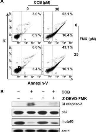
Fig. 5. Crosstalk between CCB-induced apoptotic and autopha- gic cell death. (A) MCF7-MDR cells were treated with 30 μM CCB in the absence or presence of 25 μM Z- DEVD-FMK for 24 hr, and the percentages of early and late apoptotic cells were quantified by FACS for Annexin- V and PI staining. Images shown are representative of three independent experiments. (B) Changed levels in Cl caspase-3, p62 and mutp53 of MCF7-MDR cells treated with 30 μM CCB and/or 25 μM Z-DEVD-FMK for 24 hr were determined by Western blot analysis.
CCB accelerates degradation of mutp53 protein in MDR cells, which consequently leads to down-regulate mutp53.
Effect of celecoxib on induction apoptotic and au- tophagic cell death in MDR cells
To determine the role of autophagy in CCB-induced cell death in MDR cells, we used caspase-3 inhibitor Z-DEVD- FMK to block apoptotic cell death in CCB-treated MCF7- MDR cells. The MDR cells were treated with CCB in the presence of Z-DEVD-FMK, and detection of apoptosis was measured by the staining of cells with annexin-V and PI for analysis on a flow cytometer. Treatment of MCF7-MDR cells with 30 μM CCB induced prominent apoptosis. Under this condition, Z-DEVD-FMK was not able to prevent completely CCB-induced apoptotic cell death (Fig. 5A), indicating the existence of a caspase independent type of cell death. When activation of caspase-3 was significantly blocked, but levels of p62 and mutp53 was not changed in MCF7-MDR cells treated with Z-DEVD-FMK alone. In MCF7-MDR cells co- treated with CCB and Z-DEVD-FMK, CCB-induced reduc- tion of p62 and mutp53 was sustained even if activation of caspase-3 was blocked (Fig. 5B). These results suggest that CCB can induce caspase-3-independent cell death, possibly
autophagic cell death. In addition, we determined whether the combined effect of 17-AAG and CCB on autophagy in- duction could be prevented by inhibiting autophagy. After co-treatment of MCF7-MDR cells with 17-AAG and CCB in the absence or presence of LY294002, an early stage autoph- agy inhibitor, we then examined changes in LC3-II and p62 levels. The up-regulation of LC3-II by the combined treat- ment of 17-AAG and CCB was significantly blocked by LY294002 (Fig. 6A), indicating that a combinations of 17-AAG with CCB for up-regulation of LC3B attenuated by autophagy inhibitor. We also determined whether the com- bined effect of 17-AAG and CCB on autophagy induction could be prevented by inhibiting caspase-dependent apopto- sis. After co-treatment of MCF7-MDR cells with 17-AAG and CCB in the absence or presence of Z-DEVD-FMK, we then
A
B
Fig. 6. Effect of caspase-dependent apoptosis or autophagy in- hibition on co-treatment of CCB and 17-AAG-induced autophagy in MDR cells. (A) MCF7-MDR cells were co-treated with 25 μM CCB and 10 μM 17-AAG in the presence or absence of 10 μM LY294002 for 24 hr, and changed levels of LC3-II and p62 were determined by western blot analysis. (B) MCF7-MDR cells were co- treated with 10 μM 17-AAG and 25 μM CCB in the pres- ence or absence of 25 μM Z-DEVD-FMK for 24 hr, re- spectively, and changed levels of caspase-3 and PARP cleavage (Cl caspase-3 and Cl PARP), LC3-II and p62 were determined by Western blot analysis.
examined changes in caspase-3 and PARP activity and au- tophagy-indicators LC3-II/LC-I and p62 (Fig. 6B). MCF7- MDR cells showed that an increase of LC3-II and a decrease of p62 induced by the combined treatment with17-AAG and CCB were not meaningfully altered even if activation of cas- pase-3 and subsequent PARP was blocked by Z- DEVD-FMK, showing that treatment of MDR cells with Z- DEVD-FMK failed to completely suppress activation of au- tophagy by combined treatment with 17-AAG and CCB.
These data demonstrated that cell death induced by com- bined treatment with 17-AAG and CCB is partially regulated by a caspase-3 independent, autophagy-based cell death mechanism in MDR cells. The acceleration of LC3-II increase and p62 decrease in combined treatment with 17-AAG and CCB was prevented by LY294002 and caspase-3 independent, autophagy-based cell death contributed to cell death in- duced by combined treatment with 17-AAG and CCB.
Discussion
In cancer, autophagy could serve as a pro-survival mecha- nism when cancer cells are subjected to damage by chemical or physical treatment. On the other hand, elimination of can- cer cells may also be mediated through autophagic cell death. The term autophagic cell death should be used when cell death is suppressed by the inhibition of autophagy using chemical inhibitors or genetic ablation (knockout or siRNA silencing of essential autophagy genes). If inhibiting autoph- agy does not prevent cell death, this process should not be called autophagic cell death [21].
In the present study, we found that celecoxib (CCB), one of commonly used NSAIDs, induced autophagy in MDR cells since treatment of MDR cells with CCB and led to up-regulation of LC3-II, Beclin-1 and Atg7 and down-regu- lation of p62 that plays a central role in autophagy induction and autophagy-mediated cell death, which contributed to potentiate Hsp90 inhibitor 17-AAG-induced cell death of MDR cells by CCB. Autophagy-mediated cell death is in- duced in apoptosis-resistant cells, particularly in the absence of the pro-apoptotic proteins Bax and Bak. and this type of cell death is inhibited by autophagy inhibitors or by silenc- ing autophagy genes such as Atg5 and Atg6 [20].
Our data showed that basal and CCB-induced autophagy levels were increased in HeyA8-MDR and MCF7-MDR cells possessing apoptosis- and MDR phenotype a relative to non-drug resistant parental HeyA8 and MCF-7 cells, re- spectively. Indeed, it has been reported that basic autophagy in P-glycoprotein (P-gp)-positive multidrug-resistant human leukemia K562/ADM cells was distinctly higher than that in K562 cells, which could be indicated by more cytosolic contents-packaged autophagic vacuoles, higher Beclin1 ex- pression level and LC3-II/LC3-I ratio, and lower p62 level in MDR K562/ADM cells [2], and expression level of LC3-II was higher in doxorubicin-resistant RPMI8226/DOX cells than in DOX doxorubicin sensitive RPMI8226/S cells [18].
On the other hand, emerging evidence arises that the in- crease in autophagy upon some agents could facilitate MDR reversal. Cysteamine-induced autophagy could reverse re- sistance of adriamycin-resistant MCF-7/ADR cells to doxor- ubicin in vitro and in vivo [24]. We also showed that autoph- agy-mediated chemosensitization by CCB could contribute to overcome 17-AAG resistance of HeyA8-MDR cells.
We previously found that NSAIDs such as CCB and ibu- profen induced autophagy as well as apoptosis and autopha-
gic degradation of mutp53 protein that closely associated with P-gp expressing cancer cells, and inhibition of Akt/
mTOR/p70S6K/4E-BP1 phosphorylation and p-STAT3 and Mcl-1 participates in the molecular events of CCB-induced autophagy. Although encouraging clinical responses have confirmed the potential of Hsp90 inhibitors such as 17-AAG, their therapeutic benefits were often limited by toxicity and resistance of cancer cells [17]. It has been reported that resist- ance to Hsp90 inhibitors is linked to P-gp-mediated efflux and to the induction of heat shock proteins (Hsps) [19]. We found that CCB could significantly increase sensitivity of HeyA8-MDR cells toward Hsp90 inhibitors and sensitize the MDR cells to Hsp90 inhibitors, representing CCB as a new chemosensitizer for Hsp90 inhibitors. It has been reported that autophagy promoted the destabilization of mutp53 [3].
Hsp90 is required for the post-translational stability of mutp53, and Hsp90 inhibitors promote the degradation of mutp53 [15], and accumulation of mutp53 in MDR cells are known to contribute to resistance to multiple anticancer agents including Hsp90 inhibitors [9]. It has been reported that autophagy is the main route for mutp53 degradation and autophagy controls mutp53 expression levels [4]. We present here a novel NSAID-induced strategy for killing MDR cells expressing high levels of mutp53 that offers the possibility of reducing elevated mutp53 level in MCF7-MDR cells by CCB treatment.
Induction of autophagy in cancer cells could induce cell death, which raises the possibility of developing anti-cancer strategies based on the synergistic modulations of autoph- agy and apoptosis may be regarded as a potential ther- apeutic approach [22]. Autophagic cell death occurs in- dependently of caspase activity and may be induced when the apoptosis pathway is inhibited. In the present study, treatment of MDR cells with Z-DEVD-FMK (a caspase-3 in- hibitor) failed to completely suppress CCB or combined treatment with 17-AAG and CCB-induced cell death and au- tophagy induction, indicating that apoptosis induced by CCB is partially regulated by a caspase-3 independent, au- tophagy-based cell death mechanism in MDR cells. In addi- tion, CCB-induced autophagic ability accelerated 17-AAG- induced autophagy through an increase of LC3-II level and a decrease of p62 level, which blocked by pretreating MDR cells with autophagy inhibitor LY29004, which indicates au- tophagy inhibition prevents autophagy induction by treat- ment of CCB alone or combined treatment of CCB with Hsp90 inhibitor 17-AAG.
Taken together, our present study suggest that CCB pro- mote both apoptotic and autophagic cell death and con- sequently trigger autophagy-mediated degradation of mutp53 and sensitization of MDR cells to Hsp90 inhibitor 17-AAG.
Acknowledgement
This work was supported by a 2-Year Research Grant of Pusan National University.
References
1. Chen, N. and Karantza, V. 2011. Autophagy as a therapeutic target in cancer. Cancer Bio. Thr. 11, 157-168.
2. Cheng, J., Chen, J., Xie, B. and Wei, H. L. 2013. Acquired multidrug resistance in human K562/ADM cells is asso- ciated with enhanced autophagy. Toxicol. Mech. Methods 23, 678-683.
3. Choudhury, S., Kolukula, V. K., Preet, A., Albanese, C. and Avantaggiati, M. L. 2013. Dissecting the pathways that de- stabilize mutant p53 The proteasome or autophagy? Cell Cycle 12, 1022-1029.
4. Garufi, A,, Pucci, D., D'Orazi, V., Cirone, M., Bossi, G., Avantaggiati, M. L. and D'Orazi, G. 2014. Degradation of mutant p53H175 protein by Zn(II) through autophagy. Cell Death Dis. 5, e1271.
5. Helgason, G. V., Karvela, M. and Holyoake, T. L. 2011. Kill one bird with two stones: potential efficacy of BCR-ABL and autophagy inhibition in CML. Blood 118, 2035-2043.
6. Huang, J. M., Sheard, M. A., Ji, L. Y., Sposto, R. and Keshela- va, N. 2010. Combination of vorinostat and flavopiridol is selectively cytotoxic to multidrug-resistant neuroblastoma cell lines with mutant TP53. Mol. Cancer Ther. 9, 3289-3301.
7. Kaewpiboon, C., Surapinit, S., Malilas, W., Moon, J., Phu- wapraisirisan, P., Tip-Pyang, S., Johnston, R. N., Koh, S. S., Assavalapsakul, W. and Chung, Y. H. 2014. Feroniellin A-in- duced autophagy causes apoptosis in multidrug- resistant human A549 lung cancer cells. Int. J. Oncol. 44, 1233-1242.
8. Kastan, M. B. and Berkovich, E. 2007. p53: a two-faced can- cer gene. Nat. Cell Biol. 9, 489-491.
9. Kim, H. B., Lee, S. H., Um, J. H., Oh, W. K., Kim, D. W., Kang, C. D. and Kim, S. H. 2015. Sensitization of multi- drug-resistant human cancer cells to Hsp90 inhibitors by down-regulation of SIRT1. Oncotarget 6, 36202-36218.
10. Kroemer, G., Marino, G. and Levine, B. 2010. Autophagy and the integrated stress response. Mol. Cell. 40, 280-293.
11. Li, Y. J., Lei, Y. H., Yao, N., Wang, C. R., Hu, N., Ye, W.
C., Zhang, D. M. and Chen, Z. S. 2017. Autophagy and mul- tidrug resistance in cancer. Chin. J. Cancer 36, 52.
12. Lin, K., Rockliffe, N., Johnson, G. G., Sherrington, P. D. and Pettitt, A. R. 2008. Hsp90 inhibition has opposing effects on wild-type and mutant p53 and induces p21 expression and
초록:Celecoxib의 apoptotic 및 autophagic cell death 유도에 의한 항암제 다제내성 암세포의 17-allylamino-17-demethoxygeldanamycin 감수성 증강
문현정․박소영․이수훈․강치덕*․김선희*
(부산대학교 의학전문대학원 의과학과 생화학교실)
오토파지(Autophagy, 자가포식)는 복합적인 신호과정으로, 암세포의 증식 억제 및 항암제에 대한 내성 획득의 상반적인 조절에도 관여한다. 오토파지의 암 억제 효과는 아팝토시스(apoptosis)와 상호협력으로 오토파지성세포 사멸의 유도에 기인된다. 본 연구에서는 NSAID 계열의 다기능 약물인 celecoxib (CCB)이 아팝토시스 및 오토파 지의 복합적인 유도로, 항암제 다제내성(multidrug resistant, MDR) 암세포의 Hsp90 molecular chaperone in- hibitor인 17-allylamino-17-demethoxygeldanamycin (17-AAG)에 대한 감수성을 증가시키는 활성이 있음을 밝혔 다. 17-AAG 처리에 의한 항암제 다제내성 암세포의 변이형p53 분해 및 caspase-3 활성은 CCB 처리로 촉진되었 다. MCF7-MDR세포에서 Z-DEVD-FMK 처리에 의한 caspase-3-매개의 아팝토시스 경로 차단은 CCB 유도의 세포 사멸을 완전히 차단시키지 못함을 알 수 있었으며, 또한 17-AAG과 CCB 병합 처리에 의한 오토파지 활성화는 Z-DEVD-FMK에 의해 방해되지 않는 것을 알 수 있었다. 본 연구의 결과를 토대로, CCB의 오토파지 유도 활성은 항암제 다제내성 암의 Hsp90 inhibitor에 대한 감수성 증가를 위한 약물 개발에, CCB가 효과적인 병용 약물로서 제안 될 수 있다.
cytotoxicity irrespective of p53/ATM status in chronic lym- phocytic leukaemia cells. Oncogene 27, 2445-2455.
13. Liu, M., Li, C. M., Chen, Z. F., Ji, R., Guo, Q. H., Li, Q., Zhang, H. L. and Zhou, Y. N. 2014. Celecoxib regulates apoptosis and autophagy via the PI3K/Akt signaling path- way in SGC-7901 gastric cancer cells. Int. J. Mol. Med. 33, 1451-1458.
14. Mori, M., Hitora, T., Nakamura, O., Yamagami, Y., Horie, R., Nishimura, H. and Yamamoto, T. 2015. Hsp90 inhibitor induces autophagy and apoptosis in osteosarcoma cells. Int.
J. Oncol. 46, 47-54.
15. Muller, P., Hrstka, R., Coomber, D., Lane, D. P. and Vojtesek, B. 2008. Chaperone-dependent stabilization and degradation of p53 mutants. Oncogene 27, 3371-3383.
16. Nazim, U. M. D., Moon, J. H., Lee, J. H., Lee, Y. J., Seol, J. W., Eo, S. K., Lee, J. H. and Park, S. Y. 2016. Activation of autophagy flux by metformin downregulates cellular FLICE-like inhibitory protein and enhances TRAIL-induced apoptosis. Oncotarget 7, 23468-23481.
17. Neckers, L. and Workman, P. 2012. Hsp90 molecular cha- perone inhibitors: Are we there yet? Clin. Cancer Res. 18, 64-76.
18. Pan, Y. Z., Wang, X., Bai, H., Wang, C. B., Zhang, Q. and Xi, R. 2015. Autophagy in drug resistance of the multiple myeloma cell line RPMI8226 to doxorubicin. Genet. Mol. Res.
14, 5621-5629.
19. Piper, P. W. and Millson, S. H. 2011. Mechanisms of resis- tance to Hsp90 inhibitor drugs: a complex mosaic emerges.
Pharmaceuticals (Basel) 4, 1400-1422.
20. Shimizu, S., Kanaseki, T., Mizushima, N., Mizuta, T., Araka- wa-Kobayashi, S., Thompson, C. B. and Tsujimoto, Y. 2004.
Role of Bcl-2 family proteins in a non-apoptotic program- med cell death dependent on autophagy genes. Nat. Cell Biol. 6, 1221-1228.
21. Shimizu, S., Yoshida, T., Tsujioka, M. and Arakawa, S. 2014.
Autophagic cell death and cancer. Int. J. Mol. Sci. 15, 3145- 3153.
22. Sui, X., Chen, R., Wang, Z., Huang, Z., Kong, N., Zhang, M., Han, W., Lou, F., Yang, J., Zhang, Q., Wang, X., He, C. and Pan, H. 2013. Autophagy and chemotherapy resist- ance: a promising therapeutic target for cancer treatment.
Cell Death Dis. 4, e838.
23. Takara, K., Hayashi, R., Kokufu, M., Yamamoto, K., Kitada, N., Ohnishi, N. and Yokoyama, T. 2009. Effects of nonster- oidal anti-inflammatory drugs on the expression and func- tion of P-Glycoprotein/MDR1 in Caco-2 cells. Drug Chem.
Toxicol. 32, 332-337.
24. Wan, X. M., Zheng, F., Zhang, L., Miao, Y. Y., Man, N. and Wen, L. P. 2011. Autophagy-mediated chemosensitization by cysteamine in cancer cells. Int. J. Cancer 129, 1087-1095.
25. Yoshii, S. R. and Mizushima, N. 2017. Monitoring and meas- uring autophagy. Int. J. Mol. Sci. 18, 1865.
26. Yu, C., Li, W. B., Liu, J. B., Lu, J. W. and Feng, J. F. 2018.
Autophagy: novel applications of nonsteroidal anti-inflam- matory drugs for primary cancer. Cancer Med. 7, 471-484.