이 논문은 2011년 6월 1일 접수하여 2011년 8월 1일 채택되었음.
책임저자:장준영, 삼성서울병원 방사선종양학과 Tel: 02)3410-2596, Fax: 02)3410-2619 E-mail: [email protected]
전뇌 방사선치료 시 치료방법에 따른 두피선량평가
삼성서울병원 방사선종양학과
장준영ㆍ박수연ㆍ김종식ㆍ최병기ㆍ송기원
목 적: 뇌전이 환자에게 시행하는 고전적인 헬멧(Helmet field)형태의 대향2문방사선조사는 환자두피에 과 선량을 일으키며 이 는 탈모의 원인이 된다. 이에 본 연구에서는 환자두피를 보호하기 위한 두피보호 형태(Scalp-shielding shape)의 대향 2문 조 사와 토모 치료법을 고전적 방사선치료법과 비교하여 보다 효과적인 탈모 예방의 두피선량을 정량적으로 분석하고자 한다.
대상 및 방법: 두피선량은 RANDO 팬톰을 사용하여 열형광선량계를 전두엽의 중심선에 따라서 5개를 위치시킨 후, 세 가지 치료방법(HELMET, MLC, TOMO)으로 피부선량을 측정하였고, 전산화치료계획장치(Pinnacle3, Philips Medical System, USA), 6 MV X선(Clinac 6EX, VARIAN, USA)을 이용하여 방사선치료계획을 수립한 후, DVH로 선량분포변화와 두피선량을 비교 분석하 였다.
결 과: 열형광선량계를 사용하여 두피의 표면선량을 측정한 결과 기존의 HELMET field 치료방법과 비교했을 때 MLC technique에서는 각 포인트 지점에서 평균 87.44% 두피선량이 감소하는 것으로 측정되었으며 TOMO에서는 평균 88.03% 두피 선량이 감소하는 것으로 측정되었다. 또한 세 가지 치료방법의 두피내의 과다선량 영역(Hotspot)의 존재정도를 평가하기 위해 선량체적히스토그램(Dose volume histogram, DVH)을 사용하여 처방선량의 95%, 100%, 105%가 받는 용적의 백분율(Per- centage of volume: V95, V100, V105)을 계산한 결과 MLC technique과 TOMO plan에서 과다선량 없이 Dose coverage가 우수 함을 보여주었다.
결 론: 전뇌 방사선치료를 받는 환자에게 탈모현상을 줄여주는 것은 환자의 삶의 질을 높여주는 측면에서 중요하다. 이에 본 실험결과를 바탕으로 두피보호 형태(Scalp-shielding shape)의 대향 2문 조사와 토모 치료법을 사용하여 환자의 두피를 보호함 으로써 환자의 탈모현상을 줄여주는 효과를 기대할 수 있을 것이라 사료된다.
핵심용어: 전뇌 방사선치료, 두피선량, 탈모
서 론
뇌 전이(Brain metastasis)란 뇌 바깥에서 생긴 암세포로부 터 뇌종양(Brain cancer)이 발생한 것이다.1) 뇌 전이(Brain metastasis)는 흔한 암의 부작용(Side effect) 중 하나이며 그 발생률 또한 증가하는 것으로 보인다. 또한 인간의 평균수명 이 연장되고 원발병소(Primary lesion)의 암에 대한 치료방법 이 진보됨에 따라서 암 환자의 생존기간이 연장되어 전이성 뇌종양을 다루게 되는 빈도가 점차 증가되고 있다.2) 암 환자 의 25∼30%가 병의 기간 중에 두 개강 내 전이를 일으킨다 고 생각되며 대략 전체 두 개강 내 종양의 약 10∼15%를 차 지한다. 신체의 암이 두 개강 내로 전이하는 성향은 종양에 따라 각기 다르며 두 개강 내에서도 특정부위에 전이를 잘하
는 성향이 종양에 따라 다르다. 그 중 폐암(Lung cancer)이 가장 많아 약 40∼50%를 차지하며 유방암(Breast cancer)이 다음으로 많아 약 10∼15% 정도이다.3) 이러한 뇌 전이(Brain metastasis)에 대한 치료방법에는 여러 가지가 있다. 어느 한 가지 방법에 대한 효과를 정확하게 평가하기 어려운 경우가 많아서 대개 한 가지 이상의 복합적인 치료를 한다. 그 중에 서 환자의 남은 삶의 질을 높일 수 있을 뿐만 아니라 신경학 적 증상을 완화시키고 생존기간도 연장할 수 있는4,7) 전체 뇌 에 방사선을 조사하는 치료, 즉 전체 뇌 방사선치료(WBRT) 가 대부분의 뇌전이(Brain metastasis) 환자에게 시행된다. 이 러한 전체 뇌 방사선치료(WBRT)에서 나타나는 부작용(Side effect) 중 하나인 탈모(Alopecia)는 앞서 말한 것처럼 환자의 남은 삶의 질을 높이는데 중요한 부분을 차지한다.5) 대부분 의 전체 뇌 방사선치료(WBRT)는 생존기간이 짧기 때문에 환 자의 탈모(Alopecia)를 고려하지 않은 채 치료가 진행 되고 있다.6) 하지만 환자의 남은 삶의 질을 높이는데 탈모 (Alopecia)는 중요한 부분을 차지하기 때문에 반드시 이 부분
Fig. 1. Phatograph of the clinac 6EX, Tomothearpy Hi-Art.
Fig. 2. Photograph of a humannoid phantom (Rando, USA).
을 고려하여야 한다.8,9) 이에 본 저자는 일반적으로 탈모 (Alopecia)를 고려하지 않은 채 치료를 하는 방법(HELMET field)과 멀티다엽콜리메이터(MLC)를 사용하여 환자두피 (Scalp)를 보호함으로써 탈모(Alopecia)현상을 줄여주는 방법 (MLC Technique, Tomothearpy)으로 치료계획을 세워 환자의 두피(Scalp)에 들어가는 선량을 측정하여 비교함으로써 환자 의 탈모(Alopecia)현상을 줄여주는 방법을 모색하고자 한다.
대상 및 방법 1. 사용된 장비
측정에 사용된 장비는 선형가속기(Clinac 6EX. VARIAN, USA)와 Tomothearpy Hi-Art이며 측정에너지는 6 MV X선 을 사용하였다(Fig. 1). 뇌전체가 포함된 CT 스캔을 슬라이 스두께 5 mm로 하여 3차원 치료계획용 컴퓨터(Pinnacle3, Philips Medical System, USA)로 전송하였다.
2. 두피선량 측정방법
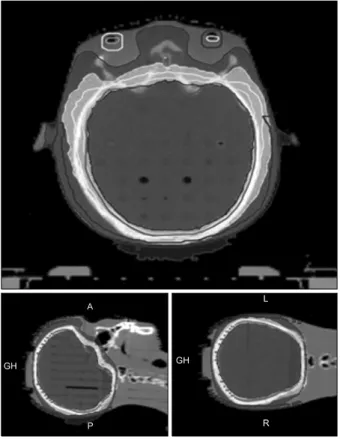
두피선량은 인체모형팬톰(Anderson Rando Phantom, Al- derson Reserch Laboratories Inc., USA)에 열형광선량계(4000, Harshaw, Solon Technologies Inc., USA)를 Midline frontal lobe를 따라서 일정간격으로 다섯 개를 위치시킨 후, 각각에 서의 측정값을 측정하였다(Fig. 2). 열형광선량계의 측정 Point는 각 치료방법에 동일하게 측정하였으며 열형광선량계 의 측정오차를 감안 통계상의 오차를 줄이기 위하여 3회 반 복하여 측정하여 평균값을 얻어 결과치를 산출하였다.
3. Contouring 및 방사선치료계획
3차원 치료계획용 컴퓨터와 Tomotherapy 치료계획 컴퓨터
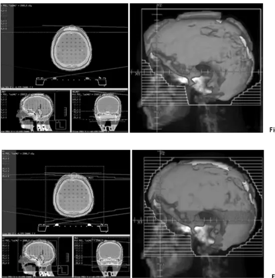
를 이용하여 세 가지 방법에 대한 임상표적체적(CTV, 전체 뇌)과 두피의 Contour를 그렸다. 6 MV X선을 사용하여 HELMET field는 대향이문조사 하였고(Fig. 3) MLC TECHNIQUE은 HELMET field와 같은 방법으로 하되 CTV margin 1.5 cm까지 MLC를 사용하였다(Fig. 4). TOMO는 TOMO therapy planning system을 사용, lens, eyeball, scalp ROI 설정하여 CTV 30 Gy, lens 5 Gy, eyeball 15 Gy, scalp 10 Gy constrain 하였다(Fig. 5).
4. 방사선치료계획에 대한 분석 및 평가
세 가지 방법에 대한 치료계획 결과를 토대로 하여 선량체 적히스토그램(Dose-volume histogram, DVH)을 분석하여 이 루어졌다. 두피내의 과다선량 영역(Hotspot)의 존재정도를
Fig. 4. MLC technique.
Fig. 3. HELMET field.
평가하기 위해 처방선량의 95%, 100%, 105%가 받는 용적의 백분율(Percentage of volume: V95, V100, V105, V110)을 계산하 였다.
결 과 1. 두피선량측정결과
HELMET field, MLC technique, TOMO therapy 세 가지 치료방법에 모두 같은 위치에 TLD를 위치시켜 놓고 측정한 결과 HELMET field와 비교했을 때 MLC technique에서는 각 포인트 지점에서 87.44%로 두피선량이 감소하는 것으로 측정되었으며 TOMO에서는 이보다 높은 88.03% 두피선량이 감소하는 것으로 측정되었다(Table 1).
2. 선량체적히스토그램(Dose-volume histogram, DVH) 분석
선량체적히스토그램(Dose volume histogram, DVH)을 사용 하여 처방선량의 95%, 100%, 105%가 받는 용적의 백분율 (Percentage of volume: V95, V100, V105)을 계산한 결과 MLC technique과 TOMO plan에서 과다선량 없이 Dose cov- erage가 우수함을 보여주었다(Fig. 6).
고안 및 결론
뇌전이(Brain metastasis)환자의 남은 삶의 질을 높일 수 있 을 뿐만 아니라 신경학적 증상을 완화시키고 생존기간도 연 장할 수 있는 전체 뇌 방사선치료(WBRT)는 대부분의 뇌전 이(Brain metastasis)환자에게 시행한다. 그러나 전체 뇌 방사 선치료(WBRT)의 부작용(Side effect) 중 하나인 탈모(Alope-
Table 1. TLD read result (cGy)
TLD reading
HELMET MLC TOMO
A B C D E
117 18.7 17.7
175.5 19.7 19.5
235.4 21.5 20.9
117.5 17.6 16
172.2 19.8 19.2
Fig. 5. TOMO technique.
Fig. 6. DVH of HELMET, MLC, TOMO.
cia)현상에 대해서는 크게 고려를 하지 않는 것이 사실이다.
하지만 남은 삶의 질을 높이는 게 목적인만큼 환자의 탈모 (Alopecia)현상을 줄여주는 것은 전체 뇌 방사선치료(WBRT) 를 받는 환자에게 중요한 부분이다. 탈모(Alopecia)현상을 줄 여주는 것은 전체 뇌 방사선 치료(WBRT) 시 두피(Scalp)에 들어가는 방사선량을 줄여줌으로써 해결할 수 있을 것이라 사료 된다. 이에 본 저자는 일반적으로 두피선량(Scalp dose) 을 고려하지 않은 HELMET field 방사선치료와 두피선량 (Scalp dose)을 줄여줄 수 있는 MLC technique과 TOMO therapy를 비교하여 두피선량(Scalp dose)차이를 알아보았다.
MLC technique과 TOMO therapy는 두피(Scalp) 주위의 산란
선(Scatter)을 줄여줌으로써 HELMET field 방사선치료와 비 교했을 때 두피선량(Scalp dose)이 현저히 떨어졌으며 이로 인해 탈모(Alopecia)가 발생하는 한도선량(Tolerance dose)을 낮춰줄 수 있었다. 그러므로 MLC Technique과 TOMO ther- apy를 사용하여 탈모(Alopecia)를 줄여줌으로써 환자의 남은 삶의 질을 높일 수 있을 것이라 사료된다.
참고문헌
1. Roberge D, Parker W, Niazi TM, Oliveres M: Treating the contents and not the container: dosimetric study of hair- sparing whole brain intensity modulated radiation therapy.
Technology in Cancer Research and Treatment 2005;4:567- 570
2. Orton N, Jaradat H, Welsh JS, Tome W: Whole scalp irra- diation using helical tomotherapy. Medical Dosimetry 2005;
30:162-168
3. Abdel-Rahman W, Seuntjens JP, Verhaegen F, Deblois F, Podgorsak EB: Validation of Monte Carlo calculated surface doses for megavoltage photon beams. Med Phys 2005;32:
286-298
4. Mehta MP, Khuntia D: Current strategies in whole-brain ra- diation therapy for brain metastases. Neurosurgery 2005;57 (Suppl 5):S33-44
5. Radiation Therapy Oncology Group: RTOG active protocols.
Available at: http://rtog.org/members/protocols/0214/0214.pdf.
Accessed July 25, 2006
6. Andrews DW, Scott CB, Sperduto PW, et al.: Whole brain radiation therapy with or without stereotactic radiosurgery boost for patients with one to three brain metastases: Phase III results of the RTOG 9508 randomised trial. Lancet
2004;363:1665-1672
7. Welsh JS, Patel RR, Ritter MA, Harari P, Mackie TR, Mehta MP: Helical tomotherapy: an innovative technology and approach to radiation therapy. Technology in Cancer Research and Treatment 2002;1:55-63
8. Welsh J, Olivera G, Hui S, et al.: Helical tomotherapy with
conformal avoidance appears superior to 3-D DRT and IMRT for treatment of complex tumor volumes. Radiology 2002;225:360
9. Mehta MP, Khuntia D: Current strategies in whole-brain ra- diation therapy for brain metastases. Neurosurgery 2005;
57(Suppl 5):S33-44
Abstract
Scalp Dose Evaluation According Radiation Therapy Technique of Whole Brain Radiation Therapy
Joon Yung Jang, Soo Yun Park, Jong Sik Kim, Byeong Gi Choi, Gi Won Song Department of Radiation Oncology, Samsung Medical Center, Seoul, Korea
Purpose: Opposing portal irradiation with helmet field shape that has been given to a patient with brain metastasis can cause excess dose in patient’s scalp, resulting in hair loss. For this reason, this study is to quantitatively analyze scalp dose for effective prevention of hair loss by comparing opposing portal irradiation with scalp-shielding shape and tomotherapy designed to protect patient’s scalp with conventional radiation therapy.
Materials and Methods: Scalp dose was measured by using three therapies (HELMET, MLC, TOMO) after five thermo-luminescence dosimeters were positioned along center line of frontal lobe by using RANDO Phantom. Scalp dose and change in dose distribution were compared and analyzed with DVH after radiation therapy plan was made by using Radiation Treatment Planning System (Pinnacle3, Philips Medical System, USA) and 6 MV X-ray (Clinac 6EX, VARIAN, USA).
Results: When surface dose of scalp by using thermo-luminescence dosimeters was measured, it was revealed that scalp dose decreased by average 87.44% at each point in MLC technique and that scalp dose decreased by average 88.03% at each point in TOMO compared with HELMET field therapy. In addition, when percentage of volume (V95%, V100%, V105% of prescribed dose) was calculated by using Dose Volume Histogram (DVH) in order to evaluate the existence or nonexistence of hotspot in scalp as to three therapies (HELMET, MLC, TOMO), it was revealed that MLC technique and TOMO plan had good dose coverage and did not have hot spot.
Conclusion: Reducing hair loss of a patient who receives whole brain radiotherapy treatment can make a contribution to improve life quality of the patient. It is expected that making good use of opposing portal irradiation with scalp-shielding shape and tomotherapy to protect scalp of a patient based on this study will reduce hair loss of a patient.
Key words: whole brain radiotherapy treatment, scalp dose, hair loss