서 론
방사선치료의 목표는 정상 조직의 장해 확률(Normal Tissue Complication Probability, NTCP)은 가능한 작 게 하고, 종양 조직에 처방된 방사선량은 증가시킴으로써 책임저자: 백민규, 창원경상대학교병원 방사선종양학과 경상남도 창원시 성산구 삼정자로11 Tel: 055-214-3211 E-mail: [email protected]
두경부암의 영상유도방사선치료에서 ExacTrac의 유용성 평가
창원경상대학교병원 방사선종양학과 백민규·김민우·하세민·채종표·조광섭·이상봉 목 적: 현대 방사선치료기술에서 종양표적위치 및 정상장기에 정확한 선량을 전달하기 위해 여러 방법의 영 상유도방사선치료(Image Guided Radiation Therapy, IGRT)가 사용되고 있으며 그 중 선형가속기에 장착된 CBCT(Cone Beam Computed Tomography, CBCT)와 이외 장치인 ExacTrac(ExacTrac X-ray System)이 있다. 두 시스템을 비교한 이전 연구들에서는 Offline-review 이용하여 후향적으로 팬텀 및 환자의 Set-up 오차를 분석하거나 X, Y, Z 축과 하나의 회전방향(Couch Rotation)으로만 연구되어졌다. 본 연구에서는 Head and Neck Cancer 환자를 대상으로 CBCT와 ExacTrac을 이용하여 한 치료중심센터에서 각각 6 DoF(Degree Of Freedom) IGRT를 시행한 후, 두 IGRT 장비에서 나타난 팬텀 및 환자의 Set-up 오차, 환자 Set-up에 걸리는 시간, 노출 방사선량의 비교를 통해 유용성을 평가하고자 한다. 대상 및 방법: Rando Phantom을 이용하여 환자 움직임을 배제한 상태의 Set-up 오차 평가와 Head and Neck Cancer 환자의 Set-up 오차 값 두 가지 경우로 나누어 획득하였다. 노출 방사선량 평가는 유리선량계 로 하였다. 환자 Set-up 후 IGRT 시행하는데 소요되는 시간을 평가하기 위해 Head and Neck Cancer 환자 11명을 대상으로 하였다. 총 치료기간동안 환자 당 평균 10회의 CBCT와 ExacTrac 영상을 동시에 얻었고, 관 심영역지정(Region Of Interest, ROI) 설정 후 6D 온라인 자동위치교정(Online Automatching) 값의 차를 6 개의 축(Translation group: SI, AP, LR; Rotation group: Pitch, Roll, Rtn)으로 각각 계산하였다. 결 과: Phantom과 환자에서 Set-up 오차는 Translation group에서 1 mm 미만, Rotation group에서 1.5° 미만의 차이가 보였으며, Rtn 값을 제외한 다른 모든 축의 RMS 값이 1 mm, 1° 미만으로 나타났다. 각 시스 템에서 최종적으로 Set-up 오차 교정까지 걸리는 시간은 CBCT를 이용한 IGRT에서는 평균 256±47.6 sec, ExacTrac을 이용 시 평균 84±3.5 sec로 각각 나타났다. 1회 치료 당 IGRT에 의한 방사선 노출선량은 Head and Neck 부위 7곳의 측정위치 중 Oral Mucosa에서 CBCT와 ExacTrac이 각각 2.468 mGy, 0.066 mGy로 상대적으로 ExacTrac에 비해 피폭선량이 37배 높게 측정되었다. 결 론: CBCT와 ExacTrac 두 시스템 간의 6D 온라인 자동위치교정을 통해 Set-up 오차는 두 시스템의 자체 적인 Systematic error 뿐 아니라, 환자 움직임(Random error)를 포함한 Set-up 오차가 1 mm, 1.02° 미만 으로 나타났다. 이는 본원에서 Head and Neck IMRT 치료 시 PTV Margin이 3 mm이라는 것을 고려했을 때, 이 오차범위는 합리적으로 사료된다. 하지만 치료기간 동안 환자체중변화로 인한 따른 표적, 손상위험장기의 변화를 고려했을 때 CBCT와 적절히 병용하여 사용하는 것이 좋을 것으로 사료된다. ▶ 핵심용어: 영상유도방사선치료, 6D 온라인 자동위치교정(6D Online Automatching), CBCT, ExacTrac종양 치유 확률 (Tumor Control Probability, TCP)을 증 가시키기 위해 방사선치료기술들은 발전되고 있다.(1)
그 중엔 세기변조방사선치료(Intensity Modulate Ra-diation Therapy, IMRT), 용적변조회전방사선치료(Vol-ume Modulate Arc Therapy, VMAT)와 같은 치료기술 이 있으며, 이는 기존의 고식적인 방사선치료보다 종양표적 에 선량분포를 최적화시켜 주기는 하지만 선량계산 오차, 환자 set-up 오차 등 여러 요인에 의해 오차가 발생하게 됨 으로써 선량 분포도가 급격하게 변함에 따라 종양표적에 선량이 불충분하게 조사되거나 정상장기에 더 많은 선량 이 조사되어 장해를 일으킬 가능성이 있다.(2) 이에 환자 Set-up 오차를 최소화하기 위해 영상유도방 사선치료 장치들의 하드웨어 및 소프트웨어 기술이 개발되 고 있다.(3,4,5)
영상유도방사선치료(Image-Guided Radiation Ther-apy: IGRT)란 1차적으로 치료실내에서 환자 피부표시 (Skin Marking)에 의한 Set-up 후 대상 부위에 X-선 영 상이나 단층영상을 획득하여 치료 전 치료계획영상과 비 교, 교정함으로써 환자 Set-up으로 인한 오차를 최소화 하고, 치료계획 영상과 최대한 일치시켜 치료의 정확성을 향상시키는 방법이다. 선량전달의 정확성을 향상시키기 위 해서는 환자 Set-up 오차와 기계적 불확실성을 최소화 하 는 것이 무엇보다 중요하다.(6,7,8) 영상유도방사선치료 장비들 중에는 Varian사의 True-beam Stx 2.5에 장착되어있는 전자포탈영상장치(Elec-tronic Portal Imaging Device, EPID), OBI(On-Board Imager), 그리고 BrainLaB사의 ExacTrac(ExacTrac X-ray System)과 같은 장치들이 있다.(9,10) OBI를 활용한 콘
빔형 전산화단층촬영(Cone Beam Computed-Tomog-raphy, CBCT)은 3차원으로 영상을 재구성하여 연부조직 을 시각화 할 수 있어 치료계획 CT영상과 직접 비교 할 수 있는 장점이 있지만, 다른 장치에 비해 피폭선량이 많고 영 상 획득 시간이 길다는 단점이 있다.(11,12,13) ExacTrac은 바
닥에 설치된 X선원에 의해 얻어진 kV 영상과 디지털재구 성영상(Digital Reconstruction Radiography, DRR)을 비교하고 Set-up 오차 값을 보상하며, 이는 CBCT에 비해 피폭선량이 적고 영상 획득 시간도 짧은 장점이 있다.(14)
Head and Neck Cancer 방사선치료는 다른 부위에 비
해 비교적 환자 Set-up 오차가 크고, 손상위험장기가 많 아 선량전달의 정확성이 요구되는 Head and Neck Can-cer 부위의 IMRT나 VMAT 치료 시 주기적으로 CBCT와 ExacTrac을 사용하고 있다.(15)
본 연구에서는 Head and Neck 대상으로 치료실 내부 에 설치된 레이저를 통해 치료중심센터에 맞추어 CBCT 및 ExacTrac 촬영을 통해 얻은 영상을 팬텀과 환자그룹으 로 나누어 Set-up 오차 값을 비교하고, 환자 Set-up 후 IGRT 시행된 소요시간, 노출 방사선량을 비교하여 Exac-Trac의 유용성을 평가하고자 한다.
대상 및 방법
1. 두 시스템의 Set-up 오차 값의 차이(Set-up Er-ror Correction Value) 및 시간측정
본 실험의 정확성을 위해서 방사선기기 취급절차서 및 품질관리절차에 따라 치료실 내의 레이저 교정을 시행하였 고, Truebeam CBCT Isocenter calibration을 통해 중심 축좌표의 허용오차 범위(< 1 mm)를 벗어나지 않도록 하였 다. ExacTrac은 Couch Isocenter Calibration을 이용하 여 ExacTrac 6D Couch 중심축을 교정하였으며, 허용오 차 범위(< 1 mm) 내에 들도록 설정하였다.
Phantom으로 두 시스템의 Set-up 오차를 계산하 기 위해 인체의 뼈와 연부조직과 유사한 원자번호 특성 을 가지고 있고, 인체해부학적 구조물과 유사하게 제작된 Alderson Rando Phantom(Alderson Research Labo-ratories, Inc., Long Island, USA)으로 실험하였으며, 치 료계획용 CT(Philips Medical System, Brilliance CT Big Bore, OH, USA)를 이용하여 FOV 50 cm, Matrix 512x512, slice Thickness 3 mm 조건으로 Vertex에서 Clavical head까지 촬영하였다.
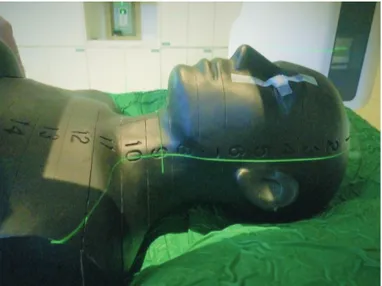
촬영영상은 종양학과 서버(Aria Networks)를 통해 전 산화치료계획시스템(Eclipse Ver.13.7, Palo Alto, USA) 으로 전송하였으며, 치료중심 좌표는 Head and Neck 치 료부위와 근사하게 설정하였다. Phantom은 치료실내의 레이저를 기준으로 치료중심좌표에 맞추고 ExacTrac과 CBCT를 번갈아 각각 10회 씩 촬영하였다(Fig. 1).
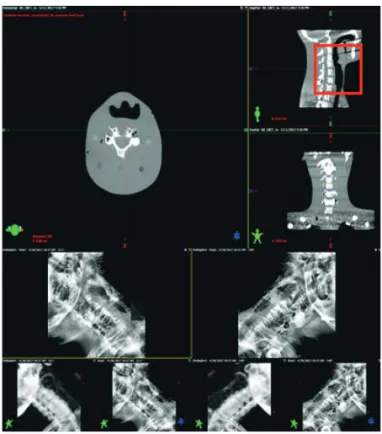
환자 데이터는 본원에서 2017년 3월부터 10월까지 Head and Neck Cancer IMRT 또는 VMAT 방사선치료 를 시행한 11명의 환자를 대상으로 하였으며, 총 109건의 영상을 얻었다. 치료계획용 CT영상은 전산화치료계획시스 템으로 전송되어 방사선종양학과 의사와 Dosimetrist가 치료계획을 설계하였다. 모든 환자는 치료종료 시까지 치 료실 내의 레이저를 기준으로 환자를 Set-up 하고, 주기적 으로 치료 직전에 CBCT와 ExacTrac 영상을 순차적으로 획득하였다(Table 1). 이 때 CBCT의 관심영역지정은 ExacTrac과 유사한 조 건으로 설정하기 위해서 Mandible, C-spine 2-7번을 포 함시키고, 6D 온라인 자동위치교정을 사용하여 디지털재 구성영상과 최대한 일치시켰다. ExacTrac은 촬영 후 별도 의 ROI 설정과정 없이 6개축의 Set-up 오차 값을 기록하 였고, 두 시스템의 각 축에 대한 Set-up 오차 값의 차를 RMS(Root Mean Square) 값으로 계산하였다(Fig. 2).
그리고 두 시스템으로 IGRT를 시행하는데 소요되는 시 간은 촬영 시 마다 독립적으로 측정하였다. 측정기준은 두 IGRT 시스템으로 kV Beam On 시점에서 영상획득 후 치 료방사선사가 최종적으로 영상을 비교하여 교정확인이 이 루어지기까지의 시간이며, 총 치료기간 동안 IGRT 시행에 소요된 평균시간과 표준편차로 산출하였다. 2. 피폭선량 측정
Rando Head and Neck Phantom 내에 부위별로 7개 씩 유리선량계를 삽입하고, CBCT와 ExacTrac의 피폭선 량을 각각 측정하였다(Fig. 3). CBCT의 촬영조건은 Head mode(full fan type, 100 kVp, 150 mAs, Diameter 26.2 cm, Scan Length 18.5 cm)로 1회 촬영하였고, Ex-acTrac은 Head and Neck protocol(100 kVp, 10 mAs)조 건으로 4회 촬영하여 평균값으로 산출하였다. 측정된 유리 선량계는 평가기관에 의뢰하여 평가하였다.
Fig. 1. Laser setup using rando phantom
Table 1. The number of CBCT and ExacTrac images ac-quired during treatment
Patient Site Fractions
Treated Imaged
1 Lt. Neck 30 18
2 Tonsil & Both Neck 30 17
3 Lt. Tonsil & Neck 20 12
4 Lt. Neck & Tonsil 20 7
5 Rt. Neck 30 13
6 Neck-Mediastinum 30 11
7 Neck LNs 20 3
8 Pyriform sinus 20 13
9 Hard palate & Lt. Neck 30 5
10 Hypopharynx 32 4
11 Tonsil 20 6
결 과
1. 두 시스템의 Set-up 오차 값의 차이(Set-up Correction Value) 비교
Phantom에서의 CBCT와 ExacTrac의 각 축에 대한 RMS 값은 Translation Group에서 Vrt, 0.05 mm ; Lng, 0.05 mm ; Lat, 0.05 mm로 나타났고, Rotation Group에 서는 Rtn, 0.07° ; Pitch, 0.07° ; Roll, 0.01°로 Transla-tion Group과 RotaTransla-tion Group의 차이가 각각 0.05 mm, 0.5° 이하로 나타났다(Table 2).
환자연구에서는 각각의 환자에서 나타나는 Set-up 오 차 값의 평균 차를 RMS 값으로 계산하였고, Transla-tion Group에서 Vrt, -0.07 mm ; Lng, -0.09 mm ; Lat, 0.05 mm, Rotation Group에서는 Rtn, 0.46° ; Pitch, -0.2° ; Roll, 0.22°로 Translation Group과 Rotation Group이 0.1 mm 이하, 0.5 미만으로 나타났다(Table 3).
Phantom 그룹과 환자 연구에서의 Set-up 오차 값의 평균을 그래프로 나타내면 아래 그림과 같다(Fig. 4).
2. CBCT, ExacTrac IGRT 소요시간
매 치료 시 마다 ㎸ Beam On에서 CBCT 영상을 plan CT와 비교하여 6D 온라인 자동위치교정 후 치료방사선사 가 교정확인이 이루어지기까지 소요되는 시간은 평균 256 ±47.6 sec, ExacTrac은 평균 84±3.5 sec로 CBCT가 약 3 배 정도 시간적인 비용이 더 많이 소요되는 것으로 나타났 다(Table 4). 3. 팬텀 실험에서 CBCT와 ExacTrac의 피폭선량 수 치 비교 Rando Phantom내에 삽입한 유리선량계의 부위 중 상 대적으로 가장 작은 차이가 난 부위는 Thymus, Thy-roid gland로 ExacTrac 1회당 각각 평균 0.035 mGy, 0.11 mGy, CBCT는 0.097 mGy, 0.64 mGy로 약 3배, 5배 Fig. 3. Absorbed dose measurement of Rando Phantom
Table 2. Differences in shift values in phantom studies
Fraction Translation(mm) Rotation(°)
AP SI LR Rtn Pitch Roll 1 -0.06 -0.05 0.06 0.19 -0.37 -0.09 2 -0.06 -0.06 0.04 0.26 -0.32 -0.25 3 -0.06 -0.06 0.05 0.27 -0.36 -0.17 4 -0.03 -0.03 0.04 0.19 -0.23 -0.39 5 -0.05 -0.06 0.06 0.23 -0.24 -0.14 6 -0.05 -0.05 0.05 0.31 -0.22 -0.13 7 -0.06 -0.06 0.05 0.34 -0.26 -0.31 8 -0.06 -0.06 0.06 0.32 -0.37 -0.26 9 -0.06 -0.07 0.05 0.27 -0.25 -0.10 10 -0.05 -0.06 0.06 0.22 -0.23 0.03 RMS 0.05 0.05 0.05 0.26 0.29 0.22 SD 0 0.01 0.03 0.07 0.07 0.01
의 차이가 있는 것으로 나타났다. 가장 많은 차이가 난 부 위는 Oral Mucosa이며, ExacTrac 1회당 평균 0.066 mGy 였던 것에 비해 CBCT는 2.468 mGy로 CBCT 피폭선량은 ExacTrac의 피폭선량에 약 37배 더 높은 수치를 보였다 (Table 5).
고안 및 결론
IGRT 기법 중 CBCT와 ExacTrac의 유용성에 관한 Chang Z. et al(16) 선행연구에서는 Spinal SBRT 치료환 Table 3. Differences in shift values in patient studiesPatient Translation(mm) Rotation(°)
AP SI LR Rtn Pitch Roll 1 -0.13 -0.13 -0.01 1.35 -0.73 0.45 2 -0.07 -0.05 0.06 1.10 -0.55 0.28 3 0.00 -0.17 0.05 0.05 -0.30 0.06 4 -0.05 -0.04 0.03 0.52 0.41 0.06 5 -0.04 -0.19 0.23 -0.50 -0.01 1.10 6 0.02 -0.04 -0.03 0.87 0.63 0.00 7 -0.01 0.03 0.12 0.21 1.23 0.40 8 -0.10 -0.14 0.00 0.88 -0.45 0.23 9 -0.03 -0.17 0.04 -0.15 -0.93 -0.84 10 -0.19 -0.10 0.08 0.18 -0.99 0.26 11 -0.13 -0.04 0.04 0.53 -0.48 0.41 RMS 0.12 0.14 0.12 1.02 0.86 0.93 SD 0.1 0.09 0.11 0.83 0.81 0.89 Table 4. Time taken for IGRT with CBCT and ExacTrac, re-spectively. Case ExacTrac CBCT 1 84±9.6 165±26.5 2 89±12.5 189±21.2 3 80±6.7 216±40.5 4 80±4.3 275±22.4 5 85±9.7 271±16.1 6 80±8.4 273±18.4 7 83±9.1 258±19 8 81±7.9 276±15.6 9 82±4.9 275±19.8 10 90±4.3 291±9.1 11 87±4.4 329±23.6 Avg±SD 84±3.5 256±47.6 Fig. 4. Phantom and patient setup errors in box and whisker graphs
⒜, ⒝ Differences in shift values in phantom studies ⒞, ⒟ Differences in shift values in patient studies
자를 대상으로 6D CBCT와 6D ExacTrac RMS의 차이 는 Translation Group에서 Vrt, 0.5 mm ; Lng, 0.9 mm ; Lat, 0.59 mm이고 Rotation Group에서는 Rtn, 0.54° ; Pitch, 0.53° ; Roll, 0.84°로 보고되어 있다.
Ma J et al(17) 연구에서는 Translation Group에서 Vrt,
1.3 mm ; Lng, 0.76 mm ; Lat, 0.71 mm이고 Rotation Group에서는 Rtn, 0.41° ; Pitch, 0.27° ; Roll, 0.4°로 보 고되었으며, Se an Oh et al(18)의 연구에서는 Translation
Group에서 Vrt, 0.43 mm ; Lng, 0.49 mm ; Lat, 0.42 mm 이고 Rotation Group에서는 Rtn, 0.4° ; Pitch, 0.53° ; Roll, 0.37°로 보고된 바 있다. 본원 장비의 6D 온라인 자동위치교정 기능에는 Pitch 와 Roll 방향이 적용되는 점을 유용하게 사용하기 위해 본 연구에서 CBCT와 ExacTrac을 순차적으로 촬영하여 나 타내는 Set-up 오차 값의 차를 RMS 값으로 나타내었고, 그 결과 두 시스템에서 나타내는 Set-up 오차 값의 차이 는 Phantom에서 Translation Group 0.1 mm Rotation Group 0.5° 미만으로 나타났고, 환자에서는 Translation Group 0.15 mm, Rotation Group 1.02° 이하로 나타났다 (Table 7). 본원의 Head and Neck IMRT 및 VMAT 치료 시 허용되는 PTV Margin 3.0 mm를 고려했을 때, 치료계 획용 CT영상과 CBCT영상이 최대한 일치된다는 가정 하 에 두 시스템 간 Set-up 오차 값의 차는 미묘하다고 사료 된다. 그러나 Rotation Group의 Margin에 대한 값은 불 분명하여 명확한 값을 제시할 순 없었다.
해부학적 구조물이 비교적 단순한 Head와는 달리 Neck 부위는 Spine과 Mandible의 유동성이 커서 Set-up 대한 재현성을 유지하는데 어려움이 있었다. 그리고 치 료기간 동안 체중변화나, 종양체적 감소 등 내부 장기의 변 화를 반영하지 않고 뼈 위주로 교정하여 반영한 값으로 CBCT으로 IGRT를 시행하였을 때, 관심영역지정을 아래 턱을 포함하는 것과 포함하지 않는 경우 두 경우 교정수 치 차이가 다소 있었다. ExacTrac으로 영상유도방사선치 료 시에는 관심영역지정이 고정되어 있어, 본 실험에서는 CBCT 영상의 ROI에 아래턱을 포함시켜서 나타난 수치를 비교했다. 이러한 결과를 바탕으로 총 치료기간 동안 누적되는 피 폭선량과 촬영시간을 고려했을 때, ExacTrac은 CBCT에 비해 피폭선량을 상당히 줄일 수 있었으며, Set-up 오차 보상 후 재확인(Verification) 과정을 거치더라도 촬영시간 은 CBCT의 1/3수준으로 적은 시간이 소요되었다. 이러한 시간적인 비용과 피폭선량도 줄여줄 수 있음에도 실제 임 Table 6. Comparison of Set-up Error Values with other papers Spine (Chang et al) Brain (Se an Oh et al) Brain (Ma et al) Head &Neck (Ours) Translation(mm) AP SI LR 0.5 0.9 0.59 0.43 0.49 0.42 1.30 0.9 0.59 0.12 0.14 0.12 Rotation(°) Rtn Pitch Roll 0.54 0.53 0.84 0.40 0.53 0.37 0.54 0.53 0.84 1.02 0.86 0.93 Translation Group(mm) Rotation Group(°) <0.5 <1.0 <0.5 <1.0 <1.0 <1.0 <0.5 ≤1.02 Table 5. Comparison of absorbed dose of ExacTrac and CBCT According to normal organ location (Unit in mGy) Site ExacTrac CBCT CBCT/ ExacTrac Thymus 0.035 0.097 2.77 Thyroid gland 0.1098 0.64 5.8 Brain 0.1813 3.001 16.5 0.168 3.196 19 Skin 0.1178 2.472 20.9 Salivary gland 0.1063 3.409 32 Submandible 0.1208 3.957 32 Oral mucosa 0.066 2.468 37
상에서는 ExacTrac이 CBCT의 2차적인 IGRT 수단으로 사용 되고 있다.
이에 본 연구를 통해 Head and Neck Cancer 환자의 영상유도방사선치료 시 CBCT와 ExacTrac의 이용을 비 교해 본 결과 ExacTrac을 사용함으로써 나타날 수 있는 Set-up 오차에 대한 불신을 CBCT를 통해 한번 더 검증 을 함으로써 두 시스템 사이에서의 차이가 경미하다는 것 을 알 수 있었고, 이는 여러 면에서 장점이 있는 ExacTrac 이 2차적인 IGRT 수단이 아니라 1차적인 수단으로써의 충 분한 장점이 있을 것으로 사료된다. 하지만 연부조직을 고 려하지 않고 뼈를 기준으로 고려하여 교정한 값으로 국한 되어 치료경과에 따른 체중감소와 종양체적변화를 영상화 할 수 있는 CBCT와 병용으로 사용하는 것이 좋을 것으로 사료된다.
본 연구는 환자 수가 작고 부위가 Head and Neck Cancer에만 국한되어 있어 추후 더 많은 환자 수로 다양 한 부위 연구가 필요하다.
참고문헌
1. Quality assurance for image-guided radiation ther-apy utilizing CT-based technologies: A report of the AAPM TG-179
2. Lee N, et al.: Int J Radiat Oncol Biol Phys. 2003: Intensity-modulated radiation therapy for head-and-neck cancer: The UCSF experience focusing on target volume delineation: 2003 Sep 1;57(1):49-60.
3. Miyabe Y, et al.: Med Phys. 2011: Positioning accu-racy of a new image-guided radiotherapy system: 2011 May;38(5):2535-41
4. Hansen EK, Larson DA, Aubin M, et al.: Image-guided radiotherapy using megavoltage cone-beam computed tomography for treatment of parasipinous tumors in the presence of orthopedic hardware: Int J Radiat Oncol Biol Phys 2006;66:323-6
5. Ryu SI, Chang SD, Kim DH, et al: Image-guided radiosurgery for the spine and pancreas: Comput Aided Surg 2000;5:278-88.
6. Murphy, M.J., Adler, J.R. Jr., Bodduluri, M. et al: Image-guided radiosurgery for the spine and pan-creas: Comput Aided Surg. 2000;5:278–288.
7. Yin, F.F., Ryu, S., Ajlouni, M. et al: Image-guided procedures for intensity-modulated spinal radio-surgery: Technical note. J Neurosurg. 2004;101:419– 424.
8. Yin, F.F., Wang, Z., Yoo, S. et al: Integration of cone-beam CT in stereotactic body radiation therapy: Technol Cancer Res Treat. 2008;7:133–139.
9. van Herk, M.: Different styles of image-guided ra-diotherapy: Semin Radiat Oncol. 2007;17:258–267.
10. Guckenberger, M., Meyer, J., Wilbert, J., Baier, K., Sauer, O., Flentje, M.: Precision of image-guided radiotherapy (IGRT) in six degrees of freedom and limitations in clinical practice: Strahlenther Onkol. 2007;183:307–313.
11. Moseley, D.J., White, E.A., Wiltshire, K.L. et al: Comparison of localization performance with im-planted fiducial markers and cone-beam computed tomography for on-line image-guided radio-therapy of the prostate: Int J Radiat Oncol Biol Phys. 2007;67:942–953.
12. Murphy, M.J., Balter, J., Balter, S. et al: The man-agement of imaging dose during image-guided ra-diotherapy: report of the AAPM Task Group 75. Med Phys. 2007;34:4041–4063.
13. Kan, M.W., Leung, L.H., Wong, W., Lam, N.: Radia-tion dose from cone beam computed tomography for image-guided radiation therapy: Int J Radiat Oncol Biol Phys. 2008;70:272–279.
14. Jin, J.Y., Yin, F.F., Tenn, S.E., Medin, P.M., Solberg, T.D.: Use of the BrainLAB ExacTrac X-ray 6D sys-tem in image-guided radiotherapy: Med Dosim. 2008;33:124–134.
15. Clemente S, et al.: Is Exactrac X-ray System an al-ternative to CBCT for Positioning Patients Head and Neck Cancers?: Med Phys. 2013 Nov ; 40(11): 111725.
16. Chang Z, et al.: 6D image guidance for spinal non-invasive stereotactic body radiation therapy: Com-parison between ExacTrac X-ray 6D with kilo-voltage cone-beam CT: Radiother Oncol. 2010 Apr;95(1):116-21.
17. Ma J, et al.: ExacTrac X-ray 6 degree-of-freedom image-guidance for intracranial non-invasive stereotactic radiotherapy: comparison with kilo-voltage cone-beam CT: Radiother Oncol. 2009 Dec;93(3):602-8.
18. Se an Oh, et al.: Evaluations of the setup discrep-ancy between BrainLAB 6D ExacTrac and cone-beam computed tomography used with the imaging guidance system Novalis-Tx for intracranial stereo-tactic radiosurgery: PLos ONE 12(5):e0177798.
Evaluation of the Usefulness of Exactrac in Image-guided
Radiation Therapy for Head and Neck Cancer
Department of Radiation Oncology, Changwon Gyengsang National University Hospital, Changwon, Korea.
Baek Min Gyu, Kim Min Woo, Ha Se Min, Chae Jong Pyo, Jo Guang Sub, Lee Sang Bong
Purpose: In modern radiotherapy technology, several methods of image guided radiation therapy (IGRT) are used to deliver accurate doses to tumor target locations and normal organs, including CBCT (Cone Beam Computed Tomography) and other devices, ExacTrac System, other than CBCT equipped with linear accelerators.
In previous studies comparing the two systems, positional errors were analysed rearwards using Offline-view or evaluated only with a Yaw rotation with the X, Y, and Z axes. In this study, when using CBCT and ExacTrac to perform 6 Degree of the Freedom(DoF) Online IGRT in a treatment center with two equipment, the difference between the set-up calibration values seen in each system, the time taken for patient set-up, and the radiation usefulness of the imaging device is evaluated.
Materials and Methods: In order to evaluate the difference between mobile calibrations and exposure radiation dose, the glass dosimetry and Rando Phantom were used for 11 cancer patients with head circumference from March to October 2017 in order to assess the difference between mobile calibrations and the time taken from Set-up to shortly before IGRT. CBCT and ExacTrac System were used for IGRT of all patients. An average of 10 CBCT and ExacTrac images were obtained per patient during the total treatment period, and the difference in 6D Online Automation values between the two systems was calculated within the ROI setting. In this case, the area of interest designation in the image obtained from CBCT was fixed to the same anatomical structure as the im-age obtained through ExacTrac.
The difference in positional values for the six axes (SI, AP, LR; Rotation group: Pitch, Roll, Rtn) between the two systems, the total time taken from patient set-up to just before IGRT, and exposure dose were measured and com-pared respectively with the RandoPhantom.
Results: the set-up error in the phantom and patient was less than 1 mm in the translation group and less than 1.5° in the rotation group, and the RMS values of all axes except the Rtn value were less than 1 mm and 1°. The time taken to correct the set-up error in each system was an average of 256±47.6 sec for IGRT using CBCT and 84±3.5 sec for ExacTrac, respectively. Radiation exposure dose by IGRT per treatment was measured at 37 times higher than ExacTrac in CBCT and ExacTrac at 2.468 mGy and 0.066 mGy at Oral Mucosa among the 7 measure-ment locations in the head and neck area.
Conclusion: Through 6D online automatic positioning between the CBCT and ExacTrac systems, the set-up er-ror was found to be less than 1 mm, 1.02°, including the patient’s movement (random erer-ror), as well as the systematic error of the two systems. This error range is considered to be reasonable when considering that the PTV Margin is 3 mm during the head and neck IMRT treatment in the present study. However, considering the changes in target and risk organs due to changes in patient weight during the treatment period, it is considered to be appropriately used in combination with CBCT.