선에 대한 별도의 연간 허용 유효선량 규준은 없으나, 갑상 선은 확률적 영향에 의해 손상 받는 장기이므로
7)방사선 치 료 시 주변선량에 의해 갑상선에 피폭되는 방사선량에 대해 주의를 기울여야 하며 막연한 추측보다는 정량적인 체계적 수치가 필요하다. 본 연구에서는 전뇌 방사선 치료 시 산란 선으로 인하여 영향을 받는 갑상선의 피폭선량을 감소시키 기 위해 차폐체를 사용하여 갑상선의 차폐 효과를 평가하고 자 한다.
대상 및 방법
1. 사용 장비 및 재료
측정에 사용된 장비는 선형가속기(Clinac iX. VARIAN, USA)이며 측정에너지는 6 MV X선을 사용하였다. 방사선량 측정용 팬텀은 인체모형팬텀(anderson rando phantom, USA)으로 갑상선의 위치에서의 입사표면선량과 심부선량 의 변화를 측정하기 위하여 사용된 차폐체는 0.5 mmPb thyroid protector(JUNGWON PRECISION IND..co.,LTD),
와 bismuth thyroid shield(F&L Medical Products co.)이고, 선 당량 0.06 mmPb를 가진다. 또한, 본원에서 1.0 mmPb을 이용해 자체 제작한 갑상선 차폐체를 사용하였다. 선량 측 정에 유리선량계(photoluminescence glass dosimeter, PLD, GD-352M, AGC techno co.,LTD)와 FGD-1000(AGC techno co.,LTD)판독기를 사용하였다.
2. 선량 측정방법
1) 갑상선의 입사표면선량 측정
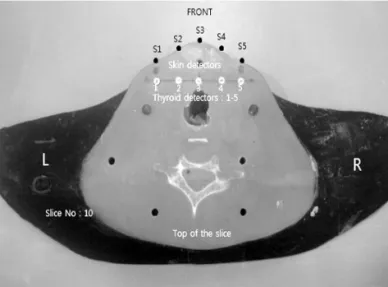
팬텀을 supine position/head first로 놓고, 전뇌 방사선 치료 자세로 위치시킨 상태에서 인체모형팬텀의 갑상선 위 치인 10번째 슬라이스 표면 midline을 중심으로 일정 간격 으로 유리선량계 다섯 개를 1.5 cm 간격으로 위치시켰다.
[그림 1, 2] 전뇌 방사선 치료 시 사용되는 방사선량은 6 MV, 300 cGy를 사용하였으며, Gantry 90도와 270도 방향 에서 좌우 이문 대향조사법을 이용하였다. 입사표면선량 측 정은 갑상선 차폐체 미사용 시, bismuth 차폐체 사용 시, 0.5 mmPb 차폐체 사용 시, 자체 제작한 1.0 mmPb 차폐체 를 사용하여 각각 5회씩 측정하여 평균값을 산출하였다.
Table 1. Phantom entrance surface dose measurements for unshielding and shielding using bismuth, 0.5 mmPb and 1.0 mmPb
S1 44.91 38.22 30.63 22.08
S2 43.29 36.41 31.98 25.12
S3 52.33 34.78 31.71 21.93
S4 41.56 34.20 30.42 22.14
S5 42.37 36.54 30.40 24.07
average 44.89 36.03 31.03 23.21
measurement site
unshielding (mGy)
bismuth shielding(mGy)
0.5 mmPb shielding(mGy)
1.0 mmPb shielding(mGy)
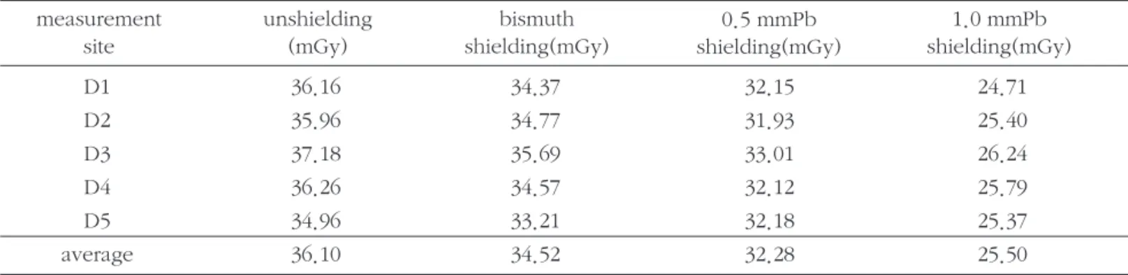
Table 2. Phantom depth 2.5 cm absorbed dose measurements for unshielding and shielding using bismuth, 0.5 mmPb and 1.0 mmPb
D1 36.16 34.37 32.15 24.71
D2 35.96 34.77 31.93 25.40
D3 37.18 35.69 33.01 26.24
D4 36.26 34.57 32.12 25.79
D5 34.96 33.21 32.18 25.37
average 36.10 34.52 32.28 25.50
measurement site
unshielding (mGy)
bismuth shielding(mGy)
0.5 mmPb shielding(mGy)
1.0 mmPb
shielding(mGy)
2) 갑상선의 2.5 cm 깊이의 심부선량 측정
갑상선의 2.5 cm 깊이의 심부선량 측정하기 위해 인체모 형팬텀의 10번째 슬라이스 표면 2.5 cm 깊이에 4 mm 구멍 을 뚫어 유리선량계 다섯 개를 1.5 cm 간격으로 위치시켰 다. [그림 2]입사표면선량 측정 조건과 동일하게 측정되었으 며, 심부선량 측정은 갑상선 차폐체 미사용 시, bismuth 차 폐체 사용 시, 0.5 mmPb 차폐체 사용 시, 자체 제작한 1.0 mmPb 차폐체를 사용하여 각각 5회씩 측정하여 평균값을 산출하였다.
Fig 1. Phantom irradiation setup. Fig 2. Placement of the PLDs in the phantom’s thyroid.
Fig 3. Dose distribution of entrance surface dose for measured sites.
Fig 5. Comparison of entrance surface dose and depth 2.5 cm absorbed dose of thyroid from phantom.
Fig 4. Dose distribution of depth 2.5 cm absorbed dose
for measured sites.
결 과
1. 갑상선의 입사표면선량 측정 결과
인체모형팬텀의 10번째 슬라이스에서의 입사표면선량은 갑상선 차폐체 미사용 시 평균 44.89 mGy로 측정되었다.
갑상선 차폐체 사용 시 bismuth 차폐체는 평균 36.03 mGy, 0.5 mmPb 차폐체는 평균 31.03 mGy, 자체 제작한 차폐체 1.0 mmPb는 평균 23.21 mGy로 측정되었다[표 1].
입사표면선량의 측정위치에 따른 선량 분포는 차폐체 미 사용 시 S3 지점에서 높게 측정되었고, bismuth 차폐체 사 용 시 S1, 0.5 mmPb 차폐체 사용 시 S2, 자체 제작한 차폐체 1.0 mmPb 사용 시 S2 지점에서 높게 측정되었다. [그림 3]
2. 갑상선의 2.5 cm 깊이의 심부선량 측정 결과
인체모형팬텀 10번째 슬라이스의 2.5 cm에서의 심부선 량은 갑상선 차폐체 미사용 시 평균 36.10 mGy로 측정되었 다. 갑상선 차폐체 사용 시 bismuth 차폐체는 평균 34.52 mGy, 0.5 mmPb 차폐체는 평균 32.28 mGy, 자체 제작한 차폐체 1.0 mmPb는 평균 25.50 mGy로 측정되었다[표 2].
심부선량의 측정위치에 따른 선량 분포는 가운데에 위치 한 D3 지점에서 모두 높게 측정되었다. [그림 4]
3. 갑상선의 입사표면선량 측정 시 차폐체 종류에 따른 감소율
갑상선의 입사표면선량 측정에서 갑상선 차폐체 미사용 시 평균 44.89 mGy로 측정되었고, 갑상선 차폐체 사용 시 bismuth 차폐체는 평균 36.03 mGy로 8.86 mGy가 감소하 였고, 0.5 mmPb 차폐체는 평균 31.03 mGy로 13.87 mGy 가 감소하였으며, 자체 제작한 1.0 mmPb 차폐체는 평균 23.21 mGy로 21.68 mGy가 감소하였다. 즉 갑상선 차폐체 를 사용하지 않았을 경우와 갑상선 차폐체를 사용했을 경우 의 감소율은 각각 bismuth 차폐체는 약 19.7%, 0.5 mmPb 차폐체는 약 30.9%, 자체 제작한 1.0 mmPb 차폐체는 약 48.3% 감소하였다[표 3].
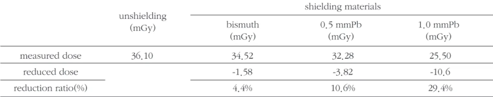
4. 갑상선의 2.5 cm 깊이의 심부선량 측정 시 차폐체 종 류에 따른 감소율
갑상선의 2.5 cm 깊이의 심부선량 측정에서 갑상선 차폐 체 미사용 시 평균 44.89 mGy로 측정되었고, 갑상선 차폐 체 사용 시 bismuth 차폐체는 평균 34.52 mGy로 1.58 mGy
가 감소하였고, 0.5 mmPb 차폐체는 평균 32.28 mGy로 3.82 mGy가 감소하였으며, 자체 제작한 1.0 mmPb 차폐체 는 평균 25.50 mGy으로, 10.6 mGy가 감소하였다. 즉 갑상 선 차폐체를 사용하지 않았을 경우와 갑상선 차폐체를 사용 했을 경우의 감소율은 각각 bismuth 차폐체는 약 4.4%, 0.5 mmPb 차폐체는 약 10.6%, 자체 제작한 1.0 mmPb 차폐체 는 약 29.4% 감소하였다[표 4].
5. 갑상선의 입사표면선량과 심부선량의 갑상선 선량 비교
갑상선의 입사표면선량은 갑상선 차폐체 미사용 시 평균 44.89 mGy로 측정되었다. 갑상선 차폐체 사용 시 bismuth 차폐체는 평균 36.03 mGy, 0.5 mmPb 차폐체는 평균 31.03 mGy, 자체 제작한 1.0 mmPb 차폐체는 평균 23.21 mGy로 측정되었다.
갑상선의 심부선량은 갑상선 차폐체 미사용 시 평균 36.10 mGy로 측정되었다. 갑상선 차폐체 사용 시 bismuth 차폐체는 평균 34.52 mGy, 0.5 mmPb 차폐체는 평균 32.28 mGy, 자체 제작한 1.0 mmPb 차폐체는 평균 25.50 mGy로 측정되었다[표 5]. [그림 5]
고 찰
방사선 치료 시 조사면 이외에 흡수되는 방사선량 즉, 주 변선량은 조사면 내에 흡수되는 선량에 비해 적은 양이나 각종 악성종양의 치료성적이 향상됨에 따라 주변선량에 의 한 부작용 또는 후유증이 점점 더 큰 문제가 되고 있다.
5)Joerg Lehmann 등8)은 조사면적의 외부에도 산란선에
의한 주변선량의 축적현상이 있음을 보고하고 있다. 특히
갑 상 선 은 다 른 부 위 보 다 방 사 선 에 민 감 한 장 기 로 써
Schneider AB 등
9)은 갑상선암의 잠재적 위험성은 방사선과
관련성이 있다고 이야기하고 있다. 방사선 조사 후 생기는
갑상선암의 발생에 있어 방사선의 작용기전은 확실하게 밝
혀지지 않았으나 두 가지의 병인론으로 설명한다. Zaina
Adnan 등
10)은 쥐를 이용한 실험에서 방사선 조사를 받은 쥐
의 갑상선이 유전자 변형을 일으키고 이 결과 세포의 탈분
화 및 종양의 성장양식(cellular dedifferentiation and
neoplastic growth pattern)을 보이는 것을 확인하였다. 이
것은 방사선이 직접 갑상선 세포에 작용하여 암을 유발함을
의미한다. 또 다른 가설은 방사선 조사를 받은 갑상선은 갑
상선 자극 호르몬(thyroid stimulating hormone,. 이하
TSH)증가를 동반한 기능저하 소견을 보이는 경우가 많은
데, 이러한 TSH의 지속적인 자극이 암의 유발요인이 될 수
있다는 가설로 Kennedy11)는 쥐 실험을 통하여 이를 주장 하였다.
경부 방사선 치료 후 갑상선암의 발생에 있어 Gerry H 등
12)
은 방사선 치료를 받은 경우가 받지 않은 대조군에 비하여 15.6배 높다고 하였으며(1.7% vs 0.07%), 전뇌 방사선 치료 후 갑상선의 2차 발암 위험이 여성이 남성보다 5.5배 증가 한다고 하였다.
13)Bonato 등
14)의 연구에 따르면 59명 중 32 명의 어린이 암 생존자들이 두경부와 전신조사(total body irradiation) 치료를 받고 난 후 생화학적인 갑상선 기능저하 증이 나타났다. 또한, 유방암 방사선 치료를 받은 생존자들 의 갑상선의 기능 screening 검사에서 갑상선 기능이 비정 상으로 나타났고
15), 수술 및 방사선 치료 후 갑상선 기능 저 하증의 빈도는 45%~62%로 보고되었다.
16,17)Shore RE
18)의 연구에서 갑상선에 전달되는 선량에 대해 갑상선에 최소 100 mGy의 선량을 받는 경우 2차적인 악성종양을 유발할 수 있다고 하였다.
방사선 치료 시 목적 부위 외에 받게 되는 불필요한 선량 을 줄여주는 것은 환자에게 중요한 부분이다. 방사선 치료 시 수반되는 주변선량의 피폭을 감소시킬 수 있는 적절한 차폐체의 활용이 필요하다. 이에 따라 갑상선에 주어지는 산란선의 차폐 효과에 대한 실험을 진행하였고, 그 결과 입 사표면선량 측정 시 갑상선 차폐체를 사용했을 경우 선량 감소율은 각각 bismuth 차폐체는 19.7%, 0.5 mmPb 차폐체 는 30.9%, 자체 제작한 1.0 mmPb 차폐체는 48.3% 의 선량 감소 효과가 있는 것으로 나타났다. 또한, 2.5 cm의 깊이에 서 선량을 측정한 결과 갑상선 차폐체를 사용했을 경우 선 량 감소율은 각각 bismuth 차폐체는 4.4%, 0.5 mmPb 차폐 체는 10.6%, 자체 제작한 1.0 mm Pb 차폐체는 29.4% 였다.
차폐체를 사용함으로써 갑상선에 피폭되는 산란선량은 감 소하였으며, 갑상선 심부보다 표면에서의 피폭선량 감소율 이 크게 나타났다. 이는 갑상선 피폭선량에 영향을 미치는 피사체 내부의 산란선, 조사부위 조직에서의 산란선보다 갠 Table 3. Reduction ratio for shielding and unshielding to kind of shielding materials in phantom entrance surface dose
shielding materials bismuth
(mGy)
0.5 mmPb (mGy)
1.0 mmPb (mGy)
measured dose 44.89 36.03 31.02 23.21
reduced dose -8.86 -13.87 -21.68
reduction ratio(%) 19.7% 30.9% 48.3%
unshielding (mGy)
Table 4. Reduction ratio for shielding and unshielding to kind of shielding materials in phantom depth 2.5cm absorbed dose shielding materials
bismuth (mGy)
0.5 mmPb (mGy)
1.0 mmPb (mGy)
measured dose 36.10 34.52 32.28 25.50
reduced dose -1.58 -3.82 -10.6
reduction ratio(%) 4.4% 10.6% 29.4%
unshielding (mGy)
Table 5. Comparision of entrance surface dose and depth 2.5 cm absorbed dose of thyroid from phantom shielding materials
bismuth (mGy)
0.5 mmPb (mGy)
1.0 mmPb (mGy)
entrance surface 44.89 36.03 31.02 23.21
depth 2.5 cm 36.1 34.52 32.28 25.50
unshielding (mGy) measured
site
트리 상부에서 발생한 누설방사선이 갑상선 표면에 크게 영 향을 미친 것으로 사료된다.
본 연구에서는 선량 측정 시 유리선량계를 이용하였는데, 기존 다른 선량계에 비해 선량 범위가 10 uGy ~ 500 Gy로 넓고, 안정성이 높아 감쇄(fading) 현상이 거의 없으며 소자 간의 균일성이 뛰어나고, 독해한 데이터의 소실이 없으며,
19)TLD와 PLD를 비교한 재현성 실험에서 TLD는 ±2%지만 PLD는 모든 소자에서 TLD보다 낮은 ±1% 이내의 값을 나 타냈다.
20)이는 PLD가 TLD보다 통계적으로 더 우수하다는 점과 기존 연구에 사용된 TLD에 비해 많은 장점을 가지고 있어 본 실험에서 유리선량계를 사용하였다. 또한, 인체 유 사팬텀 내부에 선량계를 갑상선 위치에 삽입함으로써 보다 실제 치료와 유사한 상황에서의 선량 측정연구를 할 수 있 었다.
본 연구에서 갑상선 차폐에 사용되는 기존의 상용화된 갑 상선 차폐체는 쉽게 구부러지고 착용하기에 용이하여야 하 므로 높은 당량의 납을 사용할 수 없어 차폐효율이 다소 낮 다는 단점과 환자 치료 기기의 종류 및 치료 방법에 따라서 선량 측정치가 변동사항이 있다는 제한점을 가지고 있다.
하지만 방사선 치료 시 목적 부위와 인접한 갑상선에서 2차 발암의 발생위험도가 증가한다는 사실은 변함이 없다.
전뇌 방사선 치료 시 주변선량과 산란선량에 의해 갑상선 에 전해지게 되는 선량이 미미하더라도 2차적으로 수반될 수 있는 발암의 가능성과 기능 저하, 부작용 등이 일어날 확 률이 높아진다. 따라서 갑상선 차폐체를 이용하여 주변선량 을 최소화하는 것이 고려되어져야 한다.
결 론
본 연구는 전뇌 방사선 치료 시 방사선 조사면 밖의 영역 에서 발생하는 2차 산란 및 누출 선량에 의해 영향을 받는 갑상선에 대하여 상용화된 납 갑상선 차폐체와 bismuth 갑 상선 차폐체를 이용하여 선량 감소 효과를 검토해본 결과 갑상선 심부는 약 11~30%, 갑상선 표면은 약 20~48% 정 도의 피폭선량 감소 효과가 나타났다. 갑상선은 방사선 감 수성이 예민한 인접 장기로 갑상선에 피폭되는 방사선량에 주의를 기울여야 한다.
본 연구에서 사용된 갑상선 차폐체를 활용하여 목적 치료 부위 외에 불필요한 산란선량과 주변선량을 줄여준다면 확 률적 영향에 의한 2차 갑상선암의 발생 위험과 갑상선 기능 저하, 만성 장애, 부작용 등을 상당히 줄여주고 예방할 수 있을 것이라 생각된다. 따라서 전뇌 방사선 치료 시 갑상선 차폐체를 사용함으로써 갑상선을 효과적으로 보호하며 치
료를 시행할 수 있을 것으로 사료된다.
참고문헌
1. Roberge D, Parker W, Niazi TM, Oliveres M: Treating the contents and not the container: dosimetric study of hairsparing whole brain intensity modulated radiation therapy. Technology in Cancer Research and Treatment 2005;4:567-570
2. Orton N, jaradat H. Welsh JS. Tome W: Whole scalp irradiation using helical tomotherapy. Medical Dosimetry 2005;30:162-168
3. Sasa Mutic, Jacqueline Esthappan and Eric E. Klein:
Peripheral dose distributions for a linear accelerator equipped with a secondary multileaf collimator and universal wedge. journal of applied clinical medical physics 2002;3:302-309
4. Marilyn Stovall, Sarah S. Donaldson, Rita E.
WeathersD, et al.: Genetic effects of radiotherapy for childhood cancer: Gonadal dose reconstruction.
International Journal of Radiation Oncology, Biology, Physics 2004;60:542-552
5. 이상석, 박영선, 김흥태, 고성진: 방사선 생물학. 2nd ed.
서울:정문각, 2005;211-217
6. Pubino, C., Cailleux, A. F., De vathaire, F. and Schlumberger, M.: Thyroid cancer after radiation exposure. Eur. J. Cancer 38, 2002;38:645-647
7. Niklason LT, Marx MV, Chan HP: Interventional radiologists: occupational radiation doses and risks.
Radiology 1993;187:729-733
8. Joerg Lehmann, Robin L. Stern, Thomas P. Daly, et al.:
Dosimetry for quantitative analysis of the effects of low-dose inizing radiation in radiation therapy patients. Radiation Research 2006;165(2):240-247 9. A B Schneider, E Ron, J Lubin, M Stovall, and T C
Gierlowski.: Dose-response relationships for radiation- induced thyroid cancer and thyroid nodules : evidence for the prolonged effects of radiation on the thyroid.
The Journal of Clinical Endocrinology & Metabolism 1993;77(2):362-369
10. Zaina Adnan, Eldad Arad, James Dana, Yaakov
Shendler, and Elzbieta Baron1.: Simultaneous
occurrence of medullary and papillary thyroid
microcarcinomas: a case series and review of the literature Journal of Medical case reports 2013;7:26 11. Kennedy, Ann R.: Factors that modify radiation
incuced carcinogenesis. Health Physics 2009;97:433- 445
12. Gerry H. Tan, Hossein Gharib.: Thyroid Incidentalomas:
Management approaches to nonpalpable nodules discovered incidentally on thyroid imaging Annals of internal medicine 1997;126(3):226-231
13. Kalliopi M. Kourinou, Michalis Mazonakis, Efrosini Lyraraki, john Stratakis, john Damilakis.: Scattered dose to radiosensitive organs and associated risk for cancer development from head and neck radiotherapy in pediatric patients. Physica Medica European Journal of Medical Physics 2013;29:650-655 14. Bonato C, Severino RF, Elnecave RH: Reduced
thyroid volume and hypothyroidism in survivors of childhood cancer treated with radiotherapy. Journal of Pediatric Endocrinology and Metabolism 2008;21:943-949
15. S Johansen, KV Reinertsen, K Knutstad, DR Olsenand SD Fossa.: Dose distribution in the thyroid gland following radiation therapy of breast cancer-a retrospective study. Radiation Oncology 2011;6:68 16. Posner MR, Ervin TJ, Miller D, Fabian RL, Norris CM
Jr, Weichselbaum RR, Rose C.: Incidence of hypothyroidism following multimodality treatment for advanced squamous cell cancer of the head and neck. Laryngoscope 1984;94(4):451-454
17. B. Emami, J. Lyman, Brown A, et al.: Tolerance of normal tissue to therapeutic irradiation. International journal of radiation oncology, biology, physics 1991;21:109-122
18. Shore RE.: Issues and epidemiological evidence regarding radiation-induced thyroid cancer. Radiation Research 1992;131:98-111
19. 김창규.: 자체제작 Pb 밴딩을 이용한 피폭선량 감소, The Journal of Digital Policy & Management 2013;11(6):269-273
20. 최재호, 강구준, 장서구.: DAP(Dose Area Product)를
이용한 TLD와 PLD의 선량 측정 비교. 한국콘텐츠학회
논문지 2012;12:244-250
Purpose :
To reduce the radiation dose to the thyroid that is affected to scattered radiation, the shield was used. And we evaluated the shielding effect for the thyroid during whole brain radiation therapy.Materials and Methods :
To measure the dose of the thyroid, 300cGy were delivered to the phantom using a linear accelerator(Clinac iX VARIAN, USA.)in the way of the 6MV X-ray in bilateral.To measure the entrance surface dose of the thyroid, five glass dosimeters were placed in the 10th slice’s surface of the phantom with a 1.5 cm interval. The average values were calculated by measured values in five times each, using bismuth shield, 0.5 mmPb shield, self-made 1.0 mmPb shield and unshield. In the same location, to measure the depth dose of the thyroid, five glass dosimeters were placed in the 10th slice by 2.5 cm depth of the phantom with a 1.5 cm interval. The average values were calculated by measured values in five times each, using bismuth shield, 0.5 mmPb shield, self-made 1.0 mmPb shield and unshield.
Results :
Entrance surface dose of the thyroid were respectively 44.89 mGy at the unshield, 36.03 mGy at the bismuth shield, 31.03 mGy at the 0.5 mmPb shield and 23.21 mGy at a self-made 1.0 mmPb shield. In addition, the depth dose of the thyroid were respectively 36.10 mGy at the unshield, 34.52 mGy at the bismuth shield, 32.28 mGy at the 0.5 mmPb shield and 25.50 mGy at a self-made 1.0 mmPb shield.Conclusion :
The thyroid was affected by the secondary scattering dose and leakage dose outside of the radiation field during whole brain radiation therapy. When using a shield in the thyroid, the depth dose of thyroid showed 11~30% reduction effect and the surface dose of thyroid showed 20~48% reduction effect.Therefore, by using the thyroid shield, it is considered to effectively protect the thyroid and can perform the treatment.
Myung Sic Yang・Ju Kyeong Park・Seung Hun Lee・Yang Su Kim・Sun Young Lee・Seok Yong Cha
Evaluation of usability of the shielding effect for thyroid shield for peripheral dose
during whole brain radiation therapy
Department of Radiation Oncology, Chonbuk National University Hospital
Department of Radiation Oncology, Chonbuk National University Medical School, Jeonju, Korea
Abstract