서 론
유방암은 고형암의 뇌전이 원발암 중 두 번째로 흔한 원인으로 알려져 있고 임상적으로 뇌전이율은 16%까지 보고되었다.(1,2) Purpose: To assess the incidence of brain metastasis in
patients with breast cancer receiving surgery and adjuvant radiotherapy (RT) and to evaluate subtypes associated with brain metastasis. Methods: We retrospectively reviewed the medical records of 1,000 patients with breast cancer who were treated with surgery and adjuvant RT for a cure between January 2001 and July 2005 at Samsung Medical Center.
Seventy-one patients received neoadjuvant chemotherapy before surgery. The pathological stage was I in 430 patients, II in 327, and III in 243. We divided the patients into three subtypes according to immunohistochemistry: triple negative (TN, 245 patients), human epidermal growth factor 2 (HER2) enriched (HE, 166 patients) and positive estrogen receptor or progesterone receptor without HER2 overexpression (EP, 589 patients). The median follow up time was 72 months after surgery. Results: Locoregional failure-free survival rate and distant metastasis-free survival rate at 5 years were 92.8% and 86.1%, respectively. The disease free survival rate and overall survival rate at 5 years were 84.6% and 94.7%, respectively. Thirty-nine patients had brain metasta- sis, and the brain metastasis-free survival rate at 5 years
was 97.2%. A univariate analysis showed that younger age, neoadjuvant chemotherapy, modified radical mastectomy, advanced pathological stage and the TN and HE subtypes were significant risk factors for brain metastasis. A multivari- ate analysis revealed that age, neoadjuvant chemotherapy, pathological stage and the TN and HE subtypes were sta- tistically significant factors for brain metastasis. Conclusion:
The cumulative incidence of brain metastasis was 3.9% after curative treatment. If patients have a clinically suspicious symptoms suggesting brain metastasis, clinicians should be aware that an early brain imaging work up and manage- ment are necessary. Because patients with the TN or HE subtypes accompanied by younger age and advanced patho- logical stage have increased brain metastasis (>10%), annual regular imaging follow-up may be recommended for these high risk patients.
Key Words: Breast neoplasms, Immunohistochemistry, Neoplasm metastasis, Brain, Radiotherapy
중심단어: 유방암, 면역조직화학적 아형, 뇌, 전이, 방사선치료
Incidence of Brain Metastasis and Related Subtypes in Patients with Breast Cancer Receiving Adjuvant Radiation Therapy after Surgery
Sun Hyun Bae, Doo Ho Choi, Seung Jae Huh, Do Hoon Lim, Won Park, Heerim Nam, Jung-Hyun Yang
1, Seok-Jin Nam
1, Jeong Eon Lee
1, Young-Hyuck Im
2, Jin-Seok Ahn
2, Yeon Hee Park
2Departments of Radiation Oncology, 1Surgery, 2Division of Hematology-Oncology, Department of Medicine, Samsung Medical Center, Sungkyunkwan University School of Medicine, Seoul, Korea
Breast Cancer
O R I G I N A L A R T I C L E
배선현ㆍ최두호ㆍ허승재ㆍ임도훈ㆍ박 원ㆍ남희림ㆍ양정현1ㆍ남석진1ㆍ이정언1ㆍ임영혁2ㆍ안진석2ㆍ박연희2 성균관대학교 의과대학 삼성서울병원 방사선종양학과, 1외과, 2혈액종양내과
수술 후 보조적 방사선 치료를 포함한 근치적 치료를 받은 유방암 환자들의 뇌전이 발생률과 연관된 인자 분석
책임저자: 최두호
135-710 서울시 강남구 일원동 50, 성균관대학교 의과대학 삼성서울병원 방사선종양학과
Tel: 02-3410-2436, Fax: 02-3410-2619 E-mail: [email protected]
접수일: 2010년 11월 5일 게재승인일: 2011년 2월 7일
S57
하지만 유방암 환자 1,044명의 중추 신경계 전이에 대한 부검 연 구에 의하면 부검 결과 뇌전이는 193명(18%)에서 발견되었고 중 추 신경계 전이 환자의 31%만이 사망 전에 임상적으로 전이가 진 단되었다는 것을 고려하면 실제 뇌전이율은 저평가 되었을 것으 로 생각된다.(3) 또한 최근 새로운 표적 치료제와 항암제의 발달 로 유방암 환자의 무병 기간과 생존율이 증가하였고 전이성 환자 의 예후는 증진되었지만, 이들 약제가 혈액뇌관문을 통과하지 못 하기 때문에 동시적으로 뇌전이율이 증가하여 연구에 따라 뇌전 이율을 25-36%까지 보고하였다.(4-7) 가장 높은 뇌전이율을 보 고한 Ono 등(7)에 따르면 human epidermal growth factor 2 (HER2) 과발현인 재발성 혹은 전이성 유방암 환자에서 trastu- zumab를 포함하는 항암치료제를 사용하였을 때 36.3%에서 경 과 관찰 중 뇌전이가 발견되었다.
2000년 Perou 등(8)은 유방암이 특이한 유전자 발현 정도에 따라 다양한 표현형을 나타내고 이를 여러 개의 분자학적 아형으 로 구분할 수 있다고 발표하였다. 그 뒤 분자학적 아형에 따른 예 후의 차이와 항암치료의 반응도의 차이가 보고되었다.(9-11) 최 근에는 유전자 발현 정도에 따라 원격 전이가 기관 특이적으로 나 타난다는 연구 결과들이 발표 되었고,(12-16) Smid 등(16)에 따르 면 HER2 과발현 혹은 basal subtype의 경우 뇌전이율이 다른 분자생물학적 아형보다 높다고 보고하였다. 한편, 면역조직화학적 검사를 통한 호르몬수용체 유무에 따른 연구 결과들도 발표되어 에스트로겐 수용체(estrogen receptor, ER) 음성이거나 HER2 과발현의 경우 뇌전이 위험성이 높다고 보고되었다.(17-19)
그러나 근치적 치료를 시행한 원발성 유방암과 재발성 혹은 전 이성 유방암은 생물학적 행태가 다르다는 것을 고려할 때 근치적 치료를 시행한 환자에서 뇌전이율과 이와 연관된 인자는 거의 보 고되지 않았다.(16,20) 이에 본 연구는 유방암 진단 후 수술을 시 행하고 보조적 방사선치료를 받은 환자들을 대상으로 뇌전이 발생 률을 알아보고 이와 연관된 면역조직화학적 아형을 분석하였다.
방 법
2001년 1월부터 2005년 7월까지 삼성서울병원에서 유방암으 로 진단받고 수술 후 보조적 방사선치료를 받은 환자는 총 1,240 명이었다. 유방보존수술을 시행한 환자는 모두 방사선 치료를 받 았고 변형 근치적 유방절제술을 시행한 환자는 병리학적 원발 병 소의 병기가 3기 이상 혹은 액와림프절 전이가 4개 이상인 경우 에만 시행되었다. 이 중에서 진단 시 호르몬 수용체에 대해서 면 역조직화학적 검사 결과가 보고되었고 HER2의 경우는 면역조직 화학적 검사에서 2+인 경우 형광제자리부합법을 시행하여 유전 자 증폭 여부를 확인한 환자 1,000명을 대상으로 후향적으로 분
석하였다.
환자의 연령분포는 21-80세로 중앙값은 46세였고 모든 환자 는 전신수행도(Eastern Cooperative Oncology Group 분류)가 1 이하로 연령에 상관없이 임상적 혹은 병리학적 병기에 따라 근치 적 목적의 모든 치료를 받았다. 환자는 치료 전 절제생검이나 초음 파 유도 하에서 핵생검법을 통해 유방암 진단 후 수술을 시행하였 다. 대상 환자의 특성은 Table 1에 정리하였다. 71명은 임상적
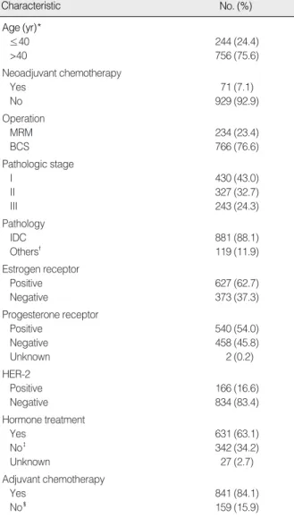
Table 1. Patients’ characteristics
Characteristic No. (%)
Age (yr)*
≤40 244 (24.4)
>40 756 (75.6)
Neoadjuvant chemotherapy
Yes 71 (7.1)
No 929 (92.9)
Operation
MRM 234 (23.4)
BCS 766 (76.6)
Pathologic stage
I 430 (43.0)
II 327 (32.7)
III 243 (24.3)
Pathology
IDC 881 (88.1)
Others� 119 (11.9) Estrogen receptor
Positive 627 (62.7)
Negative 373 (37.3)
Progesterone receptor
Positive 540 (54.0)
Negative 458 (45.8)
Unknown 2 (0.2)
HER-2
Positive 166 (16.6)
Negative 834 (83.4)
Hormone treatment
Yes 631 (63.1)
No� 342 (34.2)
Unknown 27 (2.7)
Adjuvant chemotherapy
Yes 841 (84.1)
No� 159 (15.9)
MRM=modified radical mastectomy; BCS=breast conserving surgery;
IDC=invasive ductal carcinoma.
*Median age was 46 yr; �Others are including invasive lobular carci- noma, infiltrating cribriform carcinoma, invasive micropapillary carci- noma, mucinous carcinoma, infiltrating apocrine carcinoma, meta- plastic carcinoma, medullary carcinoma and tubular carcinoma; �Eight patients refused hormone therapy; �Nine patients refused adjuvant chemotherapy; two patient could not receive chemotherapy due to neutropenia and thrombocytopenia.
원발 병소 병기가 3기 이상 혹은 액와림프절 전이가 있었던 국소 진행성 유방암으로 수술 전 선행 항암화학요법을 받았고 세 명을 제외한 환자에서 anthracycline을 포함하는 복합 항암요법을 사 용하였다. 수술방법은 원발 병소의 임상적 병기에 따라 변형 근치 적 유방절제술이 234명에서 시행되었고 유방보존수술은 766명에 서 시행되었다. 병리조직학적 유형은 침윤성 관상피암종이 881명 으로 가장 많았다. 수술 후 병리학적 병기는 제7판 American Joint Committee on Cancer (AJCC) 병기 결정 기준에 따라 분류하였고 430명이 1기, 327명이 2기, 그리고 243명이 3기였 다.(21)
수술 검체의 면역조직화학 검사에서 ER 양성은 627명, 프로게 스테론 수용체(progesterone receptor, PR) 양성은 540명, 그 리고 HER2 과발현은 166명이었다. 이들을 면역조직화학적 특 성에 따라 삼중음성군(245명), HER2 과발현군(166명), 그리고 HER2 과발현이 없으면서 ER 혹은 PR 양성인 군(589명)의 세 가지 아형으로 나누었다(Table 2).
수술 후 항암화학요법은 환자의 연령과 병리학적 병기를 고려 하여 초기에는 cyclophosphamide+methotrexate+5-fluo- rouracil 복합 항암요법을 시행하였고, 2001년도 후반부터는 주 로 anthracycline 또는 docetaxel을 포함하는 항암화학요법이 시행되었다. ER 혹은 PR 양성인 환자 중 619명은 타목시펜 혹은 아로마타제 억제제로 호르몬치료를 받았고, HER2 과발현으로 trastuzumab 치료를 받은 사람은 16명이었다.
수술 후 보조적 방사선치료는 4-6MV 광자선을 이용하여 동측 흉벽이나 남은 유방 부위에 접사면 조사(tangential technique) 로 치료하였다. 방사선 조사량은 하루에 1.8-2.0 Gy씩 총 50- 50.4 Gy를 조사하였고 유방보존수술을 시행한 환자는 수술 부위 에 하루에 2-3 Gy씩 9-10 Gy를 전자선으로 추가 조사하였다.
그리고 271명은 쇄골상부림프절 영역에 50-50.4 Gy를 조사받 았다.
재발 여부는 국소재발과 원격전이로 나누어 분석하였고 국소 재발의 경우는 동측 흉벽, 남은 유방조직, 동측 액와림프절, 동측
쇄골상부림프절과 동측 내유림프절에서 발생한 것으로 정의하였 고, 그 외 다른 부위에 재발한 경우는 전신재발로 정의하였다. 뇌 전이는 경과관찰 중 시행한 뇌 자기공명영상에서 뇌실질에 병변 이 관찰되는 경우로 정의하였다. 척수연수막 전이나 뇌실질 전이 를 동반하지 않는 경막 전이는 제외하였다.
생존율은 Kaplan-Meier 분석을 사용하였으며, 생존곡선의 비교는 단변량분석으로 log-rank test를, 다변량분석으로 Cox multivariate regression model을 사용하였고 통계학적 유의 수준은p<0.05으로 하였다. 통계 프로그램은 The SAS�System (SAS 8.0; SAS Institute Inc., Cary, USA)을 이용하였다.
결 과
추적조사기간은 수술 시행 후 7-111개월로 중앙값이 72개월이 었다. 전체 환자의 5년 국소제어율은 92.8%, 원격전이 제어율은 86.1%, 무병생존율은 84.6%, 전체생존율은 94.7%이었다.
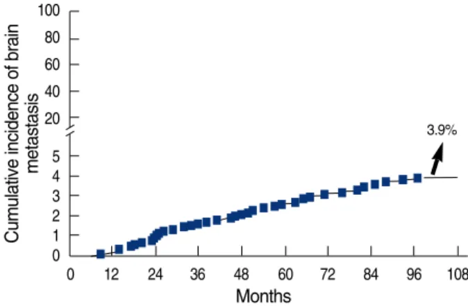
뇌전이는 근치적 목적으로 치료를 받은 1,000명의 환자 중, 39
Table 2. Immunohistochemical subtypes according to status of ER, PR, and HER2 overexpression
Subtype No. (%)
Triple negative* 245 (24.5)
HER2 enriched� 166 (16.6)
ER/PR positive�without HER2 overexpression 589 (58.9) ER=estrogen receptor; PR=progesterone receptor.
*Negative estrogen receptor (ER), negative progesterone receptor (PR) and without HER2 overexpression; �HER2 overexperssion regardless of status of ER and PR; �ER positive or PR positive or both positive.
Cumulative incidence of brain metastasis 100
80 60 40 20 5 4 3 2 1 0
0 12 24 36 48 60 72 84 96 108
Months
3.9%
97.2% at 5 yr
Figure 1. Cumulative incidence of brain metastasis.
BM free survival rate (%)
100
80
60
40
20
0
0 12 24 36 48 60 72
Months
Figure 2. Five-years brain metastasis (BM) free survival rate.
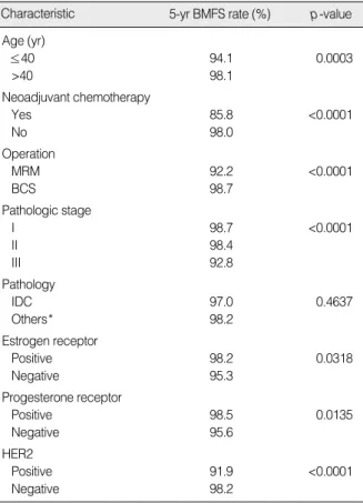
명(3.9%)의 환자에서 관찰되었고 뇌전이 발생의 중앙값은 46개 월(범위, 9-97개월)이었다(Figure 1). 전체 환자의 5년 뇌전이 무병생존율은 97.2%였다(Figure 2).
뇌전이에 영향을 미치는 인자를 알아보기 위해 단변량분석을 시행하였을 때 40세 이하, 선행 항암화학요법을 시행한 경우, 변 형 근치적 유방절제술을 시행한 경우, 병리학적 병기가 높을수록, ER 음성, PR 음성, 그리고 HER2 과발현의 경우 통계적으로 유 의하게 뇌전이의 위험성이 증가하였다(Table 3). 추가적으로 면 역조직화학적 특성에 따른 세 가지 아형에 따라서 단변량분석을 시행하였을 때, 5년 뇌전이 무병생존율이 HER2 과발현군에서 91.9%로 가장 낮았고 삼중음성군이 95.2%, HER2 과발현이 없 으면서 ER 혹은 PR 양성인 군이 99.4%으로 세 군 사이의 뇌전이 율은 통계적으로 유의하게 차이를 보였다(p<0.001) (Figure 3).
Cox multivariate regression model을 이용한 다변량분석
에서 연령, 선행 항암화학요법, 병리학적 병기 그리고 면역조직 화학적 아형이 뇌전이율과 연관하여 통계적으로 유의한 인자였 다(Table 4).
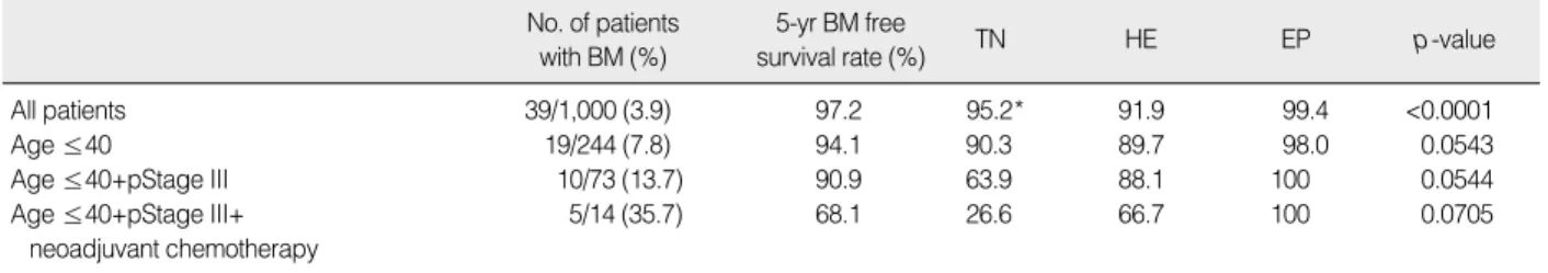
단변량분석과 다변량분석에서 모두 유의하게 나온 인자들을 가지고 뇌전이 위험성에 대해서 하위군 분석을 시행하였다(Table 5). 하위군 중에서 40세 이하이면서 병리학적 병기가 3기인 경우 뇌전이가 13.7%에서 발생하고 5년 뇌전이 무병 생존율이 90.9%, 수술 후 뇌전이 발생의 중앙값은 43.5개월이었다(범위, 14-97개 월). 40세 이하이면서 병리학적 병기가 3기, 선행 항암화학요법을 시행한 환자군에서는 35.7%에서 뇌전이가 발생하고 뇌전이 무병 생존율이 68.1%, 수술 후 뇌전이 발생의 중앙값은 32개월이었다 (범위, 14-82개월). 두 환자군 모두에서 면역조직화학적 아형에 따라 삼중 음성군이나 HER2 과발현군이 HER2 과발현이 없으 면서 ER 혹은 PR 양성인 군보다 뇌전이를 많이 하는 차이를 보 였으나 통계적으로 유의하지는 않았다(Figure 4).
Table 3. Univariate analysis of factors associated with brain metastasis free survival
Characteristic 5-yr BMFS rate (%) p-value Age (yr)
≤40 94.1 0.0003
>40 98.1
Neoadjuvant chemotherapy
Yes 85.8 <0.0001
No 98.0 Operation
MRM 92.2 <0.0001
BCS 98.7
Pathologic stage
I 98.7 <0.0001
II 98.4
III 92.8
Pathology
IDC 97.0 0.4637
Others* 98.2
Estrogen receptor
Positive 98.2 0.0318
Negative 95.3 Progesterone receptor
Positive 98.5 0.0135
Negative 95.6 HER2
Positive 91.9 <0.0001
Negative 98.2
BMFS=brain metastasis free survival; MRM=modified radical mastec- tomy; BCS=breast conserving surgery; IDC=invasive ductal carcinoma.
*Others are including invasive lobular carcinoma, infiltrating cribriform carcinoma, invasive micropapillary carcinoma, mucinous carcinoma, infiltrating apocrine carcinoma, metaplastic carcinoma, medullary car- cinoma and tubular carcinoma.
BM free survival rate (%)
100
90
40 20 0
0 12 24 36 48 60 72
Months
Figure 3. There was a statistically significant difference in brain metastasis (BM) free survival according to hormone subtype:
91.9% in HER2 enriched group, 95.2% in triple negative group and 99.4% in ER+/- PR positive without HER2 overexpression (p<0.0001).
ER=estrogen receptor; PR=progesterone receptor.
p<0.0001 ER/PR(+) without HER2 overexpression
HER2 enriched Triple negative
Table 4. Cox multivariate regression analysis of risk factors affecting brain metastasis free survival
Characteristic Hazard ratio (95% p-value confidence limits)
Age >40 yr 0.4 (0.25-0.89) 0.0220 Use of neoadjuvant chemotherapy 2.7 (1.28-5.82) 0.0088
MRM 1.8 (0.76-4.64) 0.1650
Pathologic stage III 1.4 (1.07-1.95) 0.0155 ER/PR positive* without 0.6 (0.48-0.79) 0.0002
HER2 overexpression
MRM=modified radical mastectomy; ER=estrogen receptor; PR=
progesterone receptor.
*ER positive or PR positive or both positive.
고 찰
본 연구에서 근치적 치료를 받은 유방암 환자를 후향적으로 분 석하였을 때 뇌전이 발생률은 3.9%였다. 그동안 뇌전이의 빈도 가 10-16% 정도로 인식되다가 최근 새로운 표적 치료제와 항암 제의 사용으로 25-36%까지 증가한 것을 고려하면 본 연구와 많 은 차이가 발견된다.(1-7) 그러나 지금까지 보고된 연구들의 연구 대상은 대부분 재발성 혹은 전이성 유방암 환자였고 초기 유방암 에서 20-30%가 원격전이를 한다는 것을 고려하면 전체 환자 중 에서 뇌전이 환자의 비율은 훨씬 적을 것으로 생각된다.(22) Lee 등(23)은 1990년부터 2006년까지 조직학적으로 진단된 유방암 환자 중에서 198명(2.5%)이 뇌전이가 발생하였다고 보고하였다.
Gonzalez-Angulo 등(20)은 선행 항암화학요법을 시행한 원격 전이가 없었던 국소진행성 혹은 염증성 유방암 환자 768명 중 8%
에서 중추신경계 전이가 발생하였고 이 중 80%가 뇌전이였다고 보고하였다. 따라서 원격 전이가 없는 유방암 환자에서 근치적 치
료를 시행 후 뇌전이가 발생할 위험성은 6.5% 이내로 추정되고, 한국유방암학회에서 발간한 유방암백서(24)에서 국내 조기유방 암의 비율이 2006년도에 48.8%이고 그 비율이 점차 증가하는 경 향을 보이는 것을 고려하면 우리나라에서 근치적 치료 후 뇌전이 가 발생할 위험성은 더 낮을 것으로 추정되고 본 연구 결과와 일 치하는 것으로 생각된다.
본 연구에서 뇌전이는 9-97개월 사이에 발생하였고 중앙값이 46개월로 이전의 보고된 27-32개월보다 늦게 진단되었는데 이 는 대상 환자군의 차이 때문으로 생각된다.(20,23) 본 연구에서는 1기가 43.2%, 2기가 32.7%로 조기유방암이 50% 이상을 차지한 다. Gonzalez-Angulo 등(20)은 종양의 크기가 클수록, 액와림 프절 전이가 양성일 때 그리고 병리학적 병기가 높을수록 통계적 으로 유의하게 뇌전이 위험성이 증가하고, 따라서 질병경과가 더 진행된 종양일수록 뇌전이까지 간격이 짧아진다고 하였다. 발생 시기에 있어서는 세 연구 간에 차이가 있지만 시간이 경과하더라 도 뇌전이는 계속 발생한다는 점과 5년 이상 경과한 후에도 다른
BM free survival rate (%)
100
80
60
40
20
0
0 12 24 36 48 60 72
Months
Figure 4. There was a difference in brain metastasis (BM) free survival according to immunohistochemical subtypes (A) in patients with younger age (≤40 yr) and pathologic stage III (B) in patients with younger age (≤40 yr), pathologic stage III and neoadjuvant chemotherapy.
ER=estrogen receptor; PR=progesterone receptor.
p=0.0544
BM free survival rate (%)
100
80
60
40
20
0
0 12 24 36 48 60 72
Months
p=0.0705 ER/PR(+) without HER-2 overexpression
HER2 enriched
Triple negative
ER/PR(+) without HER2 overexpression
HER2 enriched
Triple negative
Table 5. Subgroups analysis according to risk factors affecting brain metastasis free survival on both univariate and multivariate analysis
p-value EP
HE No. of patients TN
with BM (%)
5-yr BM free survival rate (%)
All patients 39/1,000 (3.9) 97.2 95.2* 91.9 99.4 <0.0001
Age ≤40 19/244 (7.8) 94.1 90.3 89.7 98.0 0.0543
Age ≤40+pStage III 10/73 (13.7) 90.9 63.9 88.1 100 0.0544
Age ≤40+pStage III+ 5/14 (35.7) 68.1 26.6 66.7 100 0.0705
neoadjuvant chemotherapy
BM=brain metastasis; TN=triple negative (negative estrogen receptor [ER], negative progesterone receptor [PR] and without HER2 overexpression);
HE=HER2 enriched (HER2 overexperssion regardless of status of ER and PR); EP=ER positive or PR positive or both positive without HER2 over- expression; pStage=pathologic stage.
*5-yr BM free survival rate.
A B
부위의 재발 없이 뇌전이가 발견되거나, 원격전이와 동반해서 함 께 혹은 추가적으로 발생하는 경우가 지속적으로 관찰된다는 점은 일치한다.
본 연구는 이미 알려져 있는 임상적, 병리학적 요인들 외에도 호르몬 수용체에 따라 ER 음성, PR 음성, HER2 과발현 그리고 면역조직화학적 아형 중 HER2 과발현군 혹은 삼중음성군 모두 가 뇌전이에 대한 단변량분석과 다변량분석에서 통계적으로 유의 한 인자임을 보고하였다. 이는 Smid 등(16)이 유전자 발현 정도 에 따라 유방암 환자를 여러 분자생물학적 아형으로 나누었을 때 HER2 과발현 혹은 기저형(basal subtype)의 경우 뇌전이율이 증가한다는 보고와 재발성 혹은 전이성 유방암 환자를 연구한 최 근의 연구들에서 HER2 과발현군의 경우나 ER 음성 혹은 PR 음 성일 경우 뇌전이가 증가한다는 보고와 일치한다.(8,17-19) 그러 나 여러 연구들에서 보고한 것처럼 HER2 과발현 군에서 tras- tuzumab로 치료할 때 뇌전이율이 증가하는 현상은 본 연구에서 HER2 과발현 환자 중에서 2002년도 후반기부터 10%만이 tras- tuzumab 치료를 받았다는 점을 고려할 때 아직 해석하기에 어 려움이 있다.
Graesslin 등(19)은 재발성 혹은 전이성 유방암 환자의 뇌전이 에 영향을 미치는 인자들을 분석하였고 연령, 종양의 분화도, ER 상태, HER2 과발현 유무, 원격 전이의 수, 그리고 진단부터 재 발까지 시간 간격이 독립적으로 중요한 인자임을 확인하였고, 각 각의 인자를 점수화 하여 계산도표를 만들고 환자 특성에 따라 뇌 전이 위험성을 예측하려고 시도하였다. Slimane 등(17)은 전이성 유방암 환자의 특성을 분석하였을 때 처음 원격 전이가 발생한 장 기가 폐이거나 호르몬수용체 음성인 경우가 그렇지 않은 환자에 비해 뇌전이의 위험성이 크기 때문에 해당 환자의 경우 뇌전이 여 부를 확인하기 위한 선별검사가 필요하고, 예방적 전뇌조사(pro- phylactic cranial irradiation) 혹은 혈액뇌관문을 통과하는 약제 사용에 대한 연구가 필요하다고 제안하였다. 근치적 치료를 시행한 유방암 환자를 대상으로 한 본 연구에서는 하위군 분석에 서 40세 이하, 병리학적 병기가 3기인 경우 뇌전이가 13.7%, 40세 이하, 병리학적 병기가 3기 그리고 선행 항암화학요법을 시행한 경우 뇌전이가 35.7%로 전체 환자군에 비해 뇌전이 위험성이 높 았다. 이들 환자군에서 면역조직화학적 아형에 따라 분석할 때 삼 중 음성군이나 HER2 과발현일 경우 그 위험성이 더 증가하는 것 이 관찰되었으나 통계적으로 유의하지는 않았는데 이는 환자수가 적어서 유의성 판단을 위해서는 더 많은 환자수가 필요하다고 생 각된다. 이러한 고위험군에서, 수술 후 14개월 이후에 지속적으로 뇌전이가 발생하는 것을 고려할 때 수술 후 1년마다 뇌전이 여부 를 확인하기 위한 선별검사를 고려할 수 있을 것으로 생각된다.
결론적으로 근치적 치료를 받은 유방암 환자에서 뇌전이 발생
률은 3.9%였다. 뇌전이는 시간이 경과하더라도 계속 발생하기 때 문에 경과관찰 중에 환자가 임상적으로 뇌전이가 의심되는 증상 (두통, 구역, 구토, 행동장애, 성격변화, 신경학적 증상 등)을 호 소하는 경우 뇌전이의 가능성을 고려하여 빠르고 적절한 검사와 처치가 필요할 것으로 생각된다. 특히 40세 이하, 병리학적 병기 가 3기인 경우 뇌전이 발생률이 10%를 넘고, 이를 면역조직화학 적 아형에 따라 분류할 때 삼중음성군이나 HER2 과발현에서 그 위험성이 증가하는 경향성을 보였다. 본 연구 결과를 고려하면 이 러한 위험군의 특성을 지닌 환자들에서는 근치적 치료 후 1년마다 뇌전이 유무를 확인하기 위한 선별검사를 고려할 수 있을 것으로 생각되지만, 본 연구가 후향적으로 분석되었고 연구 대상이 된 환 자의 표본 수가 충분하지 않아 결과적으로 뇌전이 발생수가 39예 에 불과하여 향후 대규모 환자를 대상으로 한 추가적인 연구가 필 요할 것으로 생각된다.
참고문헌
1. Zimm S, Wampler GL, Stablein D, Hazra T, Young HF. Intracerebral metastases in solid-tumor patients: natural history and results of treat- ment. Cancer 1981;48:384-94.
2. Lin NU, Bellon JR, Winer EP. CNS metastases in breast cancer. J Clin Oncol 2004;22:3608-17.
3. Tsukada Y, Fouad A, Pickren JW, Lane WW. Central nervous system metastasis from breast carcinoma. Autopsy study. Cancer 1983;52:
2349-54.
4. Lai R, Dang CT, Malkin MG, Abrey LE. The risk of central nervous system metastases after trastuzumab therapy in patients with breast carcinoma. Cancer 2004;101:810-6.
5. Lower EE, Drosick DR, Blau R, Brennan L, Danneman W, Hawley DK. Increased rate of brain metastasis with trastuzumab therapy not associated with impaired survival. Clin Breast Cancer 2003;4:114-9.
6. Clayton AJ, Danson S, Jolly S, Ryder WD, Burt PA, Stewart AL, et al. Incidence of cerebral metastases in patients treated with trastuzumab for metastatic breast cancer. Br J Cancer 2004;91:639-43.
7. Ono M, Ando M, Yunokawa M, Nakano E, Yonemori K, Matsumoto K, et al. Brain metastases in patients who receive trastuzumab-con- taining chemotehrapy for HER2-overexpressing metastatic breast cancer. Int J Clin Oncol 2009;14:48-52.
8. Perou CM, Sorlie T, Eisen MB, Van de Rijn M, Jeffrey SS, Rees CA, et al. Molecular portraits of human breast tumours. Nature 2000;406:
747-52.
9. Sørlie T, Perou CM, Tibshirani R, Aas T, Geisler S, Johnsen H, et al.
Gene expression patterns of breast carcinomas distinguish tumor sub- classes with clinical implications. Proc Natl Acad Sci U S A 2001;98:
10869-74.
10. Sørlie T, Tibshirani R, Parker J, Hastie T, Marron JS, Nobel A, et al.
Repeated observation of breast tumor subtypes in independent gene expression data sets. Proc Natl Acad Sci U S A 2003;100:8418-23.
11. Rouzier R, Perou CM, Symmans WF, Ibrahim N, Cristofanilli M, Anderson K, et al. Breast cancer molecular subtypes respond differently to preoperative chemotherapy. Clin Cancer Res 2005;11:5678-85.
12. Smid M, Wang Y, Klijn JG, Sieuwerts AM, Zhang Y, Atkins D, et al. Genes associated with breast cancer metastatic to bone. J Clin Oncol 2006;24:2261-7.
13. Kang Y, Siegel PM, Shu W, Drobnjak M, Kakonen SM, Cordon- Cardo C, et al. A multigenic program mediating breast cancer meta- stasis to bone. Cancer Cell 2003;3:537-49.
14. Deckers M, van Dinther M, Buijs J, Que I, Lowik C, Van der Pluijm G, et al. The tumor suppressor Smad4 is required for transforming growth factor beta-induced epithelial to mesenchymal transition and bone metastasis of breast cancer cells. Cancer Res 2006;66:2202-9.
15. Minn AJ, Gupta GP, Siegel PM, Bos PD, Shu W, Giri DD, et al.
Genes that mediate breast cancer metastasis to lung. Nature 2005;
436:518-24.
16. Smid M, Wang Y, Zhang Y, Sieuwerts AM, Yu J, Klijn JG, et al.
Subtypes of breast cancer show preferential site of relapse. Cancer
Res 2008;68:3108-14.
17. Slimane K, Andre F, Delaloge S, Dunant A, Perez A, Grenier J, et al.
Risk factors for brain relapse in patients with metastatic breast cancer.
Ann Oncol 2004;15:1640-4.
18. Kaal EC, Vecht CJ. CNS complications of breast cancer: current and emerging treatment options. CNS Drugs 2007;21:559-79.
19. Graesslin O, Abdulkarim BS, Coutant C, Huguet F, Gabos Z, Hsu L, et al. Nomogram to predict subsequent brain metastasis in patients with metastatic breast cancer. J Clin Oncol 2010;28:2032-7.
20. Gonzalez-Angulo AM, Cristofanilli M, Strom EA, Buzdar AU, Kau SW, Broglio KR, et al. Central nervous system metastases in patients with high-risk breast carcinoma after multimodality treatment. Cancer 2004;101:1760-6.
21. American Joint Committee on Cancer (AJCC). AJCC Cancer Staging Manual. 7th ed. New York: Springer; 2010.
22. Early Breast Cancer Trialists’Collaborative Group (EBCTCG).
Effects of chemotherapy and hormonal therapy for early breast cancer on recurrence and 15-year survival: an overview of the randomised trials. Lancet 2005;365:1687-717.
23. Lee SS, Ahn JH, Kim MK, Sym SJ, Gong G, Ahn SD, et al. Brain metastases in breast cancer: prognostic factors and management. Breast Cancer Res Treat 2008;111:523-30.
24. Korean Breast Cancer Society. 2006-2008 Breast Cancer Facts &
Figures. Seoul: Korean Breast Cancer Society; 2008. p.5-12.