두경부 영역에 발생한 선양낭성암종에서 CK7, CK19, CK20, SMA 및 Ki-67의 발현에 관한 면역조직화학적 연구
충북대학교 의과대학 이비인후과학교실,1 병리학교실2
문영은1·정우진1·이동욱1·송형근2
= Abstract =
Immunohistochemistry of CK7, CK19, CK20, SMA and Ki-67 Expression in Adenoid Cystic Carcinoma of the Head and Neck
Young Eun Moon, MD1, Woo Jin Jeong, MD1, Dong Wook Lee, MD1, Hyung Geun Song, MD2 Department of Otorhinolaryngology-Head and Neck Surgery1 and Pathology,2 College of Medicine,
Chungbuk National University, Cheongju, Korea
Objectives:The aim of this study was to investigate immunohistochemical expression of CK7, CK19, CK20, SMA and Ki-67 in Adenoid cystic carcinoma(ACC) of the Head and Neck. Material and Methods:Sixteen patients who were treated in Chungbuk National University Hospital from 1992 to 2004, were included in this study. Ten ACCs, 3 MECs, 1 Salivary duct carcinoma, 1 Adenocarcinoma(NOS), and 1 cacinoma ex pleomorphic adenoma were analyzed immunohistochemically for CK7, CK19, CK20, SMA, and Ki-67. Results:CK7 was expressed in 100% of the adenoid cystic carcinoma and 75% of the other tumors. CK19 was expressed in 75%
of the adenoid cystic carcinoma and 100% of the other tumors. CK20 was not expressed in all tumors. SMA was expressed in 88.9% of the adenoid cystic carcinoma and not expressed in the other tumors. Ki-67 was expressed in low level in the adenoid cystic carcinoma. Conclusion:The Ki-67 index could explain the natural course of tumor. Immunohistochemistry of CK7, CK19, CK20, SMA and Ki-67 expression in Adenoid cystic carcinoma may provide useful information to diagnosis.
KEY WORDS:Adenoid cystic carcinoma·Immunohistochemical staining·Head and neck.
서 론
타액선 악성종양은 태생학적 기원의 다양성으로 인해 여러 병리조직학적 소견을 보이는 종양으로 구분되며, 그 성질 및 예후 또한 각각 다르다.1) 또한 해부학적으로 복잡하고 다양 한 병리학적 형태를 나타내므로, 질병의 경과를 예측하기 힘 들어 적절한 치료 계획을 세우기 어렵다.2) 두경부 영역에서 발생하는 선양낭성암종은 비교적 드문 종양으로서 타액선 종
양의 10% 정도를 차지하고, 소타액선 종양 중에서는 가장 흔 한 악성 종양이다.
선양낭성암종은 일반적으로 성장이 완만하고 치료 후 다발 성 재발이 빈번하다. 또한, 오랜 시간이 경과한 후에도 국소 재발이나 원격전이를 잘하고 원격전이가 예측할 수 없는 형 태로 나타나지만, 원격 전이된 경우에도 높은 생존율을 보이 는 특이한 임상경과를 가진 종양이다.3,4)
암종은 조직학적 특성에 따라 관상형(tubular type), 사 상형(cribriform type), 고형형(solid type)의 세 아형으로 구분할 수 있고, 이 아형들은 종양의 악성도 및 예후에 직접 적인 영향을 미치는 것으로 알려져 있다.
이 연구의 목적은 두경부 영역의 선양낭성암종에서의 CK7, CK19, CK20, SMA, Ki-67의 면역조직화학적 표현 양상 교신저자:송형근, 361-711 충북 청주시 흥덕구 개신동 성봉로 410
충북대학교 의과대학 이비인후과학교실
전화:(043) 269-6157·전송:(043) 265-6157 E-mail:[email protected]
을 분석하는 것이다. 아울러, 선양낭성암종의 특징을 알아보 고 다른 암종과의 감별 진단에 도움을 얻고자 한다.
대상 및 방법
1. 대 상
1994년부터 2006년까지 충북대학교병원 이비인후-두경부 외과에서 두경부 영역의 선양낭성암종으로 진단받고 수술적 치 료를 받은 10명의 환자를 대상으로 하였다. 남자 7명, 여자 3 명이며 연령분포는 35세에서 73세로 평균 연령은 54.6세이었 다. 각 환자를 관상형(Tubular), 사상형(Cribriform), 고형형 (Solid)의 세 군의 조직학적 아형으로 분류하였다. 대조군으로 는 타액선암종 중 선양낭성암종을 제외한 점액표피양암종 3명, 타액선관암종 1명, 다형선종유래암종 1명, 선암종(not other- wise specified) 1명 총 6명을 대상으로 하였다(Table 1).
2. 방 법
1) 면역조직화학적 검사
각 환자의 파라핀 포매조직을 5μm의 두께로 연속절편을 만들어 avidin-biotin-immunoperoxidase complex 방법 을 이용하여 면역조직화학적 염색을 시행하였다. Xylene으로 5분간 3회 탈파라핀화 시킨후 100%, 90%, 80%, 70%의 알코올로 1분씩 처리후 증류수에 함수화 하였다. 10mM/L citrate buffer(pH 6.0)에 담근 상태에서 123℃로 15분간
autoclave한 후 내인성 과산화효소를 억제하기 위하여 3%
과산화수소수로 15분간 처리하였다. 완충식염수(trisma buf- fered saline, pH 7.6 이하 TBS로 약함)로 5분간 3회 세척 하였고 비특이적 반응을 억제하기 위해서 5% 정상 염소 혈 청에서 15분간 배양하였다. TBS로 5분간 2회 세척한 후에 일차항체인 Mouse Anti-Human CK7 단클론성 항체(Di- NonA Inc., Korea), Mouse Anti-Human CK19 단클론성 항체(DiNonA Inc., Korea), Mouse Anti-Human CK20 단클론성 항체(DiNonA Inc., Korea), Ki-67 Antigen liq- uid mouse 단클론성 항체(novocastra, United Kingdom), SMA Mouse 단클론성 항체(LAB VISION, USA)로 실온 에서 12시간 이상 반응 시켰다. 상온에서 PBS에 5분간 4회 세척하고 2차 항체인 universal immuno-peroxidase에 결 합된 anti-mouse and -rabbit immunoglobin(N-Histone, Japan)에 상온에서 10분간 반응후 TBS에 5분간 4회 세척 한 후, streptoavidin에 상온에서 10분간 반응후 TBS에 세 척한 후 3,3 diaminobenzidine tetrahydrochloride(DAB, Sigma, USA)로 10분간 발색시키고 TBS에 5분간 2회 세 척하였다. Hematoxylin으로 대조 염색한 후 탈수 및 per- mount로 봉입하여 광학현미경하에서 관찰하였다. 음성 대조 군은 일차항체 대신 TBS로 대치시키고 나머지 방법은 같은 방법으로 시행하였다.
2) 결과 분석
염색결과 판독은 병리전문의에 의하여 시행되었다. 광학현 미경 시야에서 종양세포의 세포질이 갈색으로 염색될 때 양 성으로 판독하였다. 면역조직화학적 염색에서 염색된 부분이 종양세포가 차지한 부분의 몇 %를 차지하는가에 따라 발현 정도를 정하였다. 염색이 되지 않았을 경우는 -, 염색된 부 분이 종양세포의 10% 미만일 경우는 +/-, 10%부터 50%
까지는 1+, 50%를 초과할 경우는 2+로 하였다.
결 과
1. 선양낭성암종에서 Cytokeratin들의 발현 정도
CK7은 선양낭성암종 10예 모두(100%)에서 2+를 보였으 며, 대조군에서 다형선종유래암종 1예를 제외한 5예(83.3%) 에서 2+를 나타냈다. CK19는 선양낭성암종의 8예에서 염 색하였는데, 6예(75%)에서 2+를 보였고 2예는 염색되지 않았다. 대조군에서는 6예(100%) 모두 2+로 염색되었다.
CK20은 두 군의 16예 모두에서 염색되지 않았다(Table 2).
2. 선양낭성암종에서 SMA의 발현 정도
선양낭성암종의 9예를 SMA로 염색한 결과, 8예(88.9%) 에서 2+로 나타났다. 그러나, 대조군은 모두 염색되지 않았 다(Table 2).
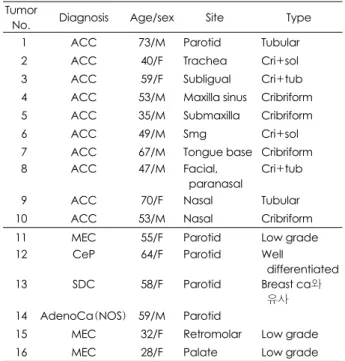
Table 1. Clinicopathologic datas of two group of adenoid cyst- ic carcinoma and other salivary gland cancer Tumor
No. Diagnosis Age/sex Site Type
01 ACC 73/M Parotid Tubular
02 ACC 40/F Trachea Cri+sol
03 ACC 59/F Subligual Cri+tub 04 ACC 53/M Maxilla sinus Cribriform
05 ACC 35/M Submaxilla Cribriform
06 ACC 49/M Smg Cri+sol
07 ACC 67/M Tongue base Cribriform 08 ACC 47/M Facial,
paranasal
Cri+tub
09 ACC 70/F Nasal Tubular
10 ACC 53/M Nasal Cribriform 11 MEC 55/F Parotid Low grade 12 CeP 64/F Parotid Well
differentiated 13 SDC 58/F Parotid Breast ca와
유사 14 AdenoCa(NOS) 59/M Parotid
15 MEC 32/F Retromolar Low grade 16 MEC 28/F Palate Low grade ACC:Adenoid cystic carcinoma, SMG:Submandibular gland, CeP:Carcinoma ex pleomorphic adenoma, SDC:Salivary duct carcinoma, AdenoCa(NOS):Adenocarcinoma(not oth- erwise specified), MEC:Mucoepidermoid carcinoma, cri:
Cribriform, sol:Solid, tub:Tubular
3. 선양낭성암종에서 Ki-67의 발현 정도
선양낭성암종 2예(20%)는 발현되지 않았으며, 2예(20%) 에서는 +/-로 발현되었다. 5예(50%)에서 1+, 1예(10%) 에서 2+로 나타났다. 대조군 중 점액표피양암종 3예 모두 Ki-67이 발현되지 않았다., 타액선관암종 +/-, 다형선종유 래암종과 NOS는 2+로 발현되었다(Table 2).
고 찰
선양낭성암종의 면역조직화학적 진단에 이용되는 것으로 알려진 것은 다음과 같다. 대부분의 연구자들은 S-100 단 백질은 선양낭성암종에서 발현된다고 하였다. 선양낭성암종 에서 keratin과 vimentin이 동시에 발현되고 muscle, myosin 에 대한 반응을 보이는 것은 암종의 근상피 분화를 나타내는 증거이다. 실제 내강을 지닌 분화 관세포는 CEA(carcinoem- bryonic antigen), EMA(epithelial membrane antigen), lactoferrin에 반응을 보이나, muscle-specific actin, vimen- tin에는 반응을 하지 않는다.
Cytokeratins(CK)은 20개의 다른 폴리펩티드를 포함한 intermediate filament에 속하는 단백질 복합체로, 양성 및 악성 상피세포에 존재한다. 하지만, 비상피세포에도 존재할 수 있다.5) CK7은 많은 정상인의 관(ductal), 샘(glandular) 과 이행(transitional)상피에서 발견되지만, 중층편평상피에 는 발견되지 않는다. 또한 CK7은 내피세포에서도 발현될 수 있다.5) CK20은 정상인 조직에서 위장상피, 요 상피와
Merkel 세포에서만 발견된다.6) CK7과 CK20의 면역표현형 의 조합은 여러 종양의 기원을 밝히는데 도움이 될 수 있어, 실제 많은 종양에서 종양의 기원을 밝히는 데 이용되고 있 다.5) 물론 종양에 대해 CK7과 CK20 면역표현형의 정확도, 민감도가 100%인 것은 아니다. 편도환(Waldeyer’s ring) 의 소타액선을 포함한 정상 타액선의 관구조에 CK7은 염색 되나, CK20은 염색되지 않는다.7) Lee 등8)은 타액선의 악성 종양에서 CK7은 모두 양성이었으나, CK20에 대해서는 모 두 음성을 보인다고 하였다. 본 연구에서도 선양낭성암종 10예 모두에서 CK7은 양성으로 결과가 나왔으며, 기타 암 종에서도 다형선종유래암종을 제외한 모든 예에서 양성으로 나왔다. CK20은 16예 모두에서 음성으로 나타났다. CK19 는 CK8나 CK18과 마찬가지로 정상 및 종양의 단순상피와 비각화성 상피를 포함하는 많은 형태의 상피에서 발현된다.
그러나, 편평상피암종에서는 발현되지 않으며 각종 선암종 에서 높게 발현되는 특징이 있다.9,10) CK19의 타액선 종양 에서의 발현 정도에 대한 기존의 연구는 거의 없었다. 본 연 구에서 선양낭성암종에서 CK19의 염색 양성율은 75%, 그 외의 암종에서는 100%의 양성을 보였다.
SMA(smooth muscle actin)의 면역조직화학적 염색은 타액선 종양의 근상피 성분의 표지자이다.11,12) Beltran 등은 선양낭성암종과 다형 저등급 선암종을 감별진단하는데 SMA 가 의미가 있다고 하였다.11,13) 또한 기저세포선암과 basaloid squamous carcinoma 에서는 SMA의 발현율이 적으며, 상 피-근상피 암종에서 높은 발현율을 나타내어 감별진단하는 데 도움을 얻을 수 있다. 본 연구에서 선양낭성암종 9예 중 8예(88.9%)에서 SMA 양성으로 나왔다. 다른 6예의 암종 에서는 모두 음성으로 나왔다
Ki-67은 세포주기의 S과 M기에 우세하게 표현되는 핵항 원이다. 여러 연구에서 이 항원은 많은 종양의 성장 분율을 추정하는 데 유용하다고 알려져 있다.14) 또한, 타액선 종양에 서도 유용한 예후인자로 알려져 있다.11,15,16) Ki-67염색 정 도는 선양낭성암종의 단기간 임상 경과의 가장 중요한 안내 자로 증명되어 왔다.17,18) 본 연구에서 선양낭성암종 중 1예 를 제외한 나머지에서 Ki-67의 발현 정도는 낮게 보고되었 다. 이는 선양낭성암종의 자연 경과가 나쁘지는 않다는 것을 간접적으로 나타내는 것이라 하겠다. 본 연구에서도 선양낭 성암종의 Ki-67의 발현 정도는, 저등급 점액표피양암종보 다는 높았으며, 그 외 암종보다는 발현정도가 낮게 나타났다.
타액선 악성종양은 일반적으로 저등급(Low grade), 중간등 급(Intermediate grade), 고등급(High grade)으로 나눌 수 있으며, 선양낭성암종은 일반적으로 중간등급에 해당한다. 본 연구에서 Ki-67 발현 정도도 이에 합당한 결과를 보였다.
그러나, 선양낭성암종중 1예는(No. 10) SMA 염색 음성을 보이고, Ki-67은 2+ 염색 양성으로 나와, 본 연구의 전반
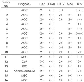
Table 2. Immunohistochemical datas of two group of adenoid cystic carcinoma and other salivary gland cancer Tumor
No. Diagnosis CK7 CK20 CK19 SMA Ki-67
01 ACC 2+ (-) (-) (+/-)
02 ACC 2+ (-) 2+ 2+ 1+
03 ACC 2+ (-) 2+ 2+ (-)
04 ACC 2+ (-) 2+ 1+
05 ACC 2+ (-) 2+ (-)
06 ACC 2+ (-) 2+ 2+ (+/-)
07 ACC 2+ (-) 2+ 2+ 1+
08 ACC 2+ (-) 2+ 2+ 1+
09 ACC 2+ (-) (-) 2+ 1+
10 ACC 2+ (-) 2+ (-) 2+
11 MEC 2+ (-) 2+ (-) (-) 12 CeP (-) (-) 2+ (-) 2+
13 SDC 2+ (-) 2+ (-) 1+
14 AdenoCa(NOS) 2+ (-) 2+ (-) 2+
15 MEC 2+ (-) 2+ (-) (-) 16 MEC 2+ (-) 2+ (-) (-) ACC:Adenoid cystic carcinoma, CeP:Carcinoma ex pleo- morphic adenoma, SDC:Salivary duct carcinoma, Adeno- Ca(NOS):Adenocarcinoma(not oth-erwise specified), MEC:
Mucoepidermoid carcinoma
Fig. 1. Pathologic findings with immunostaining of CK7(A), CK20(B) of adenoid cystic carcinoma. A:It shows high uptake of CK7 (100×). B:It shows no uptake of CK20(100×).
A B
Fig. 2. Pathologic findings with immunostaining of CK19 of adenoid cystic carcinoma(A), mucoepidermoid carcinoma(B). A:It shows high uptake of CK19(40×). B:It shows also high uptake of CK19(100×).
A B
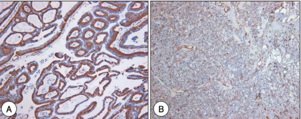
Fig. 3. Pathologic findings with immunostaining of SMA of adenoid cystic carcinoma(A), mucoepidermoid carcinoma(B). A:It shows high uptake of SMA(100×). B:It shows no uptake of SMA(40×).
A B
Fig. 4. Pathologic findings with immunostaining of Ki-67of adenoid cystic carcinoma(cribriform type)(A), adenocarcinoma(not otherwise specified)(B). A:It shows low uptake of Ki-67(40×). B:It shows high uptake of Ki-67(40×).
A B
적인 결과와는 상반된 결과를 나타냈다. 10번의 종양은 종양 특성이 일반적인 선양낭성암종과는 다른 것으로 생각된다.
선양낭성과 감별진단되어야 할 종양으로는 다형선종, 다형 저등급 선암종, 상피-근상피 암종, 기저세포선종, basaloid squamous carcinoma 등이 있다. 본 연구에서 시행한 면역 조직화학적 검사를 이용하면 선양낭성암종을 진단하는 데 도 움이 될 것으로 기대된다.
결 론
두경부 선양낭성암종은 CK7, CK19, CK20, SMA에 대한 면역조직화학적 염색 양성율은 100%, 75%, 0%, 88.9%이 었다. 두경부 선양낭성암종의 Ki-67 염색 발현정도는 높지 않아, 선양낭성암종의 자연 경과가 나쁘지 않다는 것을 간접 적으로 알 수 있었다.
위와 같은 결과는 선양낭성암종을 감별 진단하는데 면역조 직화학 염색을 활용하는 것이 가치 있을 것으로 판단된다. 그 러나, 더 많은 환자를 대상으로 한 추가적인 연구가 필요할 것 으로 사료된다.
중심 단어:
선양낭성암종·면역조직화학·두경부.References
1) Choi CS, Choi K, Song JJ, Hwang KS, Jeong KY, Choi JW. Dis- tribution of β-Catenin Expression in Adenoid Cystic Carcinoma of the Head and Neck. Korean J Otolaryngol. 2000;43(2):188- 192.
2) Kim H, Nam SY, Kim JH, Kim SY, Lee KS. Postoperative Com- plication and Prognosis of Salivary Gland Tumor. Korean J Oto- laryngol. 1997;40(2):197-203.
3) Park YY, Shim YS, Oh KK, Lee YS, Choi JH. Clinical Analysis in Adenoid Cystic Carcinoma of the Head and Neck. Korean J Otolaryngol. 1997;40(10):1398-1403.
4) Kim KH, Seong MH, Jang KH, Lee JS, Jo YS. Immunohistoche- mical Study on the Expression of p53 Protein in Adenoid Cystic Carcinoma of the Head and Neck. Korean J Otolaryngol. 1994; 37:753-759.
5) Campbell F, Herrington CS. Application of cytokeratin 7 and 20 immunohistochemistry to diagnostic pathology. Current Diagno-
stic Pathology. 2001;7:113-122.
6) Miettinen M, Keratin 20. Immunohistochemical marker for gas- trointestinal, uroepithelial and Merkel cell carcinomas. Mod Pathol. 1995;8:384-388.
7) Regauer S, Beham A, Mannweiler S. CK7 expression in carci- nomas of the Waldeyer’s ring area. Hum Pathol. 2000;31:1096- 1101.
8) Lee JH, Lee JH, Kim A, Kim I, Chae YS. Unique expression of MUC3, MUC5AC and cytokeratins in salivary gland carcinomas, Pathol Int. 2005;55:386-390.
9) Takeda T, Sugihara K, Hirayama Y, Hirano M, Tanuma JI, Sem- ba I. Immunohistological evaluation of Ki-67, p63, CK19 and p53 expression in oral epithelial dysplasias. Oral Pathol Med.
2006;35:369-375.
10) Bartek J, Bartkova J, Taylor-Papadimitriou J, Rejthar A, Kovarik J, Lukas Z, et al. Differential expression of cytokeratin 19 in hu- man epithelial tissues revealed by monospecific monoclonal an- tibodies. Histochem J. 1986;18:565-575.
11) Beltran D. Faquin WC, Gallagber G, August M. Selective immu- nohostochemical comparision of Polymorphous low-grade aden- ocarcinoma and Adenoid cystic carcinoma, J Oral Maxillofac Surg. 2006;64:415-423.
12) de Araújo VC, de Sousa SO, Carvalho YR, de Araújo NS. Appli- cation of immunohistochemistry to the disgnosis of salivary gland tumors. Appl Immunohistochem Mol Morphol. 2000;8:195.
13) Ogawa Y. Immunocytochemistry of myoepithelial cells in the salivary glands. Histochem Cytochem. 2003;38:343-426.
14) Carlinfante G, Lazzaretti M, Ferrari S, Bianchi B, Crafa P. p53, bcl-2 and Ki-67 expression in adenoid cystic carcinoma of the palate. A clinico-pathologic study of 21 cases with long-term follow-up. Pathology-Research and Practice. 2005;200:791-799.
15) Kiyoshima T, Shima K, Kobayashi I, Matsuo K, Okamura K, Ko- matsu S, et al. Exprssion of ρ53 tumor suppressor gene in ade- noid cystic and mucoepidermoid carcinomas of the salivary glands.
Oral oncology. 2001;37:315-322.
16) Alves FA, Pires FR, de Almeida OP, Lopes MA, Kowalski LP.
PCNA, Ki-67 and p53 expressions in submandibular salivary gland tumors. Int J Oral Maxillofac. Surg. 2004;33:593-597.
17) Nordgard S, Franzen G, Boysen M, Halvorsen TB. Ki-67 as a prognostic marker in adenoid cystic carcinoma assessed with the monoclonal antibody MIBI in paraffin sections, Laryngoscope.
1997;107:531-536.
18) Barnes L, Eveson JW, Reichart P, Sidransky D. Pathology &
genetics Head and Neck Tumors, WHO classification of tumors.
2002;221-231.