Influence of Interferon-τ on the Production of Prostaglandins, Cyclooxygenase-2 Expression In Vitro and Release of Progesterone in Bovine Endometrial Cells
Ji-Eun Lee1, Yong-Seung Lee1, Han-Jun Yoo3, Kyoung-Jin Lee1, Joung-Jun Park3, Hee-Tae Cheong2, Boo-Keun Yang1 and Choon-Keun Park1,*
1College of Animal Life Science, Kangwon National University, Chuncheon 200-701, Korea
2College of Veterinary Medicine, Kangwon National University, Chuncheon 200-701, Korea
3Animal Reproduction & Biotechnology Center, Hoengseung 225-807, Korea
ABSTRACT
The purpose of the present study was to investigate the effect of IFN-τ on prostaglandin synthesis, cyclooxygenase-2 (COX-2) gene expression in vitro and concentration of progesterone (P4) in endometrial cells. Epithelial and stromal cells cultured in vitro were isolated from bovine endometrium and stimulated with increasing doses of IFN-τ (0, 0.02, 0.2 and 2 ug/ml). Human chorionic gonadotropin (hCG, 1.5 IU/ml) was used as a positive control. Prostaglandin E2
and F2α levels in the culture media were analyzed by enzyme immunoassays and total RNA was extracted from the cells for RT-PCR. P4 concentrations of blood samples were assayed by chemiluminescent immuno assays system. In epithelial cells, COX-2 gene expression was increased in the presence of IFN-τ (p<0.05), but it was not significantly different in all groups of stromal cells except for 2 ug/ml IFN-τ group (p<0.05). Although IFN-τ did not affect PGE2
and PGF2α production in epithelial cells, it decreased PGE2 and PGF2α production significantly in stromal cells (p<
0.05). In vivo experiment, blood concentration of P4 was significantly increased after addition of IFN-τ (1 ug/ml). The results indicate that PG production was mediated by COX-2 expression in stromal cells but it was not affected in epithelial cells and this suggest that treatment of IFN-τ could improve the implantation environment of uterine by maintenance of high P4 concentration.
(Key words : IFN-τ, COX-2, PG, P4, bovine endometrial cell)
†This work was carried out with the support of “Cooperative Research Program for Agriculture Science & Technology Development (Project No. PJ907008)” Rural Development Administration, Republic of Korea.
*Correspondence : E-mail : [email protected]
INTRODUCTION
Even though numerous substances within organisms affect the success of implantation, the most critical regulating factor would be prostaglandin (PG) (Poyser, 1995). Among many types of PG, prostaglandin F2α (PGF2α) and prostaglandin E2 (PGE2) are the regulating substances that affect the maintenance of corpus luteum, which is important for the pregnancy success (Pratt et al., 1977). In mammals, PGF2α generally keeps repro- duction cycle regularly repeating by controlling the life span of corpus luteum, and it is produced within uterus (McCracken et al., 1999; Okuda et al., 2002). On the contrary to the most animals where PGF2α is secreted from the uterus, it is produced from the ovary in remnants (Hearn et al., 1991). PGE2, unlike PGF2α, is known to maintain the existence of corpus luteum (Magness et al., 1981; Pratt et al., 1977). PG is the physiolo- gical substance which is created from arachidonic acid, rich in
cell membranes. PG synthase as well as Cyclooxygenase (COX) also can change arachidonic acid into PG (Smith et al., 2000).
Therefore, COX is one of the important substances that regu- late the formation of PG.
COX has three isozymes, COX-1, COX-2 and COX-3. En- zymes that are useful for PG formation are COX-1 and COX-2 (Chandrasekharan et al., 2002). Previous research, however, reported that COX-1 was constitutive enzyme that affects house- keeping function and COX-2 works as induced enzyme that has various pathologic, physiological functions (Smith et al., 1996; Smith et al., 2000). mRNA amount and protein produc- tion of COX-2 changes according to the reproduction cycle while there is no change in COX-1. Therefore, COX-2 is assumed to be more closely related to the production of PG within bovine endometrial cells compared to COX-1 (Arosh et al., 2002).
Substances that induce secretion of COX-2 are many inclu-
ding oxytocin and cytokine (Asselin et al., 1997; Arosh et al., 2002). Interferon-τ (IFN-τ) is secreted from the trophoblasts of embryo within uterus of its mother (Bazer, 1992; Roberts et al., 1992). It is also called, therefore, Trophoblastic protein-1 (TP-1) and belongs to a type of cytokine such as interleukin, lymphokine. IFN-τ, about 18000~20000 Da proteins, is known to be secreted the most from the 15th day to 19th during preg- nancy. It is most essential substance to acknowledge the preg- nancy signals for cows or sheep (Bartol et al., 1985). For an example, when IFN-τ was injected into the uterine cavity of non-pregnant female sheep, the endometrial cells showed the responses which usually are shown during pregnancy (Chen et al., 2006). The main target of IFN-τ is endometrial cells not the corpus luteum. But if IFN-τ stimulates corpus luteum, PGF2α
would be produced so that for a stable implantation, appro- priate control must be made (Bazer et al., 1991). The common theory of controlling secretion of PGF is that IFN-τ restrains the increase of estrogen receptor-alpha (ERa), which usually occurs on the epithelial cells when estrus cycles ends within non-pregnant female. Low concentration of ERa restrains oxyto- cin receptor’s increase and as a result, PGF secretion from the endrometrial cells stops (Spencer et al., 2007). Even though various researches using sheep have been done, it is uncertain that how IFN-τ regulates PG production on within endometrial cells in “cows” (Robinson et al., 2001; Roberts et al., 2008).
To make the best environment for embryo to settle, during the implantation period, numerous hormones are secreted from the uterus. Progesterone (P4) is the hormone secreted from the corpus luteum to maintain pregnancy and it is highly impor- tant to keep the pregnancy safe (Bearden and Fuquay, 2000).
If pregnancy does not occur within 16~17 days, PG is pro- duced and until the removal of corpus luteum, it processes the decrease of luteum and P4 concentration also decreases. FSH secretion increases, on the contrary, to keep the normal estrus cycle to repeat (Schams et al., 1977).
Research about controlling the production of oocysts have been done for decades and mechanical methods, medicinal me- thods or hormonal methods have been developed. Among those methods introduced, method using P4 for creating oocyst was found out (Bo et al., 1993; Bo et al., 1995; Bergfelt et al., 1997; Kohram et al., 1998). By implanting P4 system into the ear to create similar uterus environment to the one during preg- nancy, 5mg of estrogen (E-17B) was injected into the muscle additionally. It was found to be effective for synchronization of estrus cycle when the method was used (Bo et al., 1995). For
the assisted reproductive technology, P4 concentration level would be the important factor for determining the success of implantation within the uterus. Research on relationship bet- ween P4 and PG once showed the result that when the sheep got an injection of P4 showed the higher PG synthase amount from the endometrial cells (Salmonsen et al., 1991). There is an enzyme called prostaglandin H synthase(PGHS) that formu- lates PG, and when it is activated, arachidonate gets stimulated to be changed into PGH2. Therefore, increase in PGHS means the increase in PG production. In the research on effect of P3 on PGHS, it was stated that when P4 was controlled in the endometrium of sheep, there was no effect on expression of PGHS-2 genes while when PGHS-2 was abundant in the uterus, it greatly stimulated the process (Wu et al., 1997).
Therefore, this research was carried out to find out what kind of relationship IFN-τ injection has on the production of P4 when IFN-τ was controlled in vitro conditions after analy- zing the expression of COX-2 gene and PG production.
MATERIALS AND METHODS
1. Harvest of Endometrial Cells
The uterus of Korean native cow(Hanwoo), no later than 30 min after being slaughtered, was moved to the ice box and tran- sported to a laboratory within 1~1.5 h. Estrous cycle of uterus, based on the previous research results (Ireland et al., 1979), was judged by the shape of ovary. Only luteal phase of day 5~10 was used. For collect cells, luteal ovary and same uterine horn were used. Cultured cells of passage 0~3 were only used.
Three uteruses, in total, were used for this experiment. Epithe- lial cells and stromal cells from bovine uterus was collected from three uteruses (Murakami et al., 2003). Uterine horn, trans- ported to the lab, was sterilized by gauze and with 70% al- cohol then sealed in the zipper bag, then moved to clean bench.
Polyvinyl catheter was injected through the tube of uterine horn and fixed by forceps. Through the catheter, HBSS (Hanks’
balanced salt solution, sterile Ca2+ - and Mg2+ -free) added with 100 IU/ml penicillin (Sigma; P3032), 100 ug/ml strep- tomycin (Sigma; S9137) and 0.1% BSA (Qbiogene; BSASG100) was injected and uterine cavity was washed 3 time. Following the completion of cleaning, the other side got bound as well.
Then, the uterine cavity got filled with 0.76% EDTA.4Na buffer - solution of 20 mM EDTA.2Na (Sigma, St.Louis, MO, USA;
E5134), 117 mM NaCl (Sigma; S5886), 2.7 mM KCl (Sigma;
P5405), 6.8 mM Na2HPO4 (Sigma; S0876) and 1.5 mM KH2PO4
(Sigma; P5379). Then, in the beaker of 0.85% saline, being kept from saline going into the uterine cavity, it was cultured for 50 min within the incubated water bath of 37℃. Forceps of uterine cavity was released, after culture, and 0.76% EDTA.
4Na buffer was recollected into 50 ml tube. HBSS, added with antibiotics and 0.1% BSA was used to was the uterine cavity out. Sterilized scissors was used to cut it to expose the endo- metrium and epithelial cells were carefully harvested by medi- cal blades and kept into the 50 ml tube with HBSS. To collect stromal cells, endometrium was slightly sliced by surgical blade and mesometrium was collected into the 50 ml tube with HBSS and it was torn into small pieces (1 mm3) by sterilized scissors.
Tubes with HBSS and harvested epithelial cells and stromal cells were made sure to be 50 ml in total. Collagenase (Wor- thington, collagenase type IV) was injected to epithelial cells (500 ul) while 1,000 ul of collagenase was injected into stromal cells. Within the incubated water bath of the temperature of 37℃, epithelial cells were cultured for 10 min and stromal cells were for 30 min. Epithelial and stromal cells, after being cul- tured, were filtered through 70 um cell strainer (BD Falcon, Cat.No. 352350) and centrifuged for 5 min at 1,500 rpm. Su- pernatant was being removed and 15 ml of HBSS was added into the remaining cell pellet, and then centrifugation at 1,500 rpm was made for 5 min. Such HBSS washing procedure was repeated for about 2~3 times and to remove the blood, 25 ml of Tris-NH4 buffer was injected after. After waiting for 10 min, centrifugation at 1,500 rpm was done for 5 min and su- pernatant was removed. Suspension was made by adding 5 ml of DMEM + 0.1% BSA solution, after removal of blood. It, again, went through centrifugation at 1,500 rpm for 5 min.
After completion of these steps, cells were divided on 4 well dish(Nunc, Cat.No. 176740) with the concentration of 1.5×10⁵ cells/well and were cultured within the incubator of 37℃, CO2
5%. Culture fluid was changed every other day until the cell confluency reaches 80% (6~7 days after culture).
2. Treatment
To investigate the effect of IFN-τ on secretion of PGF2α, PGE2 and expression of COX-2 in the epithelial cells and stro- mal from cows, cells showing more than 80% of confluency are exposed to various Recombinant ovine interferon-tau with different concentrations (0.02, 0.2 and 2 ug/ml) for 24 h. Cul- ture fluid without IFN-τ was used as the negative control, culture fluid containing hCG of 1.5 IU/ml was used as a po- sitive control. After 24 h, culture solution was recollected and
kept under 20℃ to be used in ELISA, cells were collected and homogenated in RNAiso for RT-PCR.
3. RT-PCR
RNA extraction was done by using Trizol(RNAiso Plus) and Chloroform. Cells were harvested from the 4 well dish and ex- posed to LN2 steam, to damage the cell membrane. 400 ul RNAiso Plus(TAKARA, Cat.No. 9109) was sprayed and it was exposed to react for 1 h under room temperature. Trizol and Chloroform (J.T.Baker, Cat.No. 9257-68) in the ratio of 4:1 (each 400 ul, 100 ul) were added and voltexing was done. Under the temperature of 4℃, centrifugation at 1,2000 rpm for 10 min, RNA extraction only got collected. Extracted RNA fluid was combined with the Isopropyl alcohol (DAEJUNG, Cat.No. 5035- 4405) at the equal amounts, RNA got laid down by centrifu- gation at 12,000 rpm for 10 min. After supernatant was re- moved, 70% Ethanol (DAEJUNG, Cat. No. 64-17-5) was added in the amount of 1ml. Following the pipetting, centrifugation at 12,000 rpm for 5 min was practiced and RNA was washed.
After centrifugation, supernatant got all removed and alcohol was vaporized totally. RNA was diluted by DEPC (BIONEER, Cat.No. C-9030) and measured by Nano drop 2000 c (Thermo scientific) into of 0.1~1 ug of Total RNA. After measuring Total RNA 0.1~1 ug, DEPC was used to arrange Total reac- tion volume into 20 ul. The solution was put into the RT Pre- Mix Kit (iNtRon, Cat.No. 25081), under MyGenie32 Thermal Block (BIONEER), cDNA was synthesized for 60 min under the temperature of 45℃. And RTase was deactivated for 5 min under 95℃. cDNA, reverse-transcripted during RT phase, was put under PCR by using AccuPower PCR PreMix (Bioneer, Cat.No. K-2016). B-actin, housekeeping gene within cells, was regarded as IS and the experiment continued. 10 pmol of each primer were added to the PCR tube, in addition to 1 U of Taq DNA polymerase, each deoxynucleosidetriphosphate at a con- centration of 250 um, 50 mM Tris-HCl (pH 8.3), 40 mM KCl, 1.5 mM MgCl2 and gel loading dye. The final volume was adjusted to 20 ul with distilled water. Primer sequences of genes used in the experiment are stated in the Table 1 and the other conditions are following. COX-2 was under repetition of 44 cycles; after DNA polymerase activation for 7 min at 94℃, denaturation for 1min at 94℃, annealing for 1 min. at 54℃, extension for 2 min at 72℃. B-actin went under a repetition of 29 cycles; DNA polymerase activated for 30 s at 94℃, de- naturation for 30 s at 94℃, annealing for 30 s at 60℃, exten- sion for 1 min at 72℃. Both genes were put under extension
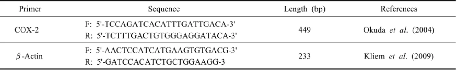
Table 1. Prier sequences used in RT-PCR analysis
Primer Sequence Length (bp) References
COX-2 F: 5'-TCCAGATCACATTTGATTGACA-3'
R: 5'-TCTTTGACTGTGGGAGGATACA-3' 449 Okuda et al. (2004)
β-Actin F: 5'-AACTCCATCATGAAGTGTGACG-3'
R: 5'-GATCCACATCTGCTGGAAGG-3 233 Kliem et al. (2009)
phase for 5 min additionally at 72℃. Each PCR product went under electrophoresis on the 2% Agarose gel (BIONEER, C- 9100) with addition of Ethidium bromide (BIONEER, Cat. No.
C-9009) with 100 bp DNA Ladder (Bioneer, Cat.No. 1030) and a photo was taken under UV light. Each band was analyzed by Multi gauge v3.0 software.
4. ELISA
Concentration levels of PGF2α and PGE2 in culture solution were analyzed through enzyme immunoassay and PGF2α EIA kit and PGE2 EIA kit from EnzoⓇ were used. Each Standard curve boundary refers to 0.003~50 ng/ml for PGF2α and 0.0391~ 2.5 ng/ml for PGE2. Each ED50 (Effective dose 50) level is 0.23 ng/ml for PGF2α and 0.166 ng/ml for PGE2.
5. Analysis of Progesterone Concentration within Blood
Considering the day of CIDR injection through vagina, and injection of GnRH (100 ug, 1 cc) as the first day (the starting day of experiment), regression of progesterone and estrus are induced by removal of CIDR and injection of PGF2α (25 mg, 5 cc) after seven days. On the 9th day, after two days from PGF2α injection, GnRH (100 ug, 1 cc) is injected and on the 10th day, seminal plasma (diluted seminal plasma 5 ml) is in- jected into ovary and on the 17th day, INF-tau injection (1 ug, 1 cc) is practiced. From the day of injection of seminal plasma into 5 of female cows, P4 concentration was analyzed over the interval of two days (10, 12, 14, 16, 18, 20, 22, 24, 26, 28 and 30). Using 15 ml vacutainer cleaned by heparin, between 10 am and 11 am, 10 ml of blood was collected from jugular vein and directly moved to the lab. Within 3 h, centrifugation at 3,000 rpm for 15 min was done and serum was frozen (20℃) until the analysis. P4 concentration in the blood was measured by the medical photometric immunological analyzer (Immulite 1000, DPC CIRRUS, Inc, USA).
6. Statistical Analysis
GLM (General Linear Models) Procedure, where various types
Fig. 1. Morphology of endometrial cells in bovine (a) epithelial, (b) stromal cells.
of ANOVA can be done in SAS (VERSION 9.1), was applied to each average value of the data collected from the experi- ments. Duncan’s multiple range test was used for statistical analysis. Significant difference was verified in the 95% range.
RESULTS
COX-2 gene expressed on the bovine endometrial cells of various levels of IFN-τ by the RT-PCR method. The images were analyzed through electrophoresis. After completion of elec- trophoresis, bands on the images were compared to the marker from the electrophoresis, it was observed at 449 bp. B-actin, also, was used as the inter-standard substance and consistent amount of bands were observed on every sample at 233 bp.
Expression of COX-2 within epithelial cells was 0.65 ± 0.06 in control group, 1.75 ± 0.04 in 0.02 ug/ml group, 1.79 ± 0.08 in IFN-τ 0.2 ug/ml group, 1.57 ± 0.11 in IFN-τ 2 ug/ml group, 1.36 ± 0.17 in hCG. Compared to the control group, every group showed the significant increase in its result (p<0.05). COX-2 expression on hCG, used as positive control group, also showed the increased result compared to the negative control group (IFN-τ 0 ug/ml) (p<0.05, Fig. 2 (A)). Expression of COX-2 on stromal cells was 1.30 ± 0.02 in control group, 1.28 ± 0.22 in 0.02 ug/ml group, 0.98 ± 0.12 in IFN-τ 0.2 ug/ml group, 0.81 ± 0.04 in IFN-τ 2 ug/ml group, 0.99 ± 0.08 in hCG. Ex- pression of COX-2 was significantly low when IFN-τ concen-
(A) (B)
Fig. 2. Effect of IFN-τ on COX-2 mRNA expression in bovine endometrium. (A) epithelial cells, (B) stromal cells, columns with different superscripts are significantly different (Mean±
SEM, p<0.05).
tration was 0.2 ug/ml (p<0.05, Fig. 2 (B)). In Fig. 3 and 4, pro- staglandin F2α and E2 produced on bovine endometrial cells for 24 h when IFN-τ was 0.02~2 ug/ml. The amount of PGF2α on the stromal cells was 25.27 ± 5.15, 17.37 ± 0.61 on IFN-τ 0.02 ug/ml, 15.69 ± 2.28 on IFN-τ 0.2 ug/ml, 14.88 ± 1.38 on IFN-τ 2 ug/ml and 22.52 ± 1.91 on hCG. Within the IFN-τ 0.2ug/ml group, the lowest amount was produced (p<
0.05). On the contrary, there was no significant difference among any kind of group on epithelial cells(Fig. 3 (B)). The amount of PGF2α on the epithelial cells was 7.14 ± 0.01 on the control group, 6.52 ± 0.39 on IFN-τ 0.02 ug/ml, 7.24 ± 0.51 on IFN-τ 0.2 ug/ml, 7.27 ± 0.40 on IFN-τ 2 ug/ml and 7.20 ± 0.92 on hCG(p<0.05). The amount of PGE2 on the stromal cells was 636.73 ± 167.99 on the control group, 144.63 ± 12.68 on IFN-τ 0.02 ug/ml, 84.11 ± 5.74 on IFN-τ 0.2 ug/ml, 83.79 ± 1.83 on IFN-τ 2 ug/ml and 275.78 ± 22.43 on hCG. Except the nega- tive control group where no influence of IFN-τ existed, every group showed the significantly low result (p<0.05). The amount of PGE2 on the epithelial cells was 1.66 ± 0.00.3 on the con- trol group, 1.06 ± 0.13 on IFN-τ 0.02 ug/ml, 4.56 ± 1.55 on IFN-τ 0.2 ug/ml, 2.21 ± 0.17 on IFN-τ 2 ug/ml and 0.83 ± 0.07 on hCG. Excluding the IFN-τ 0.2 ug/ml group, every group did not show any significant difference. Fig. 5 shows the ana- lysis of the concentration after synchronization of estrus in bovine following the injection of seminal plasma and IFN-τ.
The day when the seminal plasma was injected into the cows after synchronization of estrus appears as the day 0 on the graph. IFN-τ group got injected of IFN-τ on the 7th day after the day 0 when seminal plasma had been injected. Both
(A)
(B)
Fig. 3. Influence of IFN-τ on PGF2α production in bovine endo- metrial cells. (A) stromal cells (black bar), (B) epithelial cells (white bar), columns with different superscripts are signifi- cantly different (Mean±SEM, p<0.05).
(A)
(B)
Fig. 4. Influence of IFN-τ on PGE2 production in bovine endo- metrial cells. (A) stromal cells (black bar), (B) epithelial cells (white bar), columns with different superscripts are signifi- cantly different (Mean±SEM, p<0.05).
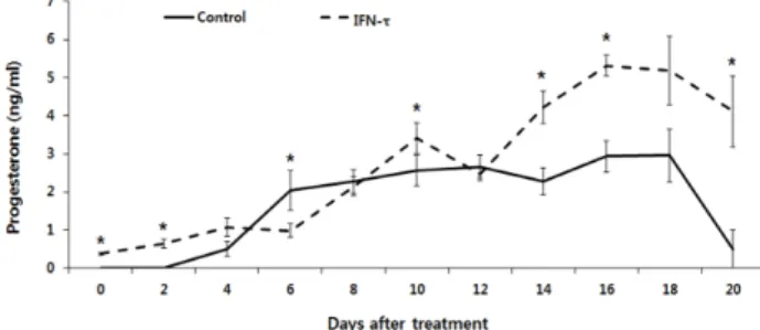
Fig. 5. Effect of IFN-τ (1 ug/ml) injection on blood progesterone concentration after synchronization of estrus in bovine, *signi- ficantly different to controls with IFN-τ (Mean±SEM, p<0.05)
groups showed the higher progesterone concentration within blood, compared to the control group, until the day 2. From day 4 to day 12, it showed the irregular pattern of concentration going up and down repeatedly compared to the control group (p<0.05). After 14 days, however, both groups showed the signi- ficant increase of progesterone concentration compared to the control group. Until the day before of second estrus cycle started, on the day 20, seminal plasma and IFN-τ group both showed the significantly high progesterone concentration (p<0.05).
DISCUSSION
Fig. 2 shows that IFN-τ works on uterine epithelial cells and stromal cells. Especially, on the epithelial cells, expression of COX-2 by IFN-τ significantly increased (p<0.05). Also, COX-2 expression on the epithelial cells did not show any significant difference according to the concentration of IFN-τ.
For the stromal cells, except the IFN-τ controlled group at the concentration of 2 ug/ml, no significant difference was shown including the control group. IFN-τ produced from the embryo stimulates the endometrial cells and restrains the formation of Oxytocin receptor and stops the production of PGF2α. There- fore, the reason why expression of COX-2 increased signifi- cantly on the epithelial cells is that the expression was stimu- lated by IFN-τ. It is a signal to produce various proteins ne- cessary for survival of embryo and it seems that it would im- prove the implantation conditions for embryo implantation. In the Fig. 3, the production of PGF2α on epithelial cells shown on the white bar graph remained low compared to the stromal cells. It would be because that IFN-τ did not play a role of stimulating the secretion of PGF2α or production of PGF2α
was not activated well on the epithelial cells. Therefore, consis- tency of production of PGF2α was observed no matter whether
IFN-τ existed or not on the endometrial epithelial cells which are the nearest to the embryo getting ready for implantation within the bovine central implantation. Since the endometrial cells from the luteal phase were used in the experiment, there was no significant difference in the produced amount of PGF2α
between in the control group and IFN-τ group. When more than 0.2 ug/ml of IFN-τ were cultured, production of PGF2α
decreased significantly because effect of PGF2α, inducing the decrease of IFN-τ to change the uterine condition into the one suitable for implantation of embryo, got reduced. PGE2 pro- duced on the epithelial cells showed similar pattern with the PGF2α production (Fig. 4). Significantly high secretion of PGE2
was observed on the 0.2 ug/ml IFN-τ cultured group but not quite high regarding the real measure of the data (p<0.05).
Excluding this specific group, other groups kept low level of secretion with no significant difference in between. Conside- ring the result from this research, epithelial cells do not show significant response from the secretion of PGF2α or PGE2 when mother accepts the pregnancy signals (IFN-τ). PGE2 is a hormone closely related to the implantation and produced from the ovary, uterus and the embryonic membrane. In humans, pregnancy signal receptor is hCG and it plays a role to divide stromal cells into Decidua. COX-2 is increased for division and it also has relation to the increase of PGE2 (Tang and Gurpide, 1993; Han et al., 1996). In rats, it was reported that COX-2 gene is regulated by the embryo implanted in the be- ginning phase of the implantation (Chakraborty et al., 1996).
Peptides such as IFN-τ (in ruminants), Placental lactogens (in rodents), estrogen and prolactin (in pigs) and hCG (in primates) have the same influence and effect of the PG regulation in the uterus before implantation. Function of PG before implantation varies to the animals but it has identical mechanism of regu- lating PGE2 through expression of COX-2 expression. In this study, amount of PGE2 produced on the stromal cells signifi- cantly decreased when IFN-τ was injected (p<0.05). It shows the opposite pattern to the results from previous researches. It is assumed to have a relation to the amount of COX-2 expressed by IFN-τ injection on the stromal cells. Even though there was no significant difference for the number of data itself, COX-2 expressed amount showed pattern of decreasing when the concentration of IFN-τ increased, and thereby the amount of PGE2 produced decreased as well. There is a possibility that inhibitor of COX-2 genes could have worked. PGE2 decrease, however, shows a significantly great difference compared to COX-2. Therefore, there must be an additional analysis to
explain an expression of COX-2 protein and secretion pattern of PGE2.
In this research, for synchronization of estrus, 24s after se- cond injection of PGF2α was set as the day 0 (estrus day) and seminal plasma was injected then. The period when embryo naturally settles down in the uterus, usually the 7th day after estrus, all groups injected with seminal plasma and IFN-τ showed the significantly high increase in P4 concentration (p<
0.05). Also, until the 20th day, the period when embryo gets ready for implantation, groups injected with seminal plasma and IFN-τ maintained the high level of P4 concentration (p<
0.05). No specifically significant difference was observed but IFN-τ controlled group that has hormone acknowledging the pregnancy signals within cows showed the higher P4 level than group with seminal plasma. P4 concentration level maintained by control of seminal plasma or IFN-τ keeps the endometrial cells be thick and form the uterine fluid, which help to in- crease the conception rate. However, group with IFN-τ injec- tion got an injection of seminal plasma on the day of 0 and additional injection of IFN-τ was made on the day 7, there must have been an effect of seminal plasma, it needs to be observed with an injection of IFN-τ only. In conclusion, expression of COX-2 on in vitro stromal cells regulate the production of PG, but it have no effect on epithelial cells. IFN-τ helps P4 con- centration level to be kept high, it seems to improve successful implantation in vivo.
REFERENCES
Arosh JA, Parent J, Chapdelaine P, Sirois J and Fortier MA.
2002. Expression of cyclooxygenase 1 and 2 and prostaglan- din E synthase in bovine endometrial tissue during the es- trous cycle. Biol. Reprod. 67:983-991.
Asselin E, Drolet P and Fortier MA. 1997. Cellular mechanisms involved during oxytocin-induced prostaglandin F2α produc- tion in endometrial epithelial cells in vitro: Role of cyclo- oxygenase-2. Endocrinology 138:4798-4805.
Bartol FF, Roberts RM, Bazer FW, Lewis GS, Godkin GD and Thatcher WW. 1985. Characterization of proteins pro- duced in vitro by periattachment bovine conceptuses. Biol.
Reprod. 32:681-693.
Bazer FW, Thatcher WW, Hansen PJ, Mirando MA, Ott TL and Plante C. 1991. Physiological mechanisms of pregnancy recognition in ruminants. J. Reprod. Fertil. Suppl. 43:39-47.
Bazer FW. 1992. Mediators of maternal recognition of pregnancy
in mammals. Proc. Soc. Exp. Biol. Med. 199:373-384.
Bearden HJ and Fuquay JW. 2000. Applied Animal Repro- duction. 5th Edition. Prentice Hall, Upper Saddle River.
New Jersey. USA. 07458.
Bergfelt DR, Bó GP, Mapletoft RJ and Adams GP. 1997. Su- perovulatory response following ablation-induced follicular wave emergence at random stages of the oestrous cycle in cattle. Anim. Reprod. Sci. 49:1-12.
Bó GA, Adams GP, Caccia M, Martinez M, Pierson RA and Mapletoft RJ. 1995. Ovarian follicular wave emergence after treatment with progestogen and estradiol in cattle. Anim.
Reprod. Sci. 39:193-204.
Bó GA, Adams GP, Nasser LF, Pierson RA and Mapletoft RJ.
1993. Effect of estradiol valerate on ovarian follicles, emer- gence of follicular waves and circulating gonadotropins in heifers. Theriogenology 40:225-39.
Chakraborty I, Das SK, Wang J and Dey SK. 1996. Develop- mental expression of the cyclo-oxygenase-1 and cyclo-oxy- genase-2 genes in the peri-implantation mouse uterus and their differential regulation by the blastocyst and ovarian steroids. J. Mol. Endocrinol. 16:107-122.
Chandrasekharan NV, Dai H, Roos KL, Evanson NK, Tomsik J, Elton TS and Simmons DL. 2002. COX-3, a cyclooxy- genase-1 variant inhibited by acetaminophen and other anal- gesic/antipyretic drugs: Cloning, structure, and expression.
Proc. Natl. Acad. Sci. 99:13926-13931.
Chen Y, Green JA, Antoniou E, Ealy AD, Mathialagan N, Wal- ker AM, Avalle MP, Rosenfeld CS, Hearne LB and Ro- berts RM. 2006. Effect of interferon-tau administration on endometrium of non-pregnant ewes: A comparison with preg- nant ewes. Endocrinology 147:2127-2137.
Han SW, Lei ZM and Rao CV. 1996. Up-regulation of cyclo- oxygenase-2 gene expression by chorionic gonadotropin du- ring the differentiation of human endometrial stromal cells into decidua. Endocrinology 137:1791-1797.
Hearn JP, Webley GE and Gidley Baird AA. 1991. Chorionic gonadotrophin and embryo-maternal recognition during the peri-implantation period in primates. J. Reprod. Fertil. 92:
497-509.
Ireland JJ, Coulson PB and Murphree RL. 1979. Follicular de- velopment during four stages of the estrous cycle of beef cattle. J. Anim. Sci. 49:1261-1269.
Kliem H, Berisha B, Meyer HHD and Schams D. 2009. Regu- latory changes of apoptotic factors in the bovine corpus luteum after induced luteolysis. Mol. Reprod. Dev. 76:220-
230.
Kohram H, Twagiramungu H, Bousquet D, Durocher J and Guilbault LA. 1998. Ovarian superstimulation after follicu- lar wave synchronization with GnRH at two different stages of the estrous cycle in cattle. Theriogenology 49:1175-86.
Magness RR, huie JM, Hoyer GL, Huecksteadt TP, Reynolds LP, Seperich GJ, Whysong G and Weems CW. 1981. Effect of chronic ipsilateral or contralateral intrauterine infusion of prostaglandin E2 (PGE2) on luteal function of unilaterally ovariectomized ewes. Prostaglandins Med. 6:389-401.
McCracken JA, Custer EE and Lamsa JC. 1999. Luteolysis: a neuroendocrine-mediated event. Physiol. Rev. 79:263-323.
Murakami S, Shibaya M, Takeuchi K, Skarzynski DJ and Okuda K. 2003. A passage and storage system for isolated bovine endometrial epithelial and stromal cells. J. Reprod. Dev.
49:531-538.
Okuda K, Kasahara Y, Murakami S, Takahashi H, Wocla- wek-Potocka I and Skarzynski DJ. 2004. Interferon-τ blocks the stimulatory effect of tumor necrosis factor-α on pros- taglandin F2α synthesis by bovine endometrial stromal cells.
Biol. Reprod. 70:191-197.
Okuda K, Miyamoto Y and Skarzynski DJ. 2002. Regulation of endometrial prostaglandin F2α synthesis during luteolysis and early pregnancy in cattle. Domest. Anim. Endocrinol.
23:255-264.
Poyser NL. 1995. The control of prostaglandin production by the endometrium in relation to luteolysis and menstruation.
Prostaglandin Leukot Essent Fatty Acids 53:147-195.
Pratt BR, Butcher RL and Inskeep EK. 1977. Antiluteolytic effect of the conceptus and of PGE2 in ewes. J. Anim. Sci.
45:784-791.
Roberts RM, Chen Y, Ezashi T and Walker AM. 2008. Inter- ferons and the maternal- conceptus dialog in mammals. Se- min. Cell Dev. Biol. 19:170-177.
Roberts RM, Leaman DW and Cross JC. 1992. Role of inter-
ferons in maternal recognition of pregnancy in ruminants.
Proc. Soc. Exp. Biol. Med. 200:7-18.
Robinson RS, Mann GE, Lamming GE and Waths DC. 2001.
Expression of oxytocin, oestrogen and progesterone recep- tors in uterine biopsy samples throughout the oestrous cycle and early pregnancy in cows. Reproduction 122:965-979.
Salamonsen LA, Hampton AL, Clements JA and Findlay JK.
1991. Regulation of gene expression and cellular localiza- tion of prostaglandin synthase by estrogen and progeste- rone in the ovine uterus. J. Reprod. Fertil. 92:393-406.
Schams D, Schallenberger E, Hoffman B and Karg H. 1977.
The oestrous cycle in a cow: Hormonal parameters and time relationships concerning oestrous ovulation, and electrical resistance of the vaginal mucus. Acta. Endocrinol. 86:180- 192.
Smith WL, DeWitt DL and Garavito RM. 2000. Cyclooxyge- nases: structural, cellular, and molecular biology. Annu.
Rev. Biochem. 69:145-182.
Smith WL, Garavito RM and Dewitt DL. 1996. Prostaglandin endoperoxide H synthases (cyclooxygenases)-1 and -2. Biol.
Chem. 271:33157-33160.
Spencer TE, Johnson GA, Bazer FW and Brughardt RC. 2007.
Fetal-maternal interactions during the establishment of preg- nancy in ruminants. Soc. Reprod. Fertil Suppl. 64:379-396.
Tang B and Gurpide E. 1993. Direct effect of gonadotropins on decidulization of human endometrial stroma cells. Am.
J. Reprod. Immunol. 26:1-4.
Wu WX, Ma XH, Zhang QI, Buchwalder L and Nathanielsz PW. 1997. Regulation of prostaglandin endoperoxide H synthase 1 and 2 by estradiol and progesterone in nonpreg- nant ovine myometrium and endometrium in vivo. Endocri- nology 138:4005-4012.
(received: 2012. 10. 18 / revised: 2012. 10. 19 / accepted: 2012. 10. 26)