September 2013 | Vol. 41 | No. 3 Korean J. Microbiol. Biotechnol. (2013), 41(3), 379–382
http://dx.doi.org/10.4014/kjmb.1305.05001 pISSN 1598-642X eISSN 2234-7305
Korean Journal of Microbiology and Biotechnology
Rhanella aquatilis 유래 당단백질과 항암제 혼합물에 의한 인체 대장 암 HT29세포에 대한 항암상승효과
박혜지, 김광현*
동의대학교자연대생명응용학과
Received : May 9, 2013 / Revised : June 3, 2013 / Accepted : June 4, 2013
오늘날다양한항암제의개발이이루어져왔으나, 대부분 의항암제는독성이강하여, 이에따른부작용으로피로, 통 증, 메스꺼움, 구토, 골수기능장애, 혈구감소, 머리털감소와 신경독소등환자에따라다양하게보고되고있다[9, 11, 14].
또한장기적으로사용되는단일항암제는다재내성세포를 출현[1, 16]시킬가능성때문에작용이다른 2종이상의약 제를사용한복합처방을행하는경우도있다[7, 8]. 그러나 복합처방역시세포독성이강한항암제로구성된경우그에 따른부작용을감수할수밖에없는실정이다. 다행히최근에 는항암제에의한부작용을최소화하고항암효과를증진시 키는물질을혼합하는항암보조제[2, 6, 10, 17]의효과가인 정되어임상에적용되고있다.
항진균제들중에는항암활성을가지는물질들이다수알 려져있으며[3, 13, 19], Rhanella aquatilis AY2000이생산 하는 당단백질성 물질인 anti-yeast substance (AYS)도
Candida albicans에항진균작용을가진다[15]. 따라서 AYS 의암세포에대한세포독성유무와 AYS를항암보조제로활 용할가능성을타진코자기존시판항암제와 AYS를혼용하 여이혼합약제가암세포에대해항암효과를증진시킬수 있는지를조사하였다. 먼저 AYS의암세포에대한세포독성 은인체의 Jurkat T세포, 마우스의 sarcoma 180세포및인 체대장암 HT20세포를대상으로하였다. 이들암세포는 10%
FBS가 함유된 RPMI1640배지를 96-well microplate에 1×10⁵ cells/ml가되도록넣고, CO2 incubator (37oC)에서 24시간동안배양시켰다. 배양된각각의암세포에서상층액
을제거시킨후일정한농도로희석된각항암물질 (AYS나
시판항암약제)을첨가하여 CO2 incubator (37oC)에서일정 한기간(1~3일) 동안배양시킨후생육된세포의수는 MTT 법[5]으로측정하였다. 또한항암활성은항암제가함유되지 않은대조군의암세포생육과비교하였으며암세포의생육 억제율(%)은 Cell growth inhibition (%) = 100−(Sample A550/ Control A550)×100로나타내었다. 그결과인체 Jurkat T 세포와마우스의 sarcoma 세포는각각 AYS (500 μg/ml) 처
리에서 20% 이하의세포생육억제로거의항암작용이나타
Synergistic Anticancer Activity of a Mixture of Anticancer Agent with Proteoglycan from Rhanella aquatilis against Human Colon Cancer Cell HT29. Park, Hae-Ji and Kwang-Hyeon Kim. Department of Life Science and Biotechnology, Dong-Eui University, Pusan 614-714, Korea
In order to investigate the anticancer activity of an anti-yeast substance (AYS), a proteoglycan produced by Rhanella aquatilis AY2000, the cytotoxicity of the AYS against cancer cells was determined in vitro. The AYS was not cytotoxic to the human Jur- kat T cell or the mouse sarcoma 180 cell, but was cytotoxic to the human colon cancer TH20 cell. The AYS was increasingly cytotoxic against human colon cancer cells in a dose-dependent manner at range from 62.5 to 500 µg/ml. Anticancer activity by combination of the AYS and an anticancer agent was also determined. The anticancer agent combined with the AYS was shown to possess greater synergistic anticancer activity against human colon cancer cells, as compared with the anticancer agent alone.
Keywords: Rhanella aquatilis, proteoglycan, human colon cancer, synergistic anticancer activity
*Corresponding author
Tel: +82-51-890-1533, Fax: +82-51-890-1532 E-mail: [email protected]
© 2013, The Korean Society for Microbiology and Biotechnology
380 Park and Kim
http://dx.doi.org/10.4014/kjmb.1305.05001
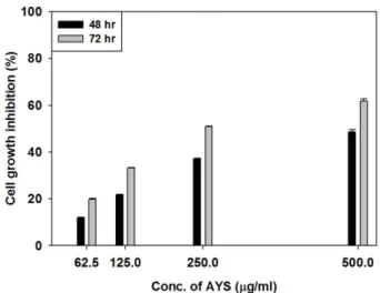
나지않았으나, 인체대장암 HT29세포는 AYS (250 μg/ml) 처리에서 24시간후에는 22.2%, 48시간후에는약 38%의세 포생육이억제되었다(Fig. 1). 따라서 AYS가인체대장암세 포에생육억제작용을나타내었으므로 AYS의농도를증가시 키고, 대장암세포의배양시간을더늘려서 AYS농도에의 존적으로대장암세포의생육을억제시키는지를검토하였다. 그결과 62.5-500 μg/ml의 AYS로처리된대장암세포들은 배양시간(48과 72 hr)에따라농도의존적으로그생육을억 제시켰다(Fig. 2). 또한 AYS와기존항암제의혼합처리에따 른대장암세포에대한병용효과를측정하기위해 Canfield
등이기술한방법[4]으로행하였다. 즉, AYS와기존항암제 가 함유된 96-well microplate에 인체 대장암 HT29 세포 (1×10⁵ cells/ml)를 접종한 후 CO2 incubator (5% CO2, 37oC)에서 3일간배양시킨후 MTT 법으로세포수를측정 하고, AYS와 기존 항암제에 대한 fractional inhibitory concentrations (FIC)과 ΣFIC(sum FIC; fractional inhibitory concentrations)를산출하여 이들 2종의약제에대한상호작 용을검토하였다. 이때사용된약제의농도는 AYS는 500 μg/
ml, etoposide는 50 μg/ml, 그 외 paclitaxel, 5-fluorouracil 과 doxorubicin은각각 20μg/ml이사용되었다. 그결과 AYS 와기존항암제의혼합물들이각각의기존항암제보다더 욱강하게대장암 HT29 세포의생육을억제시켰다(Table 1).
Fig. 1. Cytotoxicity of AYS against cancer cells.
Cytotoxicity of AYS against cancer cell was determined by MTT method. Cancer cell (1.0× 10⁵ cells/ml) was incubated in a 96-well microplate. AYS (250 µg/ml) was used to determine cytotoxicity against human colon HT29 cell. For cytotoxicity against human Jurkat T cell and mouse sarcoma 180 cell, AYS (500 µg/ml) was applied respectively. Each results represent the mean± standard deviations.
Fig. 2. Growth inhibition of human colon HT29 cell by AYS.
Human colon HT29 cell (2× 10⁵ cells/ml) was incubated with dif- ferent concentrations of AYS in a 96-well microplate for 3 days.
Growth inhibition of the HT29 cells were determined by MTT method. Each results represent the mean± standard deviations.
Table 1. Synergistic anticancer activity of anticancer agent mixed with AYS against human colon HT29 cell.
Anticancer agent IC50 (μg/ml)
Alone (A or B) Combination (A+B) FIC ΣFICa
Group 1 Etoposide (A) 11.3 6.3 0.0628 0.6203
AYS (B) 250.0 15.7 0.5575
Group 2 Fluorouracil (A) 3.2 0.4 0.0628 0.1878
AYS (B) 250.0 15.7 0.1250
Group 3 Paclitaxel (A) 0.06 0.008 0.0628 0.1961
AYS (B) 250.0 15.7 0.1333
Group 4 Doxorubicin (A) 0.6 0.1 0.0628 0.2295
AYS (B) 250.0 15.7 0.1667
aΣFIC was calculated for each well as FICA + FICB where A and B are the two drugs in the well. The FIC of each drug was calculated as (IC50 of the drug in combination)/(IC50 of the drug alone). ΣFIC was calculated for each IC50. ΣFIC < 1: synergis- tic, ΣFIC > 1: antagonistic, ΣFIC = 1: indifferent.
Synergistic Anticancer Activity by Mixture of Bacterial Proteoglycan with Anticancer Agent 381
September 2013 | Vol. 41 | No. 3
따라서이들약제를처리한대장암 HT29 세포를 3일간배
양시킨후측정된 IC50값을활용하여두가지혼합약제에 대한ΣFIC를산출하면 [AYS + Etoposide] 혼합물은 0.6203, [AYS + 5-Fluorouracil] 혼합물은 0.1878, [AYS + Paclitaxel]
혼합물은 0.1961, [AYS + Doxorubicin] 혼합물은 0.2295였 다. Canfield [4]에의하면ΣFIC 값이 <1이면서로상승작용 (synergistic)을나타내고, ΣFIC 값이 >1이면서로저해작용 (antagonistic)을나타내며, ΣFIC가 1과동일하면서로무관 하게작용한다고기술하였다. 따라서 in vitro에서 AYS는
4종의기존항암제와혼합하여사용한모든경우에ΣFIC의
값이 1 보다작았으므로이들약제를단독으로사용했을때 보다 AYS와혼합하여사용하였을때인체대장암 HT29세 포에대한항암작용이상승되었다(Table 1). 특히이들항암 제들은암세포에대한작용메카니즘에차이는있지만 AYS 와이들항암제를혼합하면항암효과가크게증진되었으므 로 AYS는일종의항암보조제로개발할가치가있다고생 각된다. 따라서 AYS와 4종의항암제를혼합물로투여시킬경 우기존항암제의량을감소시킬수있어항암제에의한부작 용도줄일수있으므로 AYS는항암보조제로서대장암치료에 화학요법제(chemotheraphy)의새로운소재로서가능성을나 타내었다. 또한일반적으로항암제의독성은생체면역세포 의기능에도영향을주어면역세포들에의한항암기능은물 론항암제투여기간동안에감염균에대한 저항력이떨어 져최악의조건으로전락할가능성도있다. 이런점을고려
하여최근에는면역요법(immunotheraphy)을행하기위한
시도도 이루어지고 있다[12, 18]. 본 AYS는 in vitro에서 hPBMC (human peripheral blood mononuclear cell)를 배 양시키면 cytokine인 IL-6, TNF-α와 IFN-r분비가유도되었
다[Data 미제출]. 특히척추동물에서면역기능은암세포증
식을억제하는데도중요한역할을하므로 AYS는 in vivo에 서면역세포를활성화시켜적어도항암제투여기간동안에 감염균에대한저항력이떨어지는부작용도다소줄일수있 을것이라고생각된다.
결론적으로 AYS는 in vitro에서농도의존적으로인체대
장암 HT29 세포의생육을억제시켰으며, 기존의항암제들과
병용하였을경우 HT29 세포에대한항암작용이상승되었다.
요 약
Rhanella aquatilis AY2000 균주가생산하는일종의당단
백질인항효모성물질 (AYS)에대한항암활성을조사하기
위해 in vitro에서암세포에대한 AYS의세포독성을조사하 였다. 그 결과 AYS는 인체의 Jurkat T 세포와 마우스의
sarcoma 180 세포에대해서는세포독성을나타내지않았으
나, 인체대장암세포인 colon cancer TH20 세포에는세포독
성을나타내었다. 또한이 AYS는 62.5에서 500μg/ml까지농 도의존적으로인체대장암세포에대해세포독성을증가시켰 다. 뿐만아니라이 AYS와시판항암제를혼합하여처리한 결과시판항암제를단독으로처리한것보다인체대장암세 포에대한항암효과가더욱상승되었다.
Acknowledgments
This work was supported by Dong-eui University Grant (2012AA098).
References
1. Abe T, Hasegawa S, Taniguchi K, Yokomizo A, Kuwano T, Ono M, et al. 1994. Possible involvement of multi-drug-resis- tance-associated protein (MRP) gene expression in spontane- ous drug resistance to vincristine, etoposide and adriamycin in human glioma cells. Int. J. Cancer. 58: 860-864.
2. Ahn JB, Shim KY, Jeung HC, Rha SY, Yoo NC, Kim NK, et al.
2001. Monthly 5-days 5-Fluorouracil and low-dose leucovorin for adjuvant chemotherapy in colon cancer. Cancer Lett. 167:
215-224.
3. Beggs WH. 1993. Anti-Candida activity of the anti-cancer drug tamoxifen. Res. Commun. Chem. Pathol. Pharmacol. 80: 125- 128.
4. Canfield CJ, Pudney M, Gutteridge WE. 1995. Interaction of Atovaquone with other antimalarial drugs against Plasmodium flaciparum in vitro. Exp. Parasitol. 80: 373-381.
5. Ciapetti G, Cenni E, Pratelli L, Pizzoferrato A. 1993. In vitro valuation of cell/biomaterial interaction by MTT assay. Bioma- terials. 14: 359-364.
6. Irving T. 2008. Current perspective adjuvant chemotheraphy after resection of liver metastases from colorectal cancer. Eur.
J. Cancer. 44: 1198-1201.
7. Jekunen AP, Christen RD, Shalinsky DR. 1994. Synergistic interaction between cisplatin and taxol in human ovarian car- cinoma cells in vitro. Br. J. Cancer. 69: 299-306.
8. Langer CJ, Leighton JC, Comis RL. 1995. Paclitaxel and car- boplatin in combination in the treatment of advanced non- small cell lung cancer: a phase II toxicity, response, and sur- vival analysis. J. Clin. Oncol. 13: 1860-1870.
9. Lee M, Yea SS, Jeon YJ. 2000. Paclitaxel causes mouse splenic lymphocytes to a state hyporesponsive to lipopolysac- charide stimulation. Int. J. Immunopharmacol. 22: 615-621.
10. Longhi A, Mariani E, Kuehn JJ. 2009. A randomized study with adjuvant mistletoe versus oral Etoposide on post relapse disease-free survival in osteosarcoma patients. Eur. J. Inte- grative Med. 1: 27-33.
11. McClendon AK, Osheroff N, 2007. DNA topoisomerase II, genotoxicity, and cancer. Mutat. Res. 623: 83-97.
12. Mihich E, Ehrke MJ. 2000. Anticancer drugs plus cytokines:
immunomodulation based therapies of mouse tumors. Int. J.
382 Park and Kim
http://dx.doi.org/10.4014/kjmb.1305.05001 Immunopharmacol. 22: 1077-1081.
13. Park BT, Na KH, Jung EC, Park JW, Kim HH. 2009. Antifungal and anticancer activities of a protein from the mushroom Cordyceps militaris. Korean J. Physiol. Pharmacol. 13: 49-54.
14. Papoian T, Lewis W. 1990. Adriamycin cardiotoxicity in vivo, Selective alterations in rat cardiac mRNAs. Am. J. Pathol.
130: 1201-1207.
15. Park HJ, Kang MJ, Lee JH, Kim KH. 2011. Action pattern of anti- yeast substance originated from Rahnella aquatilis strain AY2000. Korean J. Microbiol. 47: 163-166.
16. Shinoda C, Fujishita MM, Dohkan J, Oda H, Shinoda K, Yamada T, et al. 2005. Doxorubicin induces expression of multidrug resistance-associated protein 1 in human small cell
lung lines by the c-jun N-terminal kinase pathway. Int. J. Can- cer. 117: 21-31.
17. Suehisa H, Toyooka S. 2009. Adjuvant chemotheraphy for completely resected non-small-cell lung cancer. Acta Med.
Okayama. 63: 223-230.
18. Wang ZH, Wu BJ, Zhang XH, Xu M, Chang HM, Lu XY, et al.
2012. Purification of a polysaccharide from Boschniakia ros- sica and its synergistic antitumor effect combined with 5-fluo- rouracil. Carbohydr. Polym. 89: 31-35.
19. Zhou H, Shen T, Luo Y, Liu L, Chen W, Xu B, et al. 2010. The antitumor activity of the fungicide ciclopirox. Int. J. Cancer.
127: 2467-2477.