• 검색 결과가 없습니다.

Ground-Glass Opacity in Lung Metastasis from Breast Cancer: A Case Report

N/A
N/A
Protected

Academic year: 2021

Share "Ground-Glass Opacity in Lung Metastasis from Breast Cancer: A Case Report"

Copied!
5
0
0

로드 중.... (전체 텍스트 보기)

전체 글

(1)

Tuberc Respir Dis 2013;74:32-36

CopyrightⒸ2013. The Korean Academy of Tuberculosis and Respiratory Diseases. All rights reserved.

Ground-Glass Opacity in Lung Metastasis from Breast Cancer: A Case Report

Sae Byol Kim, M.D.1, Soohyeon Lee, M.D.1, Myoung Ju Koh, M.D.2, In Seon Lee, M.D.1, Chan Soo Moon, M.D.1, Sung Mo Jung, M.D.1, Young Ae Kang, M.D.1

Departments of 1Internal Medicine and 2Pathology, Yonsei University College of Medicine, Seoul, Korea

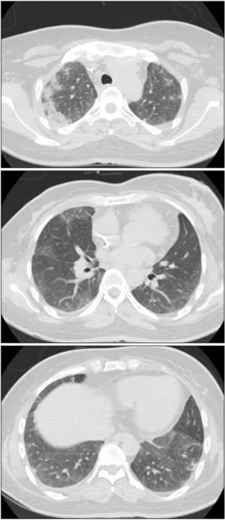
A 43-year-old woman with breast cancer who was on neoadjuvant chemotherapy presented with cough, sputum and mild fever. High-resolution computed tomography showed diffuse ground glass opacities in bilateral lungs and subpleural patchy consolidations. Initially, she was thought to have pneumonia or interstitial lung diseases such as drug-induced pneumonitis and treated with antibiotics and steroids. She subsequently got breast cancer surgery because of disease progression, and concurrent thoracoscopic lung biopsy revealed metastatic carcinoma of the lung from breast cancer. The diagnosis of suspected interstitial lung disease can be made without lung biopsy, but malignancy should always be considered and lung biopsy should be performed in the absence of a definitive clinical diagnosis.

Key Words: Neoplasm Metastasis; Lung Diseases, Interstitial; Diagnostic Imaging

Address for correspondence: Young Ae Kang, M.D.

Department of Internal Medicine, Yonsei University College of Medicine, 50, Yonsei-ro, Seodaemun-gu, Seoul 120-752, Korea

Phone: 82-2-2228-1986, Fax: 82-2-393-6884 E-mail: [email protected]

Received: Apr. 2, 2012 Revised: May 10, 2012 Accepted: Jun. 9, 2012

CCIt is identical to the Creative Commons Attribution Non-Commercial License (http://creativecommons.org/licenses/by-nc/3.0/).

Introduction

Breast cancer is one of the most prevalent cancer in women and can result in various thoracic manifestations from either treatment, its complications or tumor re- currence and metastasis

1

. Thus, the accurate diagnosis of various thoracic manifestations in patients with breast cancer is critical to guide further treatment. The lung is a common site of metastasis and it may present with solitary or multiple nodules, lymphangitic metastasis, or airspace consolidation

2

. However, ground glass opacity (GGO) is a rare form of metastatic lung involvement and can be misdiagnosed as drug-induced pneumonitis or viral infection. Here, along with a literature review,

we report the case of a 43-year-old woman with breast cancer that metastasized to the lung and presented as bilateral GGO on chest computed tomography (CT).

Case Report

A 43-year-old woman was referred to our clinic with fever and persistent cough and sputum for two weeks.

She had been diagnosed with breast cancer (invasive ductal carcinoma, estrogen receptor negative, progester- one receptor negative, and human epidermal growth factor receptor 2 negative, cT2N2M0, stage IIB) with ax- illary lymph node metastasis three months prior to pre- sentation and was on neoadjuvant chemotherapy. After completing four cycles of AC chemotherapy (doxorubi- cin 60 mg/m

2

and cyclophosphamide 600 mg/m

2

on day 1 every three weeks), she was currently on the second cycle of docetaxel/S-1 chemotherapy (docetaxel 75 mg/m

2

and S-1 30 mg/m

2

twice daily for 14 days every three weeks) as part of a clinical trial at our institution.

It was her ninth day of chemotherapy when she visited

the clinic. The patient, a non-smoker with no known

allergies, had no other past medical history, and denied

(2)

Figure 1. Chest X-ray showing a suspicious consolidation in the right upper lung field and diffuse bilateral haziness.

Figure 2. Chest computed tomography showing patchy consolidations and ground glass opacity (GGO) in the subpleural area of both upper lobes and diffuse GGO bi- laterally in the lower lobes.

hazardous substance exposure or recent travel.

She complained of cough with sputum production and mild dyspnea. She had a low-grade fever of 37.7

o

C and breath sounds were almost silent on auscultation.

The chest radiograph showed a suspicious consolidation in the right upper lung field and diffuse bilateral hazi- ness (Figure 1). Laboratory data revealed no leukocy- tosis (white blood cell [WBC] count, 5,590/μL), serum chemistry values were within normal limits, and C-re- active protein was elevated (221.18 mg/L). High-reso- lution chest CT showed patchy consolidations and GGO in the subpleural area of both upper lobes and diffuse GGO bilaterally in the lower lobes (Figure 2). The pri- mary tumor in the left breast remained stable and the metastatic lymph node in the left axilla seemed to be decreased in size after the aforementioned six cycles of chemotherapy.

Initially, she was thought to have bacterial or viral pneumonia, given immunosuppression from chemo- therapy, or drug-related interstitial pneumonitis, and was treated with antibiotics for three weeks. However, the bilateral lung GGO showed no change on serial fol- low-up CT scans. Fiberoptic bronchoscopy was done and bronchoalveolar lavage fluid from the right upper lobe was sent for analysis. It contained 1,000 WBCs/mL

with 98% macrophages and 2% lymphocytes. Cultures

revealed no specific pathogen, and cytology showed no

evidence of malignant cells. After empiric treatment

with steroids (35 mg of prednisolone) for seven days,

(3)

Figure 3. Surgical lung biopsy specimen showing meta- static carcinoma predominantly in the lymphovascular spaces (H&E stain, ×100).

her symptoms showed mild improvement, but the dif- fuse infiltrative lung lesions remained unchanged.

While she was being treated for suspected interstitial pneumonia and with the chemotherapy delayed, chan- ges in the skin color of the breast were observed.

Considered to be a clinical sign of disease progression, neoadjuvant chemotherapy was stopped after six cycles out of the initially-planned eight and she dropped out of the clinical trial. Magnetic resonance imaging of the breast showed no change in the maximum extent of the primary tumor in the left breast and the metastatic lymph node in the left axilla, but showed an increase in size of the subareolar mass with breast contraction, suggesting disease progression. Thus, she underwent modified radical mastectomy and axillary lymph node dissection for the underlying breast cancer, and thoraco- scopic lung biopsy of the right upper lobe and right middle lobe was performed concurrently for the patho- logic diagnosis of lung involvement. Postoperative path- ology of the lung revealed metastatic carcinoma from the breast, predominantly in the lymphovascular spaces (Figure 3). Diagnosed with breast cancer with lung metastasis, she is currently undergoing palliative chemo- therapy.

Discussion

Lung metastasis exhibiting diffuse GGO is a very rare pattern and can mimic interstitial lung disease (ILD).

Especially in this patient, considering the history of re- cent chemotherapy with agents with reported pulmo- nary toxicity

3-5

, diffuse lung infiltrates and GGO might lead to a diagnosis of drug-induced pneumonitis or viral infection. Thoracoscopic lung biopsy revealed meta- static carcinoma in the lymphovascular space in this case.

Breast cancer is the second most common cancer in women in Korea, and the incidence and the mortality of breast cancer have continuously increased over the last ten years

6

. Changes in thoracic imaging in patients with breast cancer can result from treatment, treat- ment-related complications, or metastases. Some pul- monary conditions may need specific treatment such as antibiotics or steroids. Also, the presence of lung meta- stasis is a critical factor in deciding treatment options for breast cancer. Thus, the accurate diagnosis of thora- cic manifestations in patients with breast cancer is of great importance.

Chemotherapy agents can cause lung injury either by direct cytotoxicity or immune-mediated reactions

7

. Clini- cal manifestations of chemotherapy-related lung injury are very nonspecific and many times it is difficult to dif- ferentiate them from other pulmonary conditions such as pulmonary edema, interstitial pneumonia, or pulmo- nary hemorrhage. Common radiologic features of drug- induced lung diseases include GGO, consolidation, in- terlobular septal thickening, and centrilobular nodules

8

. Cyclophosphamide and docetaxel are known to have pulmonary toxicity and to cause hypersensitivity pneu- monitis

3,4

, and doxorubicin-related lung injury has been reported previously

5

. However, the diagnosis is usually one of exclusion, and is based on the temporal relation- ship with chemotherapy.

Lymphangitic metastasis is the most frequently ob-

served pattern of pulmonary metastasis from breast can-

cer

9

. Lymphangitic lung metastasis of breast cancer usu-

ally appears as reticular or reticulonodular interstitial

(4)

markings, or interlobar septal thickening (Kerley B lines) on imaging

1

. Other common patterns of pulmonary metastases include multiple pulmonary nodules or a soli- tary pulmonary nodule, which results from hematoge- nous tumor spread. The tumor can spread along the al- veolar walls in a pattern like that of bronchioloalveolar carcinoma (BAC) and form airspace infiltrations that can mimic pneumonia or pneumonitis

1

. In addition, in ma- lignancies other than breast cancer, various atypical pat- terns of radiographic findings have been reported, in- cluding air-space consolidation

2

, bilateral ground glass opacities

10

, and diffuse interstitial infiltrates

11

.

Ground glass opacities of the lung can represent vari- ous entities such as pneumonia, pneumonitis, lympho- proliferative diseases, or fibrosis. Persistent nodular GGOs are known to be related to premalignant or malignant conditions including atypical adenomatous hyperplasia, BAC, and invasive adenocarcinoma

12

. In the case of dif- fuse ground glass opacities, it can be associated with various inflammatory and infectious conditions, includ- ing ILDs. One study investigated the diagnostic accuracy of high-resolution CT in diffuse lung diseases, and GGO as a predominant pattern was the most unreliable (44.2% to 75.5%) when compared with honeycombing, bronchovascular thickening (more than 90%), and cysts (80% to 89%) as a predominant pattern

13

. All of the clin- ical information, laboratory results, and radiographic studies has to be considered, but in the absence of a confident clinical diagnosis and with the possibility that definitive diagnosis by lung biopsy might change treat- ment plans, lung biopsy is recommended for the diag- nosis of ILD

14

.

Lung biopsy is considered when there are un- explained signs and symptoms with atypical radiogra- phic features that are usually progressive or rapidly deteriorating. Indications of lung biopsy in evaluating patients with suspected ILD include the followings: to prove a specific diagnosis, to exclude malignancy, or to identify certain disease entities that need specific treatments such as antibiotics or steroids. Since lung bi- opsy, either transbronchial or surgical, is associated with morbidity and mortality even in small numbers, the risks

and benefits of the procedure should be carefully weighed before pursuing it. Transbronchial lung biopsy (TBLB) is associated with 1–4% of reported complica- tions, of which pneumothorax and hemorrhage are most common, and approximately 0.1% of mortality with hemorrhage the main cause. In case of surgical lung biopsy, the complication rate is up to 7% with sur- gery related mortality rate of less than 1%

14

. With vid- eo-assisted thoracoscopic surgery increasingly used, sur- gical lung biopsy has become less invasive in recent years. Diagnostic yield of TBLB and surgical lung biop- sy is reported to be 29–79% and 37–100%, re- spectively

14

. Lung biopsy should be performed when the possible benefits of definitive histopathologic diag- nosis and treatment are expected to overweigh the po- tential risks associated with the procedure. The decision has to be made on a case by case basis.

The diagnosis of suspected ILD has always been diffi- cult but with comprehensive clinical information, labo- ratory results, and the aid of high-resolution CT, a pre- sumptive diagnosis can be made without lung biopsy in many cases

15

. However, as in this case, metastatic lung cancer can also appear as ground glass opacities, mimicking interstitial lung disease. Lung metastasis should always be considered in interstitial lung diseases, especially when the patient has a prior history of malig- nancy, and lung biopsy should be performed in the ab- sence of a confident clinical diagnosis.

We report a case of metastatic lung cancer presenting as diffuse ground glass opacities, mimicking interstitial lung disease. Malignancy should always be considered in the differential diagnosis of interstitial lung disease, and lung biopsy is required to exclude it.

References

1. Jung JI, Kim HH, Park SH, Song SW, Chung MH, Kim HS, et al. Thoracic manifestations of breast cancer and its therapy. Radiographics 2004;24:1269-85.

2. Ohnishi H, Haruta Y, Yokoyama A, Nakashima T,

Hattori N, Kohno N. Metastatic breast cancer present-

ing as air-space consolidation on chest computed to-

mography. Intern Med 2009;48:727-31.

(5)

3. Malik SW, Myers JL, DeRemee RA, Specks U. Lung tox- icity associated with cyclophosphamide use: two dis- tinct patterns. Am J Respir Crit Care Med 1996;154(6 Pt 1):1851-6.

4. Read WL, Mortimer JE, Picus J. Severe interstitial pneu- monitis associated with docetaxel administration. Cancer 2002;94:847-53.

5. Jacobs C, Slade M, Lavery B. Doxorubicin and BOOP:

a possible near fatal association. Clin Oncol (R Coll Radiol) 2002;14:262.

6. Jung KW, Park S, Kong HJ, Won YJ, Lee JY, Park EC, et al. Cancer statistics in Korea: incidence, mortality, survival, and prevalence in 2008. Cancer Res Treat 2011;43:1-11.

7. Pietra GG. Pathologic mechanisms of drug-induced lung disorders. J Thorac Imaging 1991;6:1-7.

8. Cleverley JR, Screaton NJ, Hiorns MP, Flint JD, Muller NL. Drug-induced lung disease: high-resolution CT and histological findings. Clin Radiol 2002;57:292-9.

9. Kreisman H, Wolkove N, Finkelstein HS, Cohen C, Margolese R, Frank H. Breast cancer and thoracic meta- stases: review of 119 patients. Thorax 1983;38:175-9.

10. Tokunaga T, Arakawa H, Kuwashima Y. A case of lepi- dic pulmonary metastasis from adenocarcinoma of the

gallbladder mimicking acute interstitial pneumonia.

Clin Radiol 2005;60:1213-5.

11. Shin NY, Hong YJ, Kim AH, Shim HS, Nam JE, Lee HJ, et al. Diffuse interstitial infiltrative lung metastasis of malignant melanoma: a case report. Korean J Radiol 2011;12:252-5.

12. Lee HY, Lee KS. Ground-glass opacity nodules: histo- pathology, imaging evaluation, and clinical implica- tions. J Thorac Imaging 2011;26:106-18.

13. Sundaram B, Gross BH, Martinez FJ, Oh E, Muller NL, Schipper M, et al. Accuracy of high-resolution CT in the diagnosis of diffuse lung disease: effect of predom- inance and distribution of findings. AJR Am J Roent- genol 2008;191:1032-9.

14. Bradley B, Branley HM, Egan JJ, Greaves MS, Hansell DM, Harrison NK, et al. Interstitial lung disease guide- line: the British Thoracic Society in collaboration with the Thoracic Society of Australia and New Zealand and the Irish Thoracic Society. Thorax 2008;63 Suppl 5:v1- 58.

15. Elicker B, Pereira CA, Webb R, Leslie KO. High-reso-

lution computed tomography patterns of diffuse inter-

stitial lung disease with clinical and pathological corre-

lation. J Bras Pneumol 2008;34:715-44.

수치

Figure 1. Chest X-ray showing a suspicious consolidation in the right upper lung field and diffuse bilateral haziness.
Figure  3.  Surgical  lung  biopsy  specimen  showing  meta- meta-static  carcinoma  predominantly  in  the  lymphovascular  spaces  (H&E  stain,  ×100).

참조

관련 문서

• Hormone Therapy With or Without Combination Chemotherapy in Treating Women Who Have Undergone Surgery for Node-Negative Breast Cancer (The TAILORx Trial)..

According to the result of this study, to do effective nursing that improve health promoting behaviors of early-stage lung cancer patients, nurses should

If cooling rate become fast, glass transition can be observed in liquid region in case of slow cooling

Beichel,“Automated 3-D segmentation of lungs with lung cancer in CT data using a novel robust active shape model approach,” IEEE Trans.. 저작물의

Bone is a common site of distant metastasis in prostate cancer. Association of bone and metastatic cancer cells are important in this site-specific manifestation

Anti‑cyclic citrullinated peptide antibody testing is used to diagnose rheumatoid arthritis and associated with interstitial lung disease in RA.. Herein, we

Among the various pulmonary manifestations, interstitial lung disease (ILD) is known to be associated with substantial morbidity and mortality rates in RA patients.. As RA-ILD

We report a rare case of pericatheter abscess formation after scalp acupuncture in a 25-year-old woman who had a history of meningitis and hydrocephalus, which were treated