• 검색 결과가 없습니다.

Prognostic Value of Serum Growth Differentiation Factor-15 in Patients with Chronic Obstructive Pulmonary Disease Exacerbation

N/A
N/A
Protected

Academic year: 2021

Share "Prognostic Value of Serum Growth Differentiation Factor-15 in Patients with Chronic Obstructive Pulmonary Disease Exacerbation"

Copied!
8
0
0

로드 중.... (전체 텍스트 보기)

전체 글

(1)

Prognostic Value of Serum Growth

Differentiation Factor-15 in Patients with Chronic Obstructive Pulmonary Disease Exacerbation

Miyoung Kim, M.D.

1

, Seung-Ick Cha, M.D.

1

, Keum-Ju Choi, M.D.

1

, Kyung-Min Shin, M.D.

2

, Jae- Kwang Lim, M.D.

2

, Seung-Soo Yoo, M.D.

1

, Jaehee Lee, M.D.

1

, Shin-Yup Lee, M.D.

1

, Chang-Ho Kim, M.D.

1

, Jae-Yong Park, M.D.

1

and Dong Heon Yang, M.D.

1

Departments of

1

Internal Medicine and

2

Radiology, Kyungpook National University School of Medicine, Daegu, Korea

Background: Information regarding prognostic value of growth differentiation factor 15 (GDF-15) and heart-type fatty acid-binding protein (H-FABP) in patients with chronic obstructive pulmonary disease (COPD) exacerbation is limited.

The aim of this study was to investigate whether serum levels of GDF-15 and H-FABP predict an adverse outcome for COPD exacerbation.

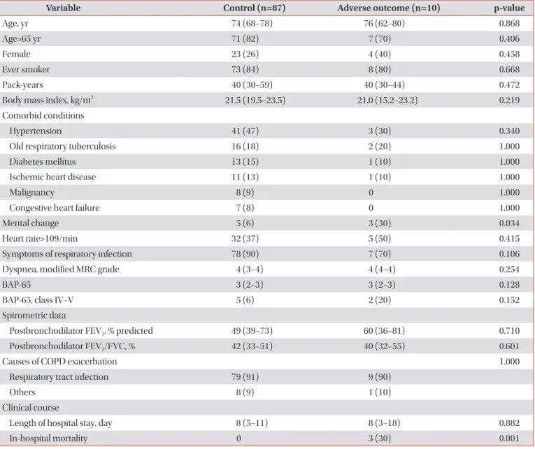
Methods: Clinical variables, including serum GDF-15 and H-FABP levels were compared in prospectively enrolled patients with COPD exacerbation that did or did not experience an adverse outcome. An adverse outcome included 30- day mortality and need for endotracheal intubation or inotropic support.

Results: Ninety-seven patients were included and allocated into an adverse outcome (n=10) or a control (n=87) group.

Frequencies of mental change and PaCO

2

>37 mm Hg were significantly higher in the adverse outcome group (mental change: 30% vs. 6%, p=0.034 and PaCO
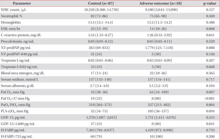
2

>37 mm Hg: 80% vs. 22%, p<0.001, respectively). Serum GDF-15 elevation (>1,600 pg/mL) was more common in the adverse outcome group (80% vs. 43%, p=0.041). However, serum H-FABP level and frequency of serum H-FABP elevation (>755 pg/mL) did not differ between the two groups. Multivariate analysis showed that an elevated serum GDF-15 and PaCO

2

>37 mm Hg were significant predictors of an adverse outcome (odds ratio [OR], 25.8; 95% confidence interval [CI], 2.7−243.8; p=0.005 and OR, 11.8; 95% CI, 1.2−115.3; p=0.034, respectively).

Conclusion: Elevated serum GDF-15 level and PaCO

2

>37 mm Hg were found to predict an adverse outcome independently in patients with COPD exacerbation, suggesting the possibility that serum GDF-15 could be used as a prognostic biomarker of COPD exacerbation.

Keywords: Pulmonary Disease, Chronic Obstructive; Disease Progression; Growth Differentiation Factor 15; FABP3 Protein, Human

Copyright © 2014 The Korean Academy of Tuberculosis and Respiratory Diseases. All rights reserved.

Address for correspondence: Seung-Ick Cha, M.D.

Department of Internal Medicine, Kyungpook National University Hospital, 130 Dongdeok-ro, Jung-gu, Daegu 700-721, Korea Phone: 82-53-200-6412, Fax: 82-53-426-2046, E-mail: [email protected]

Received: Jul. 2, 2014 Revised: Aug. 1, 2014 Accepted: Sep. 16, 2014

cc It is identical to the Creative Commons Attribution Non-Commercial License (http://creativecommons.org/licenses/by-nc/3.0/).

(2)

Introduction

Chronic obstructive pulmonary disease (COPD) is expect- ed to be the third leading cause of death in 2020

1

. COPD exac- erbation determines disease-associated morbidity and mor- tality

2

, and short-term and long-term mortalities are known to increase after COPD exacerbation

3,4

. Many factors have been reported to be associated with short-or long-term mortality in affected patients

5

, and because prognostic factors are helpful, attempts to identify new predictors are still required. Several blood biomarkers, including N-terminal-pro-B-type natriuretic peptide (NT-proBNP), troponin I or T, C-reactive protein (CRP), and procalcitonin have been proposed as prognostic factors of short-term outcome in patients with COPD exacer- bation

6-9

. Among these, NT-proBNP and troponin T elevations are strong independent predictors of early mortality, but the pathophysiological bases of these relationships are unknown

7

.

Growth differentiation factor 15 (GDF-15) is a member of the transforming growth factor-β superfamily of cytokines, and plays important roles in cell growth and differentiation

10

. Under normal conditions, GDF-15 is weakly expressed in most tissues

11

. However, circulating levels of GDF-15 are el- evated in acute coronary syndrome

12,13

, chronic heart failure

14

, acute pulmonary embolism

15

, and idiopathic pulmonary arterial hypertension

16

. These results suggest that blood GDF- 15 levels increase in response to right ventricular (RV) or left ventricular (LV) overload. On the other hand, heart-type fatty acid-binding protein (H-FABP) plays a pivotal role in myo- cardial homeostasis, because this small cytoplasmic protein facilitates the intracellular transport of insoluble fatty acids in myocardium, in which energy is mainly provided by lipid oxidation

17,18

. H-FABP can be released by myocardial injury, such as acute coronary syndrome

19

, and may be elevated by RV stress, such as that caused by pulmonary embolism

20-22

. Therefore, H-FABP is a candidate biomarker of right and left cardiac dysfunction. However, no data are available on the prognostic values of GDF-15 and H-FABP in patients with COPD exacerbation.

We hypothesized elevated serum levels of GDF-15 and H-FABP might provide prognostic information in patients with COPD exacerbation. Thus, the aims of this study were to determine whether elevated serum levels of GDF-15 and H- FABP predict an adverse outcome in these patients.

Materials and Methods

1. Study population

Between April 2011 and April 2013, all patients hospital- ized with COPD exacerbation at Kyungpook National Uni- versity Hospital (KNUH), a tertiary referral center in Daegu, South Korea, were enrolled prospectively. The Institutional

Review Board of the KNUH approved the study protocol, and informed consent was obtained from all participants. COPD was diagnosed based on available spirometric and medical records as recommended by the Global Initiative for Chronic Obstructive Lung Diseases (GOLD)

23

. COPD exacerbation was defined as acute deterioration requiring hospitalization from a stable condition

24

. On arrival at the hospital, emer- gency or internal medicine department physicians evaluated patients by history taking, physical examination, chest radiog- raphy, and routine laboratory tests. Patients with any specific causes identified during these examinations, including pneu- monia, pneumothorax, and congestive heart failure, were excluded. After exclusions, all patients underwent computed tomography (CT) within 24 hours of admission. Patients with azotemia (serum creatinine>1.5 mg/dL) or exhibiting hyper- sensitivity to contrast media were excluded. As described in a previous study

25

, the etiologies of COPD exacerbations were determined as follows: 1) pneumonia was diagnosed when consolidation or ground-glass opacity (GGO) was observed by CT; 2) tracheobronchitis if symptoms suggestive of a re- spiratory infection, including increased sputum, purulent sputum, fever, or upper respiratory infection symptoms, were identified without consolidation or GGO by CT or if centri- lobular nodules or a tree-in-bud pattern were observed on CT images, without consolidation or GGO; 3) congestive heart failure was diagnosed when CT findings indicated pulmonary edema with echocardiographic left LV dysfunction; 4) pulmo- nary embolism when a sharply delineated pulmonary arterial filling defect was located centrally within a vessel or made an acute angles at its interface with the vessel wall in at least two consecutive CT sections; and 5) undetermined etiology.

2. Clinical outcome

The 97 patients were allocated to two groups, that is, to an adverse outcome group (n=10) or to a control group (n=87).

An adverse outcome was defined as a composite end point of 30-day mortality or the need for endotracheal intubation or inotropic support.

3. Clinical data

Baseline and clinical characteristics of the patients, includ-

ing age, gender, smoking history, body mass index, comor-

bid conditions, and respiratory infection symptoms, were

checked. Dyspnea was assessed using modified Medical

Research Council (mMRC) grades

26

. For severity of COPD

exacerbation, BAP-65 classes

27

were defined as follows: class

I, patients≤65 years of age without any risk factors (blood

urea nitrogen [BUN] level≥25 mg/dL, altered mental status, or

pulse≥109 beats/min); class II, patients>65 years of age with

no risk factor; and class III, IV, and V, patients with one, two,

and three risk factors, respectively. Lengths of hospital stay

(3)

were also recorded.

Pulmonary function testing (PFT) was performed as rec- ommended by the American Thoracic Society and European Respiratory Society Guideline

28

. PFT data obtained within 6 months of index admission dates were used in the analysis.

4. Laboratory data

Blood tests included white blood cell count, erythrocyte sedimentation rate (ESR), CRP, procalcitonin, NT-proBNP, troponin I, BUN, sodium, and albumin levels. Arterial blood gas analysis data, including partial oxygen pressure in arterial blood (PaO

2

), partial carbon dioxide pressure in arterial blood (PaCO

2

), inspired oxygen fraction (FiO

2

), PaO

2

/FiO

2

ratio, and alveolar-arterial oxygen gradient (P[A-a]O

2

), were also checked.

5. Enzyme-linked immunosorbent assays for GDF-15 and H-FABP

Blood samples were obtained within 24 hours of arrival, centrifuged immediately, and stored at −80

o

C. Serum samples were registered and later provided by the National Biobank of Korea at the KNUH. Serum GDF-14 and H-FABP levels were measured using commercially available ELISA kits (Human GDF-15 Immunoassay; Quantikine, Minneapolis, MN, USA and Human H-FABP; Hycult Biotech, Uden, The Netherlands) according to the manufacturers’ protocols.

6. Echocardiography

Only 69 patients underwent echocardiography, because of operator non-availability. Transthoracic echocardiographic findings including the presence of RV dysfunction and RV systolic pressure (RVSP) were reviewed. RV dysfunction was echocardiographically defined as RV free wall hypokinesia, and RVSP was calculated using the tricuspid regurgitation flow velocity measured by Doppler echocardiography

29

. LV ejection fraction (LVEF) was determined by the Simpson bi- plane formula

30

.

7. Radiologic data

CT scans were performed using a multidetector CT with 16 or 64 detector rows: Light Speed 16 (General Electric, Mil- waukee, WI, USA) or Aquilion 64 (Toshiba Medical Systems, Tokyo, Japan). CT scans were reviewed by two radiologists (K-.

M.S. and J.L.), and diagnoses were made by consensus. The presences of consolidation or GGO, bronchiolitis (centrilobu- lar nodules or tree-in-bud pattern), bronchiectasis, bronchial wall thickening, and emphysema were checked. CT findings were defined as follows: consolidation as airspace opacifica- tion with obscuration of the underlying vasculature; GGO

as mildly increased attenuation without obscuration of the underlying vasculature; tree-in-bud pattern as centrilobular nodules with either V- or Y-shaped branching linear opaci- ties; bronchiectasis when bronchial internal diameter was greater than that of the adjacent pulmonary artery (PA); and bronchial anthracofibrosis as multiple smooth bronchial nar- rowing with adjacent calcified lymph nodes; bronchial wall thickening was assessed subjectively. As has been performed in previous studies

31,32

, diameters of main PA and ascending aorta (AA) at the level of the PA bifurcation were measured in order to obtain PA sizes and PA/AA diameter ratios. As in a previous study

33

, a PA/AA diameter ratio of >1 was selected as the cutoff value for pulmonary hypertension.

8. Statistical analysis

Statistical analyses were performed using SPSS version 12.0 (SPSS Inc., Chicago, IL, USA). Results are expressed as medians with ranges (interquartile range [IQR]) for continu- ous variables and numbers with percentages for categorical variables. The Mann-Whitney U test was used to compare continuous variables, whereas the chi-squared test or Fisher exact test was used to compare categorical variables. When continuous variables were converted to categorical variables, cut-off values were determined using receiver operating char- acteristic curves. To identify predictors of adverse outcomes, backward stepwise multiple logistic regression analysis was used using variables of p<0.1 in univariate analysis. A good- ness-of-fit test used to assess the fit of logistic regression mod- els was the Hosmer-Lemeshow test. Statistical significance was accepted for p<0.05.

Figure 1. Causes of chronic obstructive pulmonary disease exacer- bation. The most common cause was tracheobronchitis (60% [n=6]

in the adverse outcome group and 69% [n=60] in the control group,

respectively), followed by pneumonia (30% [n=3] in the adverse

outcome group and 22% [n=19] in the control group, respectively).

(4)

Results

1. Patient characteristics

Initially, 103 patients were enrolled. However, after review- ing CT scans, 6 were excluded due to underlying diseases with the potential to influence clinical features: pneumoconiosis, n=2; nonspecific interstitial pneumonia, n=1; lung cancer, n=1;

tracheal cancer, n=1; and tracheal stenosis, n=1. As a result, 97 patients were included. The adverse outcome group com- prised 10 patients; six died within 30 days after admission and four required inotropic support or endotracheal intubation.

The remaining 87 patients were allocated to the control group.

In both groups, the majority of COPD exacerbations were caused by respiratory tract infections (pneumonia or tracheo- bronchitis) (Figure 1). The frequency of mental change was significantly more common in the adverse outcome group, as compared with the control group (3 [30%] vs. 5 [6%], p=0.034) (Table 1). However, no significant intergroup difference was found for BAP-65 class or the frequency of a high BAP-65 score (class IV−V). In addition, no significant difference was evident in percent predicted value of post-bronchodilator forced expiratory volume in one second (FEV

1

) and post- bronchodilator FEV

1

/forced vital capacity between the two

Table 1. Baseline and clinical characteristics (n=97)

Variable Control (n=87) Adverse outcome (n=10) p-value

Age, yr 74 (68−78) 76 (62−80) 0.868

Age>65 yr 71 (82) 7 (70) 0.406

Female 23 (26) 4 (40) 0.458

Ever smoker 73 (84) 8 (80) 0.668

Pack-years 40 (30−59) 40 (30−44) 0.472

Body mass index, kg/m

2

21.5 (19.5−23.5) 21.0 (15.2−23.2) 0.219

Comorbid conditions

Hypertension 41 (47) 3 (30) 0.340

Old respiratory tuberculosis 16 (18) 2 (20) 1.000

Diabetes mellitus 13 (15) 1 (10) 1.000

Ischemic heart disease 11 (13) 1 (10) 1.000

Malignancy 8 (9) 0 1.000

Congestive heart failure 7 (8) 0 1.000

Mental change 5 (6) 3 (30) 0.034

Heart rate>109/min 32 (37) 5 (50) 0.415

Symptoms of respiratory infection 78 (90) 7 (70) 0.106

Dyspnea, modified MRC grade 4 (3−4) 4 (4−4) 0.254

BAP-65 3 (2−3) 3 (2−3) 0.128

BAP-65, class IV−V 5 (6) 2 (20) 0.152

Spirometric data

Postbronchodilator FEV

1

, % predicted 49 (39−73) 60 (36−81) 0.710

Postbronchodilator FEV

1

/FVC, % 42 (33−51) 40 (32−55) 0.601

Causes of COPD exacerbation 1.000

Respiratory tract infection 79 (91) 9 (90)

Others 8 (9) 1 (10)

Clinical course

Length of hospital stay, day 8 (5−11) 8 (3−18) 0.882

In-hospital mortality 0 3 (30) 0.001

Values are presented as median (interquartile range) or number (%).

MRC: Medical Research Council; FEV

1

: forced expiratory volume in one second; FVC: forced vital capacity; COPD: chronic obstructive

pulmonary disease.

(5)

groups.

2. Laboratory findings

Blood test results are presented in Table 2. Median ESR level tended to be lower in the adverse outcome group (14 mm/hr [IQR, 10−26 mm/hr] vs. 25 mm/hr [15−39 mm/hr]; p=0.068), and median serum CRP was significantly lower (1.26 mg/dL [0.35−2.92 mg/dL] vs. 3.54 mg/dL [1.33−8.27 mg/dL]; p=0.041).

Median serum procalcitonin levels did not differ significantly.

However, median serum NT-proBNP level tended to be higher in the adverse outcome group (1,799 [123−7,118] vs.

263 [69−833]; p=0.088). No significant difference was noted in plasma troponin I between the two groups. PaCO

2

>37 mm Hg was significantly more frequent in the adverse outcome group (8 [80%] vs. 19 [22%]; p<0.001), and P(A-a)O

2

tended be higher in the adverse outcome group (105 mm Hg [36−157 mm Hg]

vs. 52 mm Hg [34−75 mm Hg]; p=0.094).

Elevated serum GDF-15 levels (GDF-15>1,600 pg/mL) was significantly more common in the adverse outcome group

(8 [80%] vs. 37 [43%]; p=0.041). In contrast, median serum H-FABP and the frequency of elevated H-FABP levels (H- FABP>755 pg/mL) were not significantly different between the two groups.

3. Echocardiographic and computed tomographic find- ings

Echocardiographic parameters, including RV dysfunction, RVSP, and pulmonary hypertension, did not show any dif- ferences between the two groups (Table 3). There was no difference in LVEF between both groups. Of CT findings, no differences in PA/AA diameter ratio and signs of pulmonary hypertension (PA/AA diameter ratio>1) were observed be- tween the two groups.

4. Predictors of adverse outcomes

To identify predictors of adverse outcomes, we performed multiple logistic regression analysis. Of parameters with p<0.1

Table 2. Laboratory findings (n=97)

Parameter Control (n=87) Adverse outcome (n=10) p-value

WBC count, /μL 10,520 (8.300−14,750) 9,180 (5,043−13,090) 0.337

Neutrophil, % 82 (72−86) 75 (65−90) 0.569

Hemoglobin 13.4 (12.1−14.4) 13.2 (11.5−14.2) 0.480

ESR, mm/hr 25 (15−39) 14 (10−26) 0.068

C-reactive protein, mg/dL 3.54 (1.33−8.27) 1.26 (0.35−2.92) 0.041

Procalcitonin, ng/mL 0.05 (0.05−0.12) 0.05 (0.05−0.11) 0.529

NT-proBNP, pg/mL 263 (69−833) 1,779 (123−7,118) 0.088

NT-proBNP>840 pg/mL 21 (24) 5 (50) 0.126

Troponin I, ng/mL 0.02 (0.01−0.06) 0.03 (0.01−0.09) 0.267

Troponin I>0.02 ng/mL 33 (43) 5 (50) 0.668

Blood urea nitrogen, mg/dL 17 (13−24) 22 (10−26) 0.565

Serum sodium, mmol/L 137 (133−140) 137 (134−141) 0.717

Serum albumin, g/dL 3.7 (3.4−4.0) 3.5 (3.2−3.9) 0.104

PaCO

2

, mm Hg 32 (28−36) 64 (34−109) 0.007

PaCO

2

>37 mm Hg 19 (22) 8 (80) <0.001

PaO

2

/FiO

2

, mm Hg 318 (264−373) 337 (213−402) 0.884

P(A-a)O

2

, mm Hg 52 (34−75) 105 (36−157) 0.094

GDF-15, pg/mL 1,370 (1,007−2,815) 1,731 (1,411−4,676) 0.343

GDF-15>1,600 pg/mL 37 (43) 8 (80) 0.041

H-FABP, pg/mL 7,363 (784−8,937) 4,429 (872−8,996) 0.735

H-FABP>755 pg/mL 69 (79) 10 (100) 0.200

Values are presented as median (interquartile range) or number (%).

WBC: white blood cell; ESR: erythrocyte sedimentation rate; NT-proBNP: N-terminal-pro-B-type natriuretic peptide; GDF-15: growth

differentiation factor-15; H-FABP: heart-type fatty acid-binding protein.

(6)

in the univariate analysis, serum NT-proBNP was excluded in multivariate analysis, because both NT-proBNP and GDF-15 are surrogate markers of myocardial stretch. Consequently, mental change, PaCO

2

>37 mm Hg, P(A-a)O

2

, and GDF-15 el- evation (GDF-15>1,600 pg/mL) were selected for multivariate analysis to predict adverse outcomes in patients with COPD exacerbation (Table 4). The Hosmer-Lemeshow test indicated that the overall model fit was good (p=0.929). Multivariate analysis showed that PaCO

2

>37 mm Hg and GDF-15 eleva- tion were significant predictors of adverse outcomes (odds ra- tio [OR], 25.8; 95% confidence interval [CI], 2.7−243.8; p=0.005 and OR, 11.8; 95% CI, 1.2−115.3; p=0.034, respectively).

Discussion

The present study shows that an elevated serum GDF- 15 level and PaCO

2

>37 mm Hg are predictors of an adverse

outcome in patients hospitalized due to COPD exacerbation.

To the best of our knowledge, this is the first report to suggest that serum GDF-15 is of prognostic value in patients present- ing with COPD exacerbation. On the other hand, the present study failed to demonstrate that H-FABP predicts an adverse outcome in these patients.

GDF-15 functions as a stress-inducible cardiokine that pro- tects against pathologic myocardial remodeling in response to a pressure or volume overload

34

. Blood biomarkers of cardiac dysfunction, such as NT-proBNP and troponin T, have been previously reported to indicate a poor prognosis in patients with COPD exacerbation

7

. Similarly, our results suggest an elevated serum GDF-15 level independently predict an ad- verse outcome in these patients. However, serum GDF-15 was not found to be significantly correlated with serum NT- proBNP (data not shown), which is predominantly released by cardiac stress due to myocardial stretch and pressure or volume overload. Furthermore, median values of echocardio- graphic LVEF were similar in the two groups, suggesting that less severe LV dysfunction occurs during COPD exacerbation than during overt left heart failure. Circulating levels of GDF- 15 are known to be elevated in patients with acute pulmonary embolism

15

and in those with idiopathic pulmonary arterial hypertension

16

, which suggests that GDF-15 may respond to RV overload. However, parameters suggestive of pulmonary hypertension, such as RVSP>40 mm Hg and PA/AA diameter ratio>1 were not different in our groups, and thus, it is not clear whether elevated GDF-15 levels resulted primarily from Table 3. Echocardiographic and chest computerized tomographic findings

Parameter Control No. Adverse outcome No. p-value

Echocardiography 62 7

RV dysfunction 7 (11.3) 62 2 (28.6) 7 0.224

RVSP, mm Hg 32 (25−39) 58 22 (17−62) 4 0.257

RVSP>40 mm Hg 11 (19) 58 1 (25) 4 1.000

LV ejection fraction 57 (52-62) 51 55 (51−62) 5 0.635

CT findings 87 10

PA/AA diameter ratio 0.86 (0.77−0.97) - 0.88 (0.82−0.98) - 0.503

PA/AA diameter ratio>1 15 (17) - 1 (10) - 1.000

Emphysema 52 (60) - 8 (80) - 0.309

Bronchiectasis 21 (24) - 4 (40) - 0.276

Bronchiolitis 13 (15) - 1 (10) - 1.000

Bronchial wall thickness 78 (90) - 9 (90) - 1.000

Ground glass opacity 7 (8) - 0 (0) - 1.000

Consolidation 19 (22) - 3 (30) - 0.690

Bronchial anthracofibrosis 19 (22) - 2 (20) - 1.000

Values are presented as median (interquartile range) or number (%).

RV: right ventricle; RVSP: right ventricular systolic pressure; LV: left ventricle; CT: computerized tomography; PA/AA diameter ratio: main pulmonary artery-to-ascending aorta diameter ratio.

Table 4. Multivariate analysis for predictors of adverse outcome

Predictor Odds ratio

95% Confidence

interval p-value

GDF-15>1,600 pg/mL 11.8 1.2−115.3 0.034

PaCO

2

>37 mm Hg 25.8 2.7−243.8 0.005

GDF-15: growth differentiation factor-15.

(7)

LV or RV dysfunction. Furthermore, serum GDF-15 levels are known to be elevated in other inflammatory conditions and cancer, which leads us to speculate GDF-15 elevation might be caused by systemic inflammation associated with COPD exacerbation. However, serum GDF-15 levels were not found to be correlated with the blood levels of inflammatory mark- ers, such as ESR, CRP, or procalcitonin (data not shown). Con- sequently, we did not determine whether GDF-15 elevation was caused by RV or LV dysfunction or systemic inflamma- tion.

H-FABP is released by irreversible damage to cardiomyo- cytes, a reversible disturbance of cardiomyocyte metabo- lism, or mechanical stretching mechanisms

22

. H-FABP and troponin I are both known biomarkers of myocardial injury, but were not found to be significant predictors of an adverse outcome for severe COPD exacerbation in this study. Fur- thermore, circulating H-FABP levels were not significantly correlated with the blood levels of troponin I or NT-proBNP (data not shown). Interestingly, PaCO

2

>37 mm Hg was also identified as an independent prognostic factor of an adverse outcome in patients with COPD exacerbation, which concurs with previous reports that hypercapnia can predict short-term mortality, admission to intensive care unit, and an adverse outcome in patients with COPD exacerbation

6,7,35

.

Several study limitations need to be mentioned. First, this study was performed at a single center and the number of patients studied was inadequate. Thus, we cannot present a definitive conclusion regarding the ability of serum GDF-15 to predict an adverse outcome in patients with COPD exacerba- tion. To confirm our conclusion, larger-scale study is needed.

Second, although patients were prospectively enrolled into this study, not all patients underwent echocardiography, and thus, selection bias was not avoided. However, we do not believe echocardiographic findings influenced our main find- ings.

In conclusion, blood GDF-15 elevation and PaCO

2

>37 mm Hg were found to predict independently an adverse outcome in patients hospitalized for COPD exacerbation. These find- ings suggest that blood GDF-15 be considered to be a poten- tial novel biomarker in patients with COPD exacerbation.

Conflicts of Interest

No potential conflict of interest relevant to this article was reported.

Acknowledgments

This research was supported by Kyungpook National Uni- versity Research Fund, 2012.

References

1. Murray CJ, Lopez AD. Alternative projections of mortality and disability by cause 1990-2020: Global Burden of Disease Study. Lancet 1997;349:1498-504.

2. Anzueto A. Impact of exacerbations on COPD. Eur Respir Rev 2010;19:113-8.

3. Connors AF Jr, Dawson NV, Thomas C, Harrell FE, Jr., Des- biens N, Fulkerson WJ, et al. Outcomes following acute ex- acerbation of severe chronic obstructive lung disease. The SUPPORT investigators (Study to Understand Prognoses and Preferences for Outcomes and Risks of Treatments). Am J Respir Crit Care Med 1996;154(4 Pt 1):959-67.

4. Groenewegen KH, Schols AM, Wouters EF. Mortality and mortality-related factors after hospitalization for acute exac- erbation of COPD. Chest 2003;124:459-67.

5. Singanayagam A, Schembri S, Chalmers JD. Predictors of mortality in hospitalized adults with acute exacerbation of chronic obstructive pulmonary disease. Ann Am Thorac Soc 2013;10:81-9.

6. Stolz D, Breidthardt T, Christ-Crain M, Bingisser R, Miedinger D, Leuppi J, et al. Use of B-type natriuretic peptide in the risk stratification of acute exacerbations of COPD. Chest 2008;133:

1088-94.

7. Chang CL, Robinson SC, Mills GD, Sullivan GD, Karalus NC, McLachlan JD, et al. Biochemical markers of cardiac dysfunc- tion predict mortality in acute exacerbations of COPD. Tho- rax 2011;66:764-8.

8. Baillard C, Boussarsar M, Fosse JP, Girou E, Le Toumelin P, Cracco C, et al. Cardiac troponin I in patients with severe ex- acerbation of chronic obstructive pulmonary disease. Inten- sive Care Med 2003;29:584-9.

9. Rammaert B, Verdier N, Cavestri B, Nseir S. Procalcitonin as a prognostic factor in severe acute exacerbation of chronic obstructive pulmonary disease. Respirology 2009;14:969-74.

10. Meadows CA, Risbano MG, Zhang L, Geraci MW, Tuder RM, Collier DH, et al. Increased expression of growth differentia- tion factor-15 in systemic sclerosis-associated pulmonary arterial hypertension. Chest 2011;139:994-1002.

11. Su AI, Wiltshire T, Batalov S, Lapp H, Ching KA, Block D, et al.

A gene atlas of the mouse and human protein-encoding tran- scriptomes. Proc Natl Acad Sci U S A 2004;101:6062-7.

12. Wollert KC, Kempf T, Peter T, Olofsson S, James S, Johnston N, et al. Prognostic value of growth-differentiation factor-15 in patients with non-ST-elevation acute coronary syndrome.

Circulation 2007;115:962-71.

13. Kempf T, Bjorklund E, Olofsson S, Lindahl B, Allhoff T, Peter T, et al. Growth-differentiation factor-15 improves risk stratifica- tion in ST-segment elevation myocardial infarction. Eur Heart J 2007;28:2858-65.

14. Kempf T, von Haehling S, Peter T, Allhoff T, Cicoira M, Doeh-

ner W, et al. Prognostic utility of growth differentiation fac-

tor-15 in patients with chronic heart failure. J Am Coll Cardiol

(8)

2007;50:1054-60.

15. Lankeit M, Kempf T, Dellas C, Cuny M, Tapken H, Peter T, et al. Growth differentiation factor-15 for prognostic assessment of patients with acute pulmonary embolism. Am J Respir Crit Care Med 2008;177:1018-25.

16. Nickel N, Kempf T, Tapken H, Tongers J, Laenger F, Lehmann U, et al. Growth differentiation factor-15 in idiopathic pulmo- nary arterial hypertension. Am J Respir Crit Care Med 2008;

178:534-41.

17. Alhadi HA, Fox KA. Do we need additional markers of myo- cyte necrosis: the potential value of heart fatty-acid-binding protein. QJM 2004;97:187-98.

18. Storch J, Thumser AE. The fatty acid transport function of fat- ty acid-binding proteins. Biochim Biophys Acta 2000;1486:28- 44.

19. O’Donoghue M, de Lemos JA, Morrow DA, Murphy SA, Buros JL, Cannon CP, et al. Prognostic utility of heart-type fatty acid binding protein in patients with acute coronary syndromes.

Circulation 2006;114:550-7.

20. Dellas C, Puls M, Lankeit M, Schafer K, Cuny M, Berner M, et al. Elevated heart-type fatty acid-binding protein levels on admission predict an adverse outcome in normotensive patients with acute pulmonary embolism. J Am Coll Cardiol 2010;55:2150-7.

21. Puls M, Dellas C, Lankeit M, Olschewski M, Binder L, Geibel A, et al. Heart-type fatty acid-binding protein permits early risk stratification of pulmonary embolism. Eur Heart J 2007;28:

224-9.

22. Boscheri A, Wunderlich C, Langer M, Schoen S, Wiedemann B, Stolte D, et al. Correlation of heart-type fatty acid-binding protein with mortality and echocardiographic data in patients with pulmonary embolism at intermediate risk. Am Heart J 2010;160:294-300.

23. Rabe KF, Hurd S, Anzueto A, Barnes PJ, Buist SA, Calverley P, et al. Global strategy for the diagnosis, management, and pre- vention of chronic obstructive pulmonary disease: GOLD ex- ecutive summary. Am J Respir Crit Care Med 2007;176:532- 55.

24. Tillie-Leblond I, Marquette CH, Perez T, Scherpereel A, Zanetti C, Tonnel AB, et al. Pulmonary embolism in patients with unexplained exacerbation of chronic obstructive pul-

monary disease: prevalence and risk factors. Ann Intern Med 2006;144:390-6.

25. Choi KJ, Cha SI, Shin KM, Lee J, Hwangbo Y, Yoo SS, et al.

Prevalence and predictors of pulmonary embolism in Korean patients with exacerbation of chronic obstructive pulmonary disease. Respiration 2013;85:203-9.

26. Mahler DA, Wells CK. Evaluation of clinical methods for rat- ing dyspnea. Chest 1988;93:580-6.

27. Shorr AF, Sun X, Johannes RS, Yaitanes A, Tabak YP. Valida- tion of a novel risk score for severity of illness in acute exacer- bations of COPD. Chest 2011;140:1177-83.

28. Miller MR, Hankinson J, Brusasco V, Burgos F, Casaburi R, Coates A, et al. Standardisation of spirometry. Eur Respir J 2005;26:319-38.

29. McQuillan BM, Picard MH, Leavitt M, Weyman AE. Clinical correlates and reference intervals for pulmonary artery sys- tolic pressure among echocardiographically normal subjects.

Circulation 2001;104:2797-802.

30. Schiller NB, Shah PM, Crawford M, DeMaria A, Devereux R, Feigenbaum H, et al. Recommendations for quantitation of the left ventricle by two-dimensional echocardiography.

American Society of Echocardiography Committee on Stan- dards, Subcommittee on Quantitation of Two-Dimensional Echocardiograms. J Am Soc Echocardiogr 1989;2:358-67.

31. Wells JM, Washko GR, Han MK, Abbas N, Nath H, Mamary AJ, et al. Pulmonary arterial enlargement and acute exacerba- tions of COPD. N Engl J Med 2012;367:913-21.

32. Devaraj A, Wells AU, Meister MG, Corte TJ, Wort SJ, Hansell DM. Detection of pulmonary hypertension with multidetec- tor CT and echocardiography alone and in combination. Ra- diology 2010;254:609-16.

33. Edwards PD, Bull RK, Coulden R. CT measurement of main pulmonary artery diameter. Br J Radiol 1998;71:1018-20.

34. Shimano M, Ouchi N, Walsh K. Cardiokines: recent progress in elucidating the cardiac secretome. Circulation 2012;126:

e327-32.

35. Matkovic Z, Huerta A, Soler N, Domingo R, Gabarrus A, Tor-

res A, et al. Predictors of adverse outcome in patients hospi-

talised for exacerbation of chronic obstructive pulmonary

disease. Respiration 2012;84:17-26.

수치

Figure 1. Causes of chronic obstructive pulmonary disease exacer- exacer-bation. The most common cause was tracheobronchitis (60% [n=6]
Table 1. Baseline and clinical characteristics (n=97)
Table 2. Laboratory findings (n=97)
Table 4. Multivariate analysis for predictors of adverse  outcome Predictor Odds  ratio 95% Confidence interval p-value GDF-15&gt;1,600 pg/mL 11.8 1.2−115.3 0.034 PaCO 2 &gt;37 mm Hg 25.8 2.7−243.8 0.005 GDF-15: growth differentiation factor-15.

참조

관련 문서

Purpose: This study compares the characteristics of patients with Chronic Obstructive Pulmonary Disease (COPD) according to hospitalization frequency due to Acute Exacerbation

Effects of N-acetylcysteine on outcomes in chronic obstructive pulmonary disease (Bronchitis Randomized on NAC Cost-Utility Study, BRONCUS): a randomised placebo-controlled

Coal workers’ pneumoconiosis (CWP) and chronic obstructive pulmonary disease (COPD) are characterized as chronic inflammation of the lung in miners exposed to coal

We analyzed bacterial and viral identification rates and classified infections according to epidemiological factors, such as Global Initiative for Chronic Obstructive Lung

Exercise-induced quadriceps oxi- dative stress and peripheral muscle dysfunction in pa- tients with chronic obstructive pulmonary disease.. Rabinovich RA, Ardite E, Troosters

1) Global Initiative for Chronic Obstructive Lung Disease. Global Strategy for the Diagnosis, Management, and Prevention of Ch- ronic Obstructive pulmonary

Background: Due to the irreversible nature of chronic obstructive pulmonary disease (COPD), the treatment aim in patients with COPD is not to cure but to

Background: Although the association of hyperuricemia with an increased risk of mortality has been demonstrated in the context of acute exacerbation of chronic obstructive pulmonary