與學舍확
RAW 264.7 대 식 세 포 에 서 의 유 산 균 에 의 한 Nitric Oxide와
TNF-a의 생 성 증 가 효 과
박소희 • 정명준* . 김수동* • 백 대헌* • 강 병 용** • 하남주# 삼육대학교 대학원 약학과, *(주)쎌바이오텍,**삼육대학교 생명과학연구소
(Received September 7,2005; Revised November 11, 2005)
Effect of Lactic Acid Bacteria (Lactobacillus acidophilus, Streptococcus therm ophilus, B ifidobacterium b ifid u m ) on the Enhancement of the Production of Nitric Oxide
and TNF-a in RAW 264.7 Macrophage Cell
So Hee Park, M yung Ju n C hung*, Soo D ong K im *, Dae H eoun Baek*, Byoung Yong K ang** and N am Joo H a#
Dept, of Pharmacy, Sahmyook University, Seoul 139-742,Korea
*Cellbiotech, Co.Ltd., Seoul 157-030,Korea
^Research Institute for Life Science, Sahmyook University, Seoul 139-742,Korea
Abstract ᅳ The immune reinforcement of the probiotic lactic acid bacteria Lactobacillus acidophilus, Streptococcus ther
mophilus and Bifidobacterium bifidum was studied in RAW 264.7 cell line treated with diluted solution (dilution to 2 ) of the supernatnats of lactic acid bacteria. RAW 264.7 cell line was used as a macrophage model to assess the effects of lactic acid bacteria on the production of nitric oxide (NO) and cytokine tumor necrosis factor (TNF)-a and cell morphological changes.
The production of NO and TNF-a were largely affected by lactic acid bacteria in dose-dependent manner in 24 or 48 hr cul
tures and cell morphological changes were also largely affected by lactic acid bacteria. Especially, Bifidobacterium bifidum differentially stimulated the production of NO and TNF-a. NO production was increased by Bifidobacterium bifidum 25 \d/
ml more than LPS (20 ng/ml) control, and TNF-a by Bifidobacterium bifidum 6.25 \d/mL more than LPS (10 ng/m/) control.
The in vitro approaches employed here should be useful in further characterization of the effects of lactic acid bacteria on systemic immunity.
Keywords □ lactic acid bacteria, macrophage, nitric oxide, TNF-a
유산균(lactic acid bacteria)은탄수화물을분해하고이를이용 하여유산을만드는세균으로서, 산소가적은곳에서 잘증식하 는통성 혐기성균또는편성 혐기성균이다.^
유산균은현재까지 5개의속으로구성되어 있는것으로알려 져 있는데, Streptococcus spp., Lactobacillus spp.,Leuconostoc spp., Bifidobacteria spp. 및Pediococcus spp으로 분류되어 있다. 이들중에서사람의장내에가장많은유산균(lactic acid bacteria) 은 편성 혐기성 유산균(lactic acid bacteria)인 Bifidobacteria spp•로,Streptococcus spp. 및Lactobacillus spp.와같은통성 혐 기성유산균보다 약 1백배 내지 1천배 이상더많이 존재하는
#본 논문에 관한 문의는 저자세게로 (전화) 02-3399-3653 (팩스) 02-948-5370 (E-mail) [email protected]
것으로알려져 있다. 유산균은장내 Cbstridium spp.과같은유 해균의 번식을 억제하고설사와변비를 개선시킬 뿐아니라, 비 타민합성, 항암작용,혈청콜레스테롤저하등의 역할을수행하 고있다. 특히, 유산균은장의점막과상피세포에 강하게 결합할 수있는목정단백질을가지고있어유해세균의 성장을막는정 장작용에 많은도움을준다. 또한,유산균은대식세포의 증식을
즉진하여 대식세포(macrophage)의장내유해세균에 대한인지
능력,살균능력 등을 강화시키고,면역관련물질의분비를 촉진 하며 면역증강효과를나타내는것으로알려져 있다.2)
위장관내의 상피세포에는 영양분의 흡수를위해 광범위한표 면이 존재하고위장관을통해잠재된수많은 외부 항원이 존재 한다. 잠재적인외부항원의 제거는위장관의 면역 체계에 의해 매개된다. 장내 상피세포는또한다양한 cytokine을분비 할수 있는많은 수의 lymphocyte를포함한다.3’4) 그러므로 유산균이
460 박소희 • 정명준 • 김수동 • 백대헌 . 강병용 • 하금주
나유산균제품들은이러한 lymphocyte를직접적으로활성화시
키고 면역반응을 자극한다. 게다가 유산균은 non-specific 과 receptor-mediated 기작을통해 우리 몸에이용될수있다. 몇 몇유산균의 in y&n?에서장내상피 세포에부착하고in vivo6]]
서장내점막에부착하는능력은 잘기록되어 있다.5,6) 유산균은 macrophage나 T cell과같은 immunocompetent cell과상호작 용하며면역세포와비면역세포모두에게많은영향을미치는다 양한 cytokine을분비한다.7)
이러한유산균중B ifi(M acterium속 세균들은면역기능의 활
성화에 특히 좋은 기능을 나타내어 대식세포(macrophage)와
natural killer cell과같은면역세포의활성화및항암효과까지 있 는것으로보고되고있다.8〉
체내 이물질에 대해 초기 면역 반응을담당하는대식세포는 활성화와함께 일련의면역반응을유도한다. 대식세포는in vitro 및in 야W 에서 여러 가지 자극물 (stimuli) 에의해 활성화되는 데,지금까지 보고된 대식세포 활성인자로는 그람음성균의 lipopolysaccharide(LPS), adjuvant,9) M ycoplasm a spp.,10) lym- phokine류,11) interferon,12) 및 tumor-cell membrane13) 등이 있
다. 특히 cytokine 가운데 가장 일반적인 대식세포활성인자는
IFN-y이며, LPS 역시 대식세포를활성화하여 종양세포나 체내
기생체에 대항할수 있는면역 활성을유발시키는가장잠재력 있는활성물질로밝혀졌다.14) Cui et 은활성화된 대식세포 가암세포를인식하고파괴시킬수있는능력이있다고보고하였다. 활성화된 대식세포는표적세포살해뿐만아니라,세포독성능 이 있는 TN꾹16〉IL-1,17) R O I,18) NO19) 및 cytolytic protease20) 등과같은물질을분비한다. 특히 대식세포에 의해 생성된 nitric
oxide(NO)는종양세포나세균, 곰팡이 및기생충의성장을 억제
하거나살해시킬수있는것으로보고되었다.21)
Nitric oxide(NO)는 NOS에 의해 생성된다. Constituted neuronal NOS(nNOS)와 endothelial NOS(eNOS)와는 다르게 inducible isoform NOS(iNOS)를 통해 활성화된 염증세포들은 nitric oxide를 생성한다.22) 낮은농도에서의 NO는혈관을조절
하고면역 체계를 조절하는중요한역할을한다.23) 면역세포에
서 iNOS에의해 생성된 NO는다량으로외부의자극에 의해유
전자수준에서 발현되고주로 침입한미생물이나종양세포에 대 해독성을갖는방어물질로서 작용하는것으로 알려져 있다.24)
대식세포는또한 tumor necrosis factors(TNF)-a와같은몇몇
mediator의생성을증가시키는등면역기능을조절한다.25)
이러한 mediator들은적혈구 생성, 임파구생성, 혈소판생성
을 조절하고,26) 항상성 조절을 하는데 중요한 역할을 함으로
써,27-29) 이러한mediator의조절은 정상적인 생리 면역 상태를
위해 결정적인 역할을한다.
이연구에서우리는 NO와 TNF-a 생성에따른 유산균(lactic acid bacteria)의효과를알아보기위하여 murine macrophage 모
델로서 RAW 264.7 cell을사용하여 유산균(lactic acid bacteria)
투여에 의한 NO와 TNF-a 생성등에 대한영향을조사하여 이
를보고하고자하였다.
실험 방법
시약
DMEM(Dulbecco's modified Eagle's medium) 배지 및 fetal bovine serum(FBS), penicillin(10000 U/m/)/streptomycin(10000 U/m/)(P/S), LPS(Escherichia coli, 0127: B8 Westphal type)는 Sigam Chemical Co(St. Louis, Mo. USA)에서구입하였다.
균배양
유산균인 Lactobacillus acidophilus, Streptococcus thermophilus,
B ifidobacterium bifidum은 (주)쎌바이오텍으로부터 분양받았다. 균체의배양용배지는 Nissui Pharm. Co. Ltd.CJapan)의 General Anaerobic Medium(GAM) 배지를사용하였고,Bifidobacterium bifidum 은: Bactron Anaerobic Chamber(Sheldon Manufacturing Inc., USA)를 이용하여 혐기 배양하였으며, Lactobacillus acidophilus오} Streptococcus therm ophilus는 호기 배양하였다. 세 가지 균체를 24시간동안배양한후에이를원심분리하여상등 액을얻은후, 그상등액을 syringe filter를 이용하여 여과멸균 하여본실험에이용하였다.
RAW 264.7 세포
RAW 264.7 대식세포주는삼육대학교약학과대학원의 생화
학실험실로부터 분양받아서 이를 nitrogen tank에보관하였고, 실험에 사용할 세포는 10% fetal bovine serum(FBS), 1%
streptomycin(10000 U/m/) 및 penicillin(10000 U/m/)을 첨가한 Dulbecco's Modified Eagle Medium(DMEM)을이용하여 37°C C 02 incubator에서 배양하였다.
대식세포 활성
RAW 264.7 cell을 sterile glass-slide chamber(Nunk) 에서 1 x 10³ cells/well이되도록분주하여 48시간동안배양하였다. 48 시간 배양 후배지를 제거하고, LPS(100ng/m/),Lactobacillus acidophilus, Streptococcus thermophilus 및 Bifidobacterium
bifidum의 세균주를각농도별로처리하여 48시간배양하였다.
처리후상등액을 제거하여 세포를고정시킨후, Diff-Quick
Solution(Sysmex corproration, Japan)으로 염색하여 이를현미 경으로관찰하였다.
N O (N itric Oxide) ^정
대식세포가생성하는 NO는 6〜8초간존재하며 그후자발적
J. Pharm. Soc. Korea
RAW 264.7 대식세포에서의 유산균에 의한 Nitric Oxide 와 TNF-a 의 생성 중가 효과 461
Cell LPS 1 2 4 8 16 32
concentration (serial dilution to 2s)
□ Bifidobacterium bifidum B Streptococcus therm op hi I us □ Lactobacillus acidophilus
이있는것으로판정하였다.
결과및고찰
유산균(Latic acid bacteria)의 N O (N itric oxide) 생성능 NO는대식세포의 중요한 mediator이기 때문에,NO 생성에미 치는 유산균의 영향은 Griess assay를통해 확인하였다. Raw
264.7 세포에서 NO 생성에 미치는유산균의 효과를 조사한결
과, Fig. 1에서 보는바와같이 배지만처리한 group에서는 NO
를거의 방출하지 않았음을확인할수 있다. 이 연구에서 LPS
는대식세포 활성의 positive control로서 이용되었다. 세가지 유 산 균 (L actobacillus acidophilus, Streptococcus therm ophilus, B ifidobacterium bifidu m)의상등액을처리한 group은■ Raw 264.7
세포에배지만처리한 control group과비교하였을 때상대적으
Cell LPS 1 2 4 8 16 32
con centration(serial dilution to 2s)
□ Bifidobacterium bifidum Q Streptococcus thermophilus ED Lactobacillus acidophilus
Fig. 1 - Effect of supernatants of Lactobacillus acidophilus, Streptococcus thermophilus, Bifidobacterium bifidum on NO production in the LPS (lipopolysaccharide)- stimulatd RAW 264.7 cells. The cultures were incubated with 20 ng/m/ of LPS in the presence of supernatants of Lactobacillus acidophilus,Streptococcus thermophilus, Bifidobacterium bifidum. NO production by supernatants of Lactobacillus acidophilus, Streptococcus thermophilus, Bifidobacterium bifidum were assessed by Griess reaction, (a) without LPS
*p<0.05, compared with cells, (b) with LPS *p<0.05, compared with LPS.
으로산화되어 NO抖 N 0 3 상태로전환되어축적된다. 따라서생 성되는 reactive nitrogen intermediate(RNI)잉은 NO;를횐원요 소로전환시켜야정확하지만보통 N0 7 f 대부분이기때문에 이 를발색시켜 Stuehr and Nathan의방법에 따라 간접적으로정 량하는방법을이용한다.30)
이 실험을 위하여 96well, LPS(20 ng/m/), Lactobacillus acidophilus, Streptococcus therm ophilus, Bifidobacterium bifidum , DM EM 배지, Griess reagent(stock-I: 0.2% naphylenediamine HCI, stock-II: 2% sulfanilamide in 5% 가사용되었다.
실험방법은 대식세포를 24시간 배양 후 1X10⁶ cells/m/의 농도로 96-well plate 에 200 너씩 넣고 LPS를 20 ng/m/ 농도로 첨가하고 syringe filter로 여과한 Lactobacillus acidophilus, Streptococcus therm ophilus 및B ifidobacterium bifidum 을 2배씩 희석하여상등원액에서 2s까지의 희석하여 희석액을첨가한후,
이를 18시간동안 5% C 0 2 배양기에서 3 7 X로배양하였다. 18시간배양후상등액 100 [J를새로운 plate로옮기고여기 에동량의 Griess reagent를가하여 실온에서 10분동안방치시 킨후에, 이를 ELISA reader를이용해서 540 nm의파장에서흡 광도를측정하였다.
NO^의농도는 sodium nitrate를희석시킨 용액에대한흡광도 를측정하여, 이를토대로표준곡선을작성하여그결과를산출 하였다.
TNF-a 정량
RAW 264.7 세포주에 세가지 균주(Lactobacillus acidophilus, Streptococcus thermophilus, Bifidobacterium bifidum)를 각각의 농도별로처리한후에,이를 48시간동안배양시켰다.
배양 후배양액에 분비된 TNF-a를정량하기 위해서 Mouse
TNF-a Immunoassay Kit(BioSource International. Inc., Camarillo, California, USA)를이용하였다.
Lactobacillus acidophilus, Streptococcus thermophilus 및 Bifidobacterium bifidum%;농도별로처리한 RAW 264.7 세포의 배양액을수확하고,수확한배양액과 standard를각각의 well에 100 너씩첨가한후에 biotin conjugate를 50 \\1첨가하여 실온에 서 90분동안배양시켰다.
4번에 걸쳐 세척한 후에, Streptatividin-HRP Working Solution을 100 p/ 첨가하여 실온에서 30분동안 정치시켰다. 그 후 4차례더세척한후에 stabilized chromogen 100 p/을첨가하 여다시 30& 동안암소에서실온상태로정치시키고,여기에 stop solution 100 n/을가한후에 ELISA 분석을수행하였다.
통계처리
연구결과는각군간의 유의성 검증을위해 Student's t-test 를사용하였으며, 유의수준이 P<0.05일경우그결과에 유의성
7 4 1 8 5 2 9 2
2
2
1
1
1
( 53 9 SI Z
(2n)9J!Jlz
pX.*
*
*
ᅳ
*
J I
*
l
u
JL (a)
462 박소희 • 정명준 • 김수동 • 백대헌 • 강병용 . 하남주
cell L P S 1 2 4 8 16 32
concentration(serial dilution to 2s)
□ Bifidobacterium bifidum 目 Streptococcus thermophilus D1 Lactobacillus acidophilus
cell L P S 1 2 4 8 16 32
concentration (serial dilution to 2s)
□ Bifidobacterium bifidum 5 Streptococcus thermophilus D1 Lactobacillus acidophilus
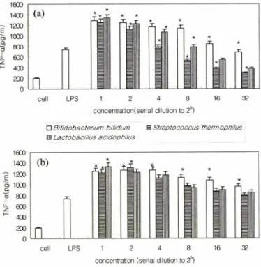
Fig. 2 - Effect of supernatants of Lactobacillus acidophilus, Strepto
coccus thermophilus, Bifidobacterium bifidum on TNF-a production in the LPS (lipopolysaccharide)- stimulatd RAW 264.7 cells, (a) without LPS *p<0.05, compared with cells, (b) with LPS. *p<0.05, compared with LPS.
Fig. 3 - Characterization of RAW 264.7 cell change in response to supernatants of Lactobacillus acidophilus, Streptococcus thermophilus and Bifidobacterium bifidum. The RAW 264.7 cells were cultured on cover slips in the presence of different concentration of supernatants of Lactobacillus acidophilus, Streptococcus thermophilus and Bifidobacterium bifidum. (a) only cells (b) with LPS (10 ng/m/). (c) Bifidobacterium bifidum (diluted solution to 22). (d) Lactobacillus acidophilus (diluted solution to 23). (e) Stereptococcus thermophilus (diluted solution to 2 )• (f) Bifidobacterium bifidum (diluted solution to 2 ) with LPS (10 ng/m/)
(e) (f)
(c) (d)
세
로 많은 양의 NO를 생성하였는데,특히 세 가지 유산균 중 Bifidobacterium bifidum 처리군에서 NO 생성이가장많았던것 으로 ^ 타났다.
Raw 264.7 세포의 배양액에처리한각각의 유산균농도를달
리하였을 때 Fig. 1과같이 NO 생성이 농도의존적으로증가 하였다. 22배희석한B ifidobacterium bifidum을 처리하였을 때 positive control group인 LPS(20 ng/m/)만 첨가한 group 보다 NO의양이 현저히 많이 생성되었음을확인할수있었다. LPS 와 세 가지 유 산 균 (Bifidobacterium bifidum , Lactobacillus acidophilus, Streptococcus thermophilusyi: 함께 처리한 group은 대식세포의 NO 생성에서상승효과(synergic effect)를나타내어 세가지 균주(Bifidobacterium bifidum , Lactobacillus acidophilus, Streptococcus thermophilus}각각을 처리한 group보다더많은양 의 NO가생성됨을확인할수 있었다.
TNF-a 정량
Raw 264.7 세포에의한 TNF-a의생성에서 유산균이미치는 효괴를조사하기위하여 LPS의존재에따라각 균주(Lactobacillus acidophilus, Streptococcus therm ophilus’ Bifidobacterium bifidum )
를 Raw 264.7세포에농도별로처리하여 배양시켰으며, 그결과
를 Fig. 2에나타내었다.
본연구결과,LPS를처리하지않은세포에서의 TNF-a의생 성이 가장낮았으며 오4배 희석한B ifi(M a d e riu m bifidum을 처 리한 group에서부터는 positive control로써 LPS(10 ng/m/)를처 리한 group보다 TNF-oc의생성이 농도의존적으로 점차증가하 는 것으로 나타났다. Lactobacillus acidophilus 오]■ Streptococcus therm ophilus 역시 22배희석한농도에서는 LPS(10 n^m/)만처 리한 group보다 TNF-a 생성이 증가된것을확인할수있었다.
유산균과 LPS(10 ng/m/)를함께처리한세포는유산균만처리한
세포들보다 TNF-oc의생성이크게증가하였다.
. I
(a) (b)
1 60 0 1 40 0 1 20 0 1 00 0 8 00 6 00 4 00
E/ecoB-
u-
Ni
■ i s
s i
i
i
llllllllllll
J. Pharm. Soc. Korea
RAW 264.7 대식세포에서의 유산균에 의한 Nitric Oxide 와 TNF-oc 의 생성 중가 효과 463
세포 형태 변호KCell m orphological change)
배지만처리한정상적인 RAW 264.7 세포는표면에 아무것도
나타나지 않은 Round 형태로 나타났으며 (Fig. 3a), 10 ng/m/의 LPS(Fig. 3b)를처리한 group과세 가지 유 산 균(Lactobacillus acidophilus, Streptococcus thermophilus,Bifidobacterium bifidum)
의상등액만처리한 group에서는 세포형태에서약간의 변화가
관찰되었다. 이들은 배지만처리한정상적인 RAW 264.7세포와 비교하였을때, 그크기가더크고 round 형태가아닌매끄럽지 못하고거친 표면을가진 것으로확인되었다. LPS와유산균을 같이 처리한 group은 LPS나유산균만처리한 group에비해서
RAW 264.7 세포의 형태가더커지고더거칠어져서 이들이 더
많이활성화되었음을짐작할수있었다.
결 론
Bifidobacteria룰: 포함한 유산균은 면역기능을 활성화 시키
며,31ᅵ33) 이러한활성은대식세포와 임파구의 활성을포함한몇 가지 면역기능을향상시키는것으로 나타났다.34-36)
대식세포는면역 반응의 초기 반응과 비특이적 면역반응을
담당하며,LPS 및 IFN-a와같은물질에 의하여특이적 활성이
유도된다. 본연구는유산균의 대식세포 활성화여부를확인하 고자혈관확장,신경 전달및숙주방어와같은다양한기능을 갖는 NO의생성정도와 대식세포의 mediator인 TNF-ot의생성 정도를조사하였다.
IFN-y와 LPS 등의 물질들은 대식세포를활성화하여 배지내
에 N 02를생성하는것으로알려졌으나37) 유산균역시 대식세포
를활성화시켜서 NO 생성과 TNF-a의생성을촉진하는것으로 났다.
Bifidobacterium bifidum, Lactobacillus acidophilus 및Sterep- tococcus thermophilus으] 세가지 유산균 모두 농도의존적으로
NO와 TNF-a의생성증가에 관여함을확인할수있었고,RAW
264.7 세포의 형태 역시 농도의존적으로 활성화되어 형태가
변화된것을관찰할수 있었다. 식세포의 활성인자인그람음성 균의 LPS만 첨가한 group보다 유산균 첨가군에서는 N O와
TNF-a의생성양이 현저히 많았으며, LPS와세 가지 유산균
{Bifidobacterium bifidum, Lactobacillus acidophilus,Streptococcus thermophilusyi 함께 처리한 group은대식세포의 NO와 TNF-a
생성에 상승 효과를 나타내어,세 가지 균 주(Bifidobacterium bifidum, Lactobacillus acidophilus, Streptococcus thermophilus) 만처리한 group보다많은양의 NO와 TNF-a를생성하는것으 로나타났다.
이와 같은 결과는,Lactobacillus acidophilus, Streptococcus thermophilus 및Bifidobacterium bifidumS] 세가지유산균이 대
식세포를자극하여 NO와 TNF-a를생성시켜서 면역기능을증
진시킨다는사실을시사하고있는것으로사료된다.
문 헌
1) Vandamme, E, Pot, B.,Gillis, M .,de Vos, R, Kersters, K. and Swings, J. : Polyphasic taxonomy, a consensus approach to bacterial systematics. Micro. Reviews 60,407 (1996).
2) Perdigon, G., de Macias, M. E., Alvarez, S., Oliver, G. and de Ruiz Holgado, A. A. : Effect of perorally administered lactobacilli on macrophage activation in mice. Infect. Immun.
53,404 (1986).
3) Lefrancois, L. : Basic aspects of intraepithelial lymphocyte immnobiology. In Handbook of Mucosal Immunology eds R L.
Ogra, J. Mestecky, M. E. Lamm, W. Strober, J. R. McGhee and J. Bienenstock, Academic Press, San Diego pp. 287 (1994).
4) Taguchi, T., Aicher, W. K.,Fujihashi, K., Yamamoto, M.,
McGhee, J. R., Bluestone, J. A. and Kiyono, H .: Novel function for intestinal intraepithelial lymphocytes: murine CD3+, y/8 TCR+ T cells produce IFN-y and IL-5. Joural of Immunology 147,3736 (1991).
5) Johansson, M. L.,Molin, G.’ Jeppsson, B.,Nobaek, S., Ahrne, S. and Bengmark, S .: Administration of different Lactobacillus strains in fermented oatmeal soup: in vivo colon isation of human intestinal mucosa and effect on the indigenous flora.
Applied and Environmental Microbiology 59,15 (1993).
6) Schiffrin, E. J., Brassart, D.,Servin, A., Rochat, F. and Dommet Hughes, A. : Immune modulation of blood leukocytes in humans by lactic acid bacteriarcriteria for strain selection.
American Journal of Clinical Nutrition 66,515S (1997).
7) Nussler,A. K. and Thomson, A. W. : Immunomodulatory agents in the laboratory and clinic. Parasitology 105, S5. (1992) 8) Sekine, K.,Watanabe-Sekine, E., Ohta, J., Toida, T., Tatsuki, T.,
Kawashima, T. and Hashimoto, Y. : Induction and activation of tumoricidal cells in vivo and in vitro by the bacterial cell wall of Bifidobacterium infantis. Bifidobact. MicrofL 13,65 (1994).
9) Hibbs, J. B.,Lambert, L. H. and Remington, J. S. : In vtiro nonimmunological destruction of cells with abnormal growth characteristics by adjuvant activated macrophages. Proc. Soc.
Exp. Biol. 139,1049 (1972).
10) Loewenstein, J., Rottem, S. and Gallily, R. : Induction of macrophage -mediated cytolysis of neoplastic cells by my coplasmas. Cell Immunol. 77,290 (1983).
11) Pissens, W. E, Churchill, W. H. and David, J. R. : Macrophages activated in vitro with lymphocyte mediators kill neoplastic but not normal cells. J. Immunol. 114,293 (1975).
12) Schultz, R. M .,Chirigos, M. A. and Heine, U. I. : Functional and morphological haracteristics of interferon-treated macrophage.
Cell. Immunol 35,84 (1978).
13) Adams, D. O. and Marini, R A. : Evidence for a multistep
464 박소희 • 정명준 • 김수동 • 백대헌 • 강병용 • 하순주
mechanism of cytolysis by G-activated macrophages: the interrelationship between the capacity for cytolysis, target binding, and secretion of cytolytic factor. J. Immunol. 126,981 (1981).
14) Carswell, E. A., Old, L. J., Kassel, R. L., Green, S., Fiore, N.
and Williamson, B. : An endotoxin-induced serum factor that causes necrosis of tumors. Proc. Natl Acad. Sci. USA 72,3666 (1975).
15) Cui, S., Jonathan, S., Reichner, Romeo, B., Mateo, and Albina, J. E .: Activated murine macrophage induce apoptosis in tumor cells through ntric oxide-dependent or independent mechanism.
Cancer Res. 54,2462 (1994).
16) Ichinose, Y., Bakouche, 0., Tsao, J. Y. and Fildler, I. J. : Tumor necrosis factor and IL-1 associated with plasma membrane of activated human monocytes lyse monokine-sensitive but not monokine-resistant tumor cells whereas viable activated monocytes lyse both. J. Immunol. 141, 512 (1988).
17) Suttles, J., Giri, J. G. and Mizel, S. B. : IL-1 secretion by macrophages. Enhancement of IL-1 secretion and processing by calcium inophores. J. Immunol. 144, 175 (1990).
18) Halliwell, B. and Cutteridge, J. M. C .: Oxygen toxicity, Oxygen radicals, transition metals and disease. Biochemical J. 219,1 (1984).
19) Moncada, S. and Higgs, A .: The arginine-nitric oxide pathway.
New Engl J. Med. 329,2002 (1993).
20) Johnson, W. J., Somers, S. D. and Adams, D. O. : Activation of macrophage for tumor cytotoxicity. Contemp. Top. Immunobiol.
14, 127 (1983).
21) Lorsbach, R. B., Murphy, W. J., Lowenstein, C. J., Snyder, S. H.’
and Russell, S. W. : Expression of the nitric oxide synthase gene in mouse macrophages activated for tumor cell killing.
Molecular basis for the synerge between interferon-gamma and lipopolysaccharide. J. Biol Chem. 268, 1908 (1993).
22) Bor-Sen Wang, Jia-Huey Chen, Yu-Chih Liang and Pin-Der Duh : Effects of Welsh onion on oxidation of low-density lipoprotein and nitric oxide production in macrophage cell line RAW 264.7. Food Chemistry 91,147 (2005).
23) Md, S., Moochhala, S. M. and Siew-Yang, K. L. : The role of inducible nitric oxide synthase inhibitor on the arteriolar hyporesponsiveness in hemorrhagic-shocked rats. Life Science 73’ 1825 (2003).
24) Oshima, H. and Bartsch, H. : Chronic infections and inflammatory processes as cancer risk factors; possible role of nitric oxide in carcinogenesis. Mutal. Res. 305,253 (1994).
25) Marin, M. L., Lee, J. H., Murtha, J., Ustunol, Z. and Pestka, J.
J. : Differential cytokine production in clonal macrophage and T-cell lines cultured with Bifidobacteria. J. Dairy Sci. 80,2713
(1997).
26) Ulch, T. T., Shin, S. S. and del Castillo, J. : Haematologic efffects of TNE J. Res. Immunol. 144,347 (1993).
27) Fukuo, K.,Inoue, T., Morimoto, S. Nakahashi, T., Yasuda, O.’
Kitano, S., Sasada, R. and Ogigara, T. : Nitric oxide mediates cytotoxicity and basic fibroblast growth factor release in cultured vascular smooth muscle cells. A possible mechanism of neo vascularization in atherosclerotic plaques./. Clin. Invest.
95,668 (1995).
28) Laskin, D. L. and Pendino, J. : Macrophages and inflammatory mediators in tissue injury. Annu. Rev. Pharmacol. Toxicol. 35, 655 (1995).
29) Sarih, M .,Souvannavong, V and Adam, A. : Nitric oxide synthase induces macrophage death by apoptosis. Biochem.
Biophys. Res. Commun. 191, 503 (1993).
30) Stuehr,D. J. and Nathan, C. F. : Nitric oxide, a macrophage product responsible for cytostasis and respiratory inhibition in tumor target cells. J. Exp. Med. 169,1543 (1989).
31) Lee, J., Amentani, A., Enomoto, A., Sato, Y., Motoshima, H.,
Ike, F. and Kaminogawa, S. : Screening for the immunopo- tentiating activity of food microorganism and enhancement of the immune response by Bifidobacterium adolescentis M101- 4. Siosci. Biotech. Biochem. 57,2127 (1993)
32) Kado-Oka, Y., Fujiwara, S. and Hirota, T. : Effects of bifidobacterial cells on mitogenic response of splenocytes and several functions of phagocytes. Milchwissenshaft 46, 626 (1991).
33) Gomez, E., Melgar, M. M., Silva, G. R, Portoles, A. and Gil, I . : Exocellular products from Bifidobacterium adolescentis as immunomodifiers in the lymphoproliferative response of mouse splenocytes. FEMS Microbiol. Lett. 56,47 (1988).
34) Hatcher, G. E. and Lambrecht, R. S. : Augmentation of macrophage phagocytic activity by cell-free extracts of selected lactic acid- producing bacteria. J. Dairy Sci. 76,2485 (1993).
35) Sekine, K., Watanabe-Sekine, E.,Ohta, J., Toida, T., Tatsuki, T.
K.,Kawashima, T. and Hashimoto, Y .: Induction and activation of tumoricidal cells in vivo and in vitro by the bacterial cell wall of Bifidobacterium infantis. Bifidobact. Microfl. 13,65 (1994).
36) Sekine, K., Watanabe-Sekine, E., Toida, T., Kasashima, T.
and Hashimoto, Y. : Adjuvant activity of the cell wall of Bifidobacteirium infantis for in vivo immune responses in mice. Immunopharmacol. Immunotoxicol. 16,589 (1994).
37) Chun, Q. C.,Assreuy, J.,Xu, D., Charles, I., Liew, F. Y. and Moncada, S. : Repeated induction of nitric oxide synthetase and leishmanicidal activity in murine macrophage. Eur. J.
Immunol. 23,1385 (1993).
J. Pharm. Soc. Korea